Giant cell tumor of bone: Difference between revisions
No edit summary |
No edit summary |
||
| Line 11: | Line 11: | ||
==Historical Perspective== | ==Historical Perspective== | ||
*In 1818, Cooper and Travers first described giant cell tumor (GCT) of bone.<ref name="pmid18322700">{{cite journal| author=Balke M, Schremper L, Gebert C, Ahrens H, Streitbuerger A, Koehler G et al.| title=Giant cell tumor of bone: treatment and outcome of 214 cases. | journal=J Cancer Res Clin Oncol | year= 2008 | volume= 134 | issue= 9 | pages= 969-78 | pmid=18322700 | doi=10.1007/s00432-008-0370-x | pmc= | url=https://www.ncbi.nlm.nih.gov/entrez/eutils/elink.fcgi?dbfrom=pubmed&tool=sumsearch.org/cite&retmode=ref&cmd=prlinks&id=18322700 }} </ref> | |||
*In 1845, Par H. Lebert described the first microscopic observations of multinucleated giant cells and fusiform cells as 'tumeur fiblastique'.<ref name="pmid7004712">{{cite journal| author=McCarthy EF| title=Giant-cell tumor of bone: an historical perspective. | journal=Clin Orthop Relat Res | year= 1980 | volume= | issue= 153 | pages= 14-25 | pmid=7004712 | doi= | pmc= | url=https://www.ncbi.nlm.nih.gov/entrez/eutils/elink.fcgi?dbfrom=pubmed&tool=sumsearch.org/cite&retmode=ref&cmd=prlinks&id=7004712 }} </ref> | *In 1845, Par H. Lebert described the first microscopic observations of multinucleated giant cells and fusiform cells as 'tumeur fiblastique'.<ref name="pmid7004712">{{cite journal| author=McCarthy EF| title=Giant-cell tumor of bone: an historical perspective. | journal=Clin Orthop Relat Res | year= 1980 | volume= | issue= 153 | pages= 14-25 | pmid=7004712 | doi= | pmc= | url=https://www.ncbi.nlm.nih.gov/entrez/eutils/elink.fcgi?dbfrom=pubmed&tool=sumsearch.org/cite&retmode=ref&cmd=prlinks&id=7004712 }} </ref> | ||
*In 1854, Sir James Paget provided the first description of the giant cell tumor in English literature.<ref name="pmid19970672">{{cite journal| author=Jaffe HL, Lichtenstein L| title=Benign Chondroblastoma of Bone: A Reinterpretation of the So-Called Calcifying or Chondromatous Giant Cell Tumor. | journal=Am J Pathol | year= 1942 | volume= 18 | issue= 6 | pages= 969-91 | pmid=19970672 | doi= | pmc=2032980 | url=https://www.ncbi.nlm.nih.gov/entrez/eutils/elink.fcgi?dbfrom=pubmed&tool=sumsearch.org/cite&retmode=ref&cmd=prlinks&id=19970672 }} </ref> | *In 1854, Sir James Paget provided the first description of the giant cell tumor in English literature.<ref name="pmid19970672">{{cite journal| author=Jaffe HL, Lichtenstein L| title=Benign Chondroblastoma of Bone: A Reinterpretation of the So-Called Calcifying or Chondromatous Giant Cell Tumor. | journal=Am J Pathol | year= 1942 | volume= 18 | issue= 6 | pages= 969-91 | pmid=19970672 | doi= | pmc=2032980 | url=https://www.ncbi.nlm.nih.gov/entrez/eutils/elink.fcgi?dbfrom=pubmed&tool=sumsearch.org/cite&retmode=ref&cmd=prlinks&id=19970672 }} </ref> | ||
*In early 1900, Bloodgood a surgeon at Johns Hopkins University, was credited with coining the term "giant-cell tumor" in his publication on radiographic features, conservative treatment, and use of bone grafts.<ref name="pmid17862876">{{cite journal| author=Bloodgood JC| title=II. The Conservative Treatment of Giant-Cell Sarcoma, with the Study of Bone Transplantation. | journal=Ann Surg | year= 1912 | volume= 56 | issue= 2 | pages= 210-39 | pmid=17862876 | doi= | pmc=1407379 | url=https://www.ncbi.nlm.nih.gov/entrez/eutils/elink.fcgi?dbfrom=pubmed&tool=sumsearch.org/cite&retmode=ref&cmd=prlinks&id=17862876 }} </ref> | *In early 1900, Bloodgood a surgeon at Johns Hopkins University, was credited with coining the term "giant-cell tumor" in his publication on radiographic features, conservative treatment, and use of bone grafts.<ref name="pmid17862876">{{cite journal| author=Bloodgood JC| title=II. The Conservative Treatment of Giant-Cell Sarcoma, with the Study of Bone Transplantation. | journal=Ann Surg | year= 1912 | volume= 56 | issue= 2 | pages= 210-39 | pmid=17862876 | doi= | pmc=1407379 | url=https://www.ncbi.nlm.nih.gov/entrez/eutils/elink.fcgi?dbfrom=pubmed&tool=sumsearch.org/cite&retmode=ref&cmd=prlinks&id=17862876 }} </ref> | ||
*In 1940, Jaffe and Lichtenstein described the clinical-radiographic-histologic identity of giant cell tumor.<ref name="pmid15595466">{{cite journal| author=Icihikawa K, Tanino R| title=Soft tissue giant cell tumor of low malignant potential. | journal=Tokai J Exp Clin Med | year= 2004 | volume= 29 | issue= 3 | pages= 91-5 | pmid=15595466 | doi= | pmc= | url=https://www.ncbi.nlm.nih.gov/entrez/eutils/elink.fcgi?dbfrom=pubmed&tool=sumsearch.org/cite&retmode=ref&cmd=prlinks&id=15595466 }} </ref><ref name="pmid21089702">{{cite journal| author=Gortzak Y, Kandel R, Deheshi B, Werier J, Turcotte RE, Ferguson PC et al.| title=The efficacy of chemical adjuvants on giant-cell tumour of bone. An in vitro study. | journal=J Bone Joint Surg Br | year= 2010 | volume= 92 | issue= 10 | pages= 1475-9 | pmid=21089702 | doi= | pmc= | url=https://www.ncbi.nlm.nih.gov/entrez/eutils/elink.fcgi?dbfrom=pubmed&tool=sumsearch.org/cite&retmode=ref&cmd=prlinks&id=21089702 }} </ref> | *In 1940, Jaffe and Lichtenstein described the clinical-radiographic-histologic identity of giant cell tumor.<ref name="pmid15595466">{{cite journal| author=Icihikawa K, Tanino R| title=Soft tissue giant cell tumor of low malignant potential. | journal=Tokai J Exp Clin Med | year= 2004 | volume= 29 | issue= 3 | pages= 91-5 | pmid=15595466 | doi= | pmc= | url=https://www.ncbi.nlm.nih.gov/entrez/eutils/elink.fcgi?dbfrom=pubmed&tool=sumsearch.org/cite&retmode=ref&cmd=prlinks&id=15595466 }} </ref><ref name="pmid21089702">{{cite journal| author=Gortzak Y, Kandel R, Deheshi B, Werier J, Turcotte RE, Ferguson PC et al.| title=The efficacy of chemical adjuvants on giant-cell tumour of bone. An in vitro study. | journal=J Bone Joint Surg Br | year= 2010 | volume= 92 | issue= 10 | pages= 1475-9 | pmid=21089702 | doi= | pmc= | url=https://www.ncbi.nlm.nih.gov/entrez/eutils/elink.fcgi?dbfrom=pubmed&tool=sumsearch.org/cite&retmode=ref&cmd=prlinks&id=21089702 }} </ref> | ||
==Pathophysiology== | ==Pathophysiology== | ||
The | *The exact [[pathogenesis]] of giant cell tumor (GCT) is not fully understood.<ref>{{cite book | last = Peabody | first = Terrance | title = Orthopaedic oncology : primary and metastatic tumors of the skeletal system | publisher = Springer | location = Cham | year = 2014 | isbn = 9783319073224 }}</ref> | ||
=== | *Various theories have been proposed concerning the [[pathogenesis]] of giant cell tumor: | ||
Giant cell tumor of bone typically occur | **Over-expression in RANK/RANKL signalling pathway with resultant over-proliferation of osteoclasts.<ref name="pmid15607894">{{cite journal| author=Liao TS, Yurgelun MB, Chang SS, Zhang HZ, Murakami K, Blaine TA et al.| title=Recruitment of osteoclast precursors by stromal cell derived factor-1 (SDF-1) in giant cell tumor of bone. | journal=J Orthop Res | year= 2005 | volume= 23 | issue= 1 | pages= 203-9 | pmid=15607894 | doi=10.1016/j.orthres.2004.06.018 | pmc= | url=https://www.ncbi.nlm.nih.gov/entrez/eutils/elink.fcgi?dbfrom=pubmed&tool=sumsearch.org/cite&retmode=ref&cmd=prlinks&id=15607894 }} </ref> | ||
**The stromal cells are believed to be the major neoplastic and proliferative component of giant cell tumor. | |||
**The stromal cells often make osteoids, thus have the potential to differentiate into osteoblastic cells. | |||
: | **Stromal cells in GCT secrete various chemokines including monocyte chemoattractant protein (MCP)-1 and SDF-1, which attract blood monocytes and stimulate their migration into tumor tissues. | ||
*Giant cell tumor of bone typically occur in the epiphyses of long bones. <ref name="ShrivastavaNawghare2008">{{cite journal|last1=Shrivastava|first1=Sandeep|last2=Nawghare|first2=Shishir P|last3=Kolwadkar|first3=Yogesh|last4=Singh|first4=Pradeep|title=Giant cell tumour in the diaphysis of radius – a report|journal=Cases Journal|volume=1|issue=1|year=2008|pages=106|issn=1757-1626|doi=10.1186/1757-1626-1-106}}</ref> | |||
*The bones often involved by epiphyseal GCT are distal femur, proximal tibia, and distal radius.<ref name="pmid1728946">{{cite journal| author=Bridge JA, Neff JR, Mouron BJ| title=Giant cell tumor of bone. Chromosomal analysis of 48 specimens and review of the literature. | journal=Cancer Genet Cytogenet | year= 1992 | volume= 58 | issue= 1 | pages= 2-13 | pmid=1728946 | doi= | pmc= | url=https://www.ncbi.nlm.nih.gov/entrez/eutils/elink.fcgi?dbfrom=pubmed&tool=sumsearch.org/cite&retmode=ref&cmd=prlinks&id=1728946 }} </ref> | |||
=== | ===Genetics=== | ||
* | *Mutation in the histone 3.3 gene H3F3A (p.Gly34)is seen in approximately 92% of giant cell tumours of bone.<ref name="pmid28505000">{{cite journal| author=Amary F, Berisha F, Ye H, Gupta M, Gutteridge A, Baumhoer D et al.| title=H3F3A (Histone 3.3) G34W Immunohistochemistry: A Reliable Marker Defining Benign and Malignant Giant Cell Tumor of Bone. | journal=Am J Surg Pathol | year= 2017 | volume= 41 | issue= 8 | pages= 1059-1068 | pmid=28505000 | doi=10.1097/PAS.0000000000000859 | pmc=5510691 | url=https://www.ncbi.nlm.nih.gov/entrez/eutils/elink.fcgi?dbfrom=pubmed&tool=sumsearch.org/cite&retmode=ref&cmd=prlinks&id=28505000 }} </ref><ref name="pmid26457357">{{cite journal| author=Cleven AH, Höcker S, Briaire-de Bruijn I, Szuhai K, Cleton-Jansen AM, Bovée JV| title=Mutation Analysis of H3F3A and H3F3B as a Diagnostic Tool for Giant Cell Tumor of Bone and Chondroblastoma. | journal=Am J Surg Pathol | year= 2015 | volume= 39 | issue= 11 | pages= 1576-83 | pmid=26457357 | doi=10.1097/PAS.0000000000000512 | pmc= | url=https://www.ncbi.nlm.nih.gov/entrez/eutils/elink.fcgi?dbfrom=pubmed&tool=sumsearch.org/cite&retmode=ref&cmd=prlinks&id=26457357 }} </ref><ref name="pmid8640728">{{cite journal| author=McComb EN, Johansson SL, Neff JR, Nelson M, Bridge JA| title=Chromosomal anomalies exclusive of telomeric associations in giant cell tumor of bone. | journal=Cancer Genet Cytogenet | year= 1996 | volume= 88 | issue= 2 | pages= 163-6 | pmid=8640728 | doi= | pmc= | url=https://www.ncbi.nlm.nih.gov/entrez/eutils/elink.fcgi?dbfrom=pubmed&tool=sumsearch.org/cite&retmode=ref&cmd=prlinks&id=8640728 }} </ref> | ||
*A subset of the mutations can be easily detected using a G34W mutation specific antibody. | |||
== | ==Causes== | ||
There are no established causes for giant cell tumor of the bone.<ref>{{cite book | last = Peabody | first = Terrance | title = Orthopaedic oncology : primary and metastatic tumors of the skeletal system | publisher = Springer | location = Cham | year = 2014 | isbn = 9783319073224 }}</ref> | |||
==Differentiating Giant cell tumor of bone from other Diseases== | ==Differentiating Giant cell tumor of bone from other Diseases== | ||
| Line 58: | Line 45: | ||
*[[Brown tumor]] of hyperparathyroidism | *[[Brown tumor]] of hyperparathyroidism | ||
*Non-ossifying fibroma | *Non-ossifying fibroma | ||
===Gross Pathology=== | |||
*Macroscopically, giant cell tumors are variable in appearance, depending on amount of [[hemorrhage]], presence of co-existent [[aneurysmal bone cyst]], and degree of presence [[fibrosis]]. | |||
===Microscopic Pathology=== | |||
*Giant cell tumor of bone is characterized by the presence of numerous Cathepsin-K producing, CD33 +, CD14 - multinucleated osteoclast-like giant cells and plump spindle-shaped stromal cells that represent the main proliferating cell population. | |||
*The spindle-shaped mononuclear cells are believed to represent the neoplastic population and are characterized at the cytogenetic level by telomeric associations and a peculiar telomere-protecting capping mechanism. | |||
*Areas of regressive change such as necrosis or fibrosis as well as extensive hemorrhage are frequently present. | |||
*Frequent mitotic figures in the mononuclear cells may be observed, especially in pregnant women or those on the [[oral contraceptive pill]] (due to increased hormone levels). | |||
==Epidemiology and Demographics== | |||
*In the United States and Europe, giant cell tumors(GCT) represent approximately 5% of all primary bone tumors and 21% of all benign bone tumors.<ref name="pmid12579271">{{cite journal |author=Gamberi G, Serra M, Ragazzini P, Magagnoli G, Pazzaglia L, Ponticelli F, Ferrari C, Zanasi M, Bertoni F, Picci P, Benassi MS |title=Identification of markers of possible prognostic value in 57 giant cell tumors of bone |journal=[[Oncology Reports]] |volume=10 |issue=2 |pages=351–6 |year=2003 |pmid=12579271 |doi= |url=http://www.spandidos-publications.com/or/10/2/351 |accessdate=2012-01-18}}</ref> | |||
*Giant cell tumors occur most commonly in the third decade of life; less than 5% of GCTs occur in patients who are skeletally immature.<ref name="pmid3805057">{{cite journal| author=Campanacci M, Baldini N, Boriani S, Sudanese A| title=Giant-cell tumor of bone. | journal=J Bone Joint Surg Am | year= 1987 | volume= 69 | issue= 1 | pages= 106-14 | pmid=3805057 | doi= | pmc= | url=https://www.ncbi.nlm.nih.gov/entrez/eutils/elink.fcgi?dbfrom=pubmed&tool=sumsearch.org/cite&retmode=ref&cmd=prlinks&id=3805057 }} </ref> | |||
*GCTs usually occur in patients older than 19 years. <ref name="pmid1609086">{{cite journal| author=Kransdorf MJ, Sweet DE, Buetow PC, Giudici MA, Moser RP| title=Giant cell tumor in skeletally immature patients. | journal=Radiology | year= 1992 | volume= 184 | issue= 1 | pages= 233-7 | pmid=1609086 | doi=10.1148/radiology.184.1.1609086 | pmc= | url=https://www.ncbi.nlm.nih.gov/entrez/eutils/elink.fcgi?dbfrom=pubmed&tool=sumsearch.org/cite&retmode=ref&cmd=prlinks&id=1609086 }} </ref> | |||
*Women are more commonly affected than men, with a 1.5:1 ratio.<ref name="pmid6833323">{{cite journal| author=Picci P, Manfrini M, Zucchi V, Gherlinzoni F, Rock M, Bertoni F et al.| title=Giant-cell tumor of bone in skeletally immature patients. | journal=J Bone Joint Surg Am | year= 1983 | volume= 65 | issue= 4 | pages= 486-90 | pmid=6833323 | doi= | pmc= | url=https://www.ncbi.nlm.nih.gov/entrez/eutils/elink.fcgi?dbfrom=pubmed&tool=sumsearch.org/cite&retmode=ref&cmd=prlinks&id=6833323 }} </ref> | |||
*There is no racial predilection to giant cell tumor. | |||
==Natural History, Complications and Prognosis== | ==Natural History, Complications and Prognosis== | ||
Revision as of 16:16, 27 December 2018
For patient information, click here
Editor-In-Chief: C. Michael Gibson, M.S., M.D. [1];Associate Editor(s)-in-Chief: Rohan A. Bhimani, M.B.B.S., D.N.B., M.Ch.[2]
Synonyms and keywords: Osteoclastoma; Giant cell myeloma; Giant cell tumor; Giant cell tumor of the bone
Overview
Giant cell tumor of bone is a relatively uncommon tumor of the bone. It is characterized by the presence of multinucleated giant cells (osteoclast-like cells). Giant cell tumor of bone accounts for 4-5% of primary bone tumors and 18.2% of benign bone tumors.[1] Giant cell tumor of bone almost invariably (97-99%) occur when the growth plate has closed and are therefore typically observed in early adulthood, with 80% of cases reported between the ages of 20 and 50, with a peak incidence between 20 and 30.[2] Giant cell tumor of bone typically occur as single lesions. They usually prefers the epiphyses of long bones. Although any bone can be affected, the most common sites are distal femur, proximal tibia, and distal radius. The progression to giant cell tumor of bone usually involves the over-expression in RANK/RANKL signalling pathway with resultant over-proliferation of osteoclasts. On gross pathology, hemorrhage, presence of co-existent aneurysmal bone cyst, and fibrosis are characteristic findings of giant cell tumor of bone. On microscopic histopathological analysis, prominent and diffuse osteoclastic giant cells and mononuclear cells with frequent mitotic figures are characteristic findings of giant cell tumor of bone. Symptoms of giant cell tumor of bone include localized pain, localized swelling, and decreased range of motion. Physical examination findings will depend on the location of the giant cell tumor. Common physical examination findings of giant cell tumor are localized swelling and tenderness at the site of the tumor. Giant cell tumor of bone must be differentiated from aneurysmal bone cyst, chondroblastoma, simple bone cyst, osteoid osteoma, osteoblastoma, osteosarcoma, and brown tumor of hyperparathyroidism. X-ray may be helpful in the diagnosis of giant cell tumor of bone. Common complications of giant cell tumor include malignant transformation, recurrence, and metastasis. Findings on x-ray suggestive of giant cell tumor include metaepiphyseal location of mass and grow to the articular surface of the involved bone with narrow zone of transition.[3] Surgery is the mainstay of treatment for giant cell tumor.
Historical Perspective
- In 1818, Cooper and Travers first described giant cell tumor (GCT) of bone.[4]
- In 1845, Par H. Lebert described the first microscopic observations of multinucleated giant cells and fusiform cells as 'tumeur fiblastique'.[5]
- In 1854, Sir James Paget provided the first description of the giant cell tumor in English literature.[6]
- In early 1900, Bloodgood a surgeon at Johns Hopkins University, was credited with coining the term "giant-cell tumor" in his publication on radiographic features, conservative treatment, and use of bone grafts.[7]
- In 1940, Jaffe and Lichtenstein described the clinical-radiographic-histologic identity of giant cell tumor.[8][9]
Pathophysiology
- The exact pathogenesis of giant cell tumor (GCT) is not fully understood.[10]
- Various theories have been proposed concerning the pathogenesis of giant cell tumor:
- Over-expression in RANK/RANKL signalling pathway with resultant over-proliferation of osteoclasts.[11]
- The stromal cells are believed to be the major neoplastic and proliferative component of giant cell tumor.
- The stromal cells often make osteoids, thus have the potential to differentiate into osteoblastic cells.
- Stromal cells in GCT secrete various chemokines including monocyte chemoattractant protein (MCP)-1 and SDF-1, which attract blood monocytes and stimulate their migration into tumor tissues.
- Giant cell tumor of bone typically occur in the epiphyses of long bones. [12]
- The bones often involved by epiphyseal GCT are distal femur, proximal tibia, and distal radius.[13]
Genetics
- Mutation in the histone 3.3 gene H3F3A (p.Gly34)is seen in approximately 92% of giant cell tumours of bone.[14][15][16]
- A subset of the mutations can be easily detected using a G34W mutation specific antibody.
Causes
There are no established causes for giant cell tumor of the bone.[17]
Differentiating Giant cell tumor of bone from other Diseases
Giant cell tumor of bone must be differentiated from:
- Aneurysmal bone cyst
- Chondroblastoma
- Simple bone cyst
- Osteoid osteoma
- Osteoblastoma
- Osteosarcoma
- Giant cell reparative granuloma
- Brown tumor of hyperparathyroidism
- Non-ossifying fibroma
Gross Pathology
- Macroscopically, giant cell tumors are variable in appearance, depending on amount of hemorrhage, presence of co-existent aneurysmal bone cyst, and degree of presence fibrosis.
Microscopic Pathology
- Giant cell tumor of bone is characterized by the presence of numerous Cathepsin-K producing, CD33 +, CD14 - multinucleated osteoclast-like giant cells and plump spindle-shaped stromal cells that represent the main proliferating cell population.
- The spindle-shaped mononuclear cells are believed to represent the neoplastic population and are characterized at the cytogenetic level by telomeric associations and a peculiar telomere-protecting capping mechanism.
- Areas of regressive change such as necrosis or fibrosis as well as extensive hemorrhage are frequently present.
- Frequent mitotic figures in the mononuclear cells may be observed, especially in pregnant women or those on the oral contraceptive pill (due to increased hormone levels).
Epidemiology and Demographics
- In the United States and Europe, giant cell tumors(GCT) represent approximately 5% of all primary bone tumors and 21% of all benign bone tumors.[1]
- Giant cell tumors occur most commonly in the third decade of life; less than 5% of GCTs occur in patients who are skeletally immature.[18]
- GCTs usually occur in patients older than 19 years. [19]
- Women are more commonly affected than men, with a 1.5:1 ratio.[20]
- There is no racial predilection to giant cell tumor.
Natural History, Complications and Prognosis
Complications
Common complications of giant cell tumor include:
- Malignant transformation
- Malignant transformation is far more common in men (M:F of ~3:1)
- Sarcomatous transformation is observed, especially in radiotherapy treated inoperable tumors.
- Recurrence
- Metastasis
- Giant cell tumor of bone may occasionally metastasize to vital organs such as the lung.[21] Hence, this entity has been called benign metastasising giant cell tumor.
Prognosis
- The prognosis of giant cell tumor is generally excellent.
Diagnosis
History and Symptoms
- Patients usually present with pain and limited range of motion caused by tumor's proximity to the joint space.
- There may be swelling as well, if the tumor has been growing for a long time.
- Some patients may be asymptomatic until they develop a pathologic fracture at the site of the tumor.
Physical Examination
Physical examination findings will depend on the location of the giant cell tumor. Most giant cell tumors are located in the long bone of extremities.
Extremities
A palpable firm non tender or tender mass may be appreciated on physical examination. The assessment of giant cell tumor during physical examination include:
X Ray
X-ray may be helpful in the diagnosis of giant cell tumor of bone. Findings on x-ray suggestive of giant cell tumor include:
- Metaphyseal location and grow to the articular surface of the involved bone
- Narrow zone of transition: a broader zone of transition is observed in more aggressive giant cell tumors
- No surrounding sclerosis: 80-85%
- Overlying cortex is thinned, expanded or deficient
- Periosteal reaction is only observed in 10-30% of cases
- Soft tissue mass is not infrequent
- Pathological fracture may be present
- No matrix calcification/mineralisation
(Images courtesy of RadsWiki)
-
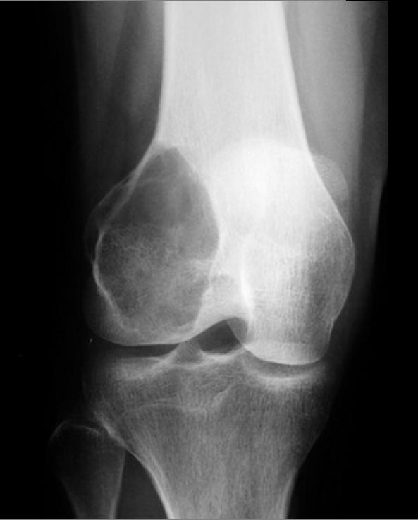
Giant cell tumor: Distal part of the femur
-
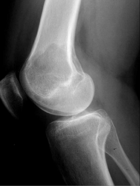
Giant cell tumor: Distal part of the femur


MRI
Typical signal characteristics on MRI of giant cell tumor of bone include:
T1:
- Low to intermediate solid component
- Low signal periphery
- Solid components enhance, helping distinguish giant cell tumor with aneurysmal bone cyst from pure aneurysmal bone cyst
- Some enhancement may also be observed in adjacent bone marrow
T2:
- Heterogenous high signal with areas of low signal intensity (variable) due to haemosiderin or fibrosis
- If an aneurysmal bone cyst component present, then fluid-fluid levels can be observed
- High signal in adjacent bone marrow thought to represent inflammatory edema
T1 C+ (Gd):
- Solid components will enhance, helping differentiate from aneurysmal bone cyst
Scintigraphy: Bone Scan
- Most giant cell tumors demonstrate increased uptake on delayed images, especially around the periphery, with a central photopenic region (doughnut sign).
- Increased blood pool activity is also observed, and can be observed in adjacent bones due to generalised regional hyperaemia.
Treatment
The treatment of giant cell tumor is directed towards local control without sacrificing joint function.[22] Surgery is the mainstay of treatment for giant cell tumor.
Surgery
- Classically, treatment is with curettage and packing with bone chips or polymethylmethacrylate (PMMA).
- Local recurrence is from the periphery of the lesion and has historically occurred in up to 40-60% of cases.
- Newer intraoperative adjuncts such as thermal or chemical treatment of the resection margins have lowered the recurrence rate to 2.5-10%.
- Wide local excision is associated with a lower recurrence rate, but has greater morbidity.
References
- ↑ 1.0 1.1 Gamberi G, Serra M, Ragazzini P, Magagnoli G, Pazzaglia L, Ponticelli F, Ferrari C, Zanasi M, Bertoni F, Picci P, Benassi MS (2003). "Identification of markers of possible prognostic value in 57 giant cell tumors of bone". Oncology Reports. 10 (2): 351–6. PMID 12579271. Retrieved 2012-01-18.
- ↑ Giant cell tumor of bone.Dr Henry Knipe and Dr Behrang Amini et al.Radiopaedia.org 2015.http://radiopaedia.org/articles/giant-cell-tumour-of-bone.Accessed on March 11, 2016
- ↑ Murphey MD, Nomikos GC, Flemming DJ, Gannon FH, Temple HT, Kransdorf MJ (2001). "From the archives of AFIP. Imaging of giant cell tumor and giant cell reparative granuloma of bone: radiologic-pathologic correlation". Radiographics : a Review Publication of the Radiological Society of North America, Inc. 21 (5): 1283–309. PMID 11553835. Retrieved 2012-01-18.
- ↑ Balke M, Schremper L, Gebert C, Ahrens H, Streitbuerger A, Koehler G; et al. (2008). "Giant cell tumor of bone: treatment and outcome of 214 cases". J Cancer Res Clin Oncol. 134 (9): 969–78. doi:10.1007/s00432-008-0370-x. PMID 18322700.
- ↑ McCarthy EF (1980). "Giant-cell tumor of bone: an historical perspective". Clin Orthop Relat Res (153): 14–25. PMID 7004712.
- ↑ Jaffe HL, Lichtenstein L (1942). "Benign Chondroblastoma of Bone: A Reinterpretation of the So-Called Calcifying or Chondromatous Giant Cell Tumor". Am J Pathol. 18 (6): 969–91. PMC 2032980. PMID 19970672.
- ↑ Bloodgood JC (1912). "II. The Conservative Treatment of Giant-Cell Sarcoma, with the Study of Bone Transplantation". Ann Surg. 56 (2): 210–39. PMC 1407379. PMID 17862876.
- ↑ Icihikawa K, Tanino R (2004). "Soft tissue giant cell tumor of low malignant potential". Tokai J Exp Clin Med. 29 (3): 91–5. PMID 15595466.
- ↑ Gortzak Y, Kandel R, Deheshi B, Werier J, Turcotte RE, Ferguson PC; et al. (2010). "The efficacy of chemical adjuvants on giant-cell tumour of bone. An in vitro study". J Bone Joint Surg Br. 92 (10): 1475–9. PMID 21089702.
- ↑ Peabody, Terrance (2014). Orthopaedic oncology : primary and metastatic tumors of the skeletal system. Cham: Springer. ISBN 9783319073224.
- ↑ Liao TS, Yurgelun MB, Chang SS, Zhang HZ, Murakami K, Blaine TA; et al. (2005). "Recruitment of osteoclast precursors by stromal cell derived factor-1 (SDF-1) in giant cell tumor of bone". J Orthop Res. 23 (1): 203–9. doi:10.1016/j.orthres.2004.06.018. PMID 15607894.
- ↑ Shrivastava, Sandeep; Nawghare, Shishir P; Kolwadkar, Yogesh; Singh, Pradeep (2008). "Giant cell tumour in the diaphysis of radius – a report". Cases Journal. 1 (1): 106. doi:10.1186/1757-1626-1-106. ISSN 1757-1626.
- ↑ Bridge JA, Neff JR, Mouron BJ (1992). "Giant cell tumor of bone. Chromosomal analysis of 48 specimens and review of the literature". Cancer Genet Cytogenet. 58 (1): 2–13. PMID 1728946.
- ↑ Amary F, Berisha F, Ye H, Gupta M, Gutteridge A, Baumhoer D; et al. (2017). "H3F3A (Histone 3.3) G34W Immunohistochemistry: A Reliable Marker Defining Benign and Malignant Giant Cell Tumor of Bone". Am J Surg Pathol. 41 (8): 1059–1068. doi:10.1097/PAS.0000000000000859. PMC 5510691. PMID 28505000.
- ↑ Cleven AH, Höcker S, Briaire-de Bruijn I, Szuhai K, Cleton-Jansen AM, Bovée JV (2015). "Mutation Analysis of H3F3A and H3F3B as a Diagnostic Tool for Giant Cell Tumor of Bone and Chondroblastoma". Am J Surg Pathol. 39 (11): 1576–83. doi:10.1097/PAS.0000000000000512. PMID 26457357.
- ↑ McComb EN, Johansson SL, Neff JR, Nelson M, Bridge JA (1996). "Chromosomal anomalies exclusive of telomeric associations in giant cell tumor of bone". Cancer Genet Cytogenet. 88 (2): 163–6. PMID 8640728.
- ↑ Peabody, Terrance (2014). Orthopaedic oncology : primary and metastatic tumors of the skeletal system. Cham: Springer. ISBN 9783319073224.
- ↑ Campanacci M, Baldini N, Boriani S, Sudanese A (1987). "Giant-cell tumor of bone". J Bone Joint Surg Am. 69 (1): 106–14. PMID 3805057.
- ↑ Kransdorf MJ, Sweet DE, Buetow PC, Giudici MA, Moser RP (1992). "Giant cell tumor in skeletally immature patients". Radiology. 184 (1): 233–7. doi:10.1148/radiology.184.1.1609086. PMID 1609086.
- ↑ Picci P, Manfrini M, Zucchi V, Gherlinzoni F, Rock M, Bertoni F; et al. (1983). "Giant-cell tumor of bone in skeletally immature patients". J Bone Joint Surg Am. 65 (4): 486–90. PMID 6833323.
- ↑ Muheremu, Aikeremujiang; Niu, Xiaohui (2014). "Pulmonary metastasis of giant cell tumor of bones". World Journal of Surgical Oncology. 12 (1): 261. doi:10.1186/1477-7819-12-261. ISSN 1477-7819.
- ↑ Puri, Ajay; Agarwal, Manish (2007). "Treatment of giant cell tumor of bone: Current concepts". Indian Journal of Orthopaedics. 41 (2): 101. doi:10.4103/0019-5413.32039. ISSN 0019-5413.