Candida vulvovaginitis pathophysiology: Difference between revisions
m (Robot: Automated text replacement (-\<youtube v=(.+)\/\> +{{#ev:youtube|\1}})) |
No edit summary |
||
| Line 1: | Line 1: | ||
__NOTOC__ | |||
{{Candidiasis}} | {{Candidiasis}} | ||
{{CMG}} | {{CMG}} | ||
==Overview== | ==Overview== | ||
==Pathophysiology== | |||
<div align="left"> | <div align="left"> | ||
| Line 51: | Line 50: | ||
</div> | </div> | ||
'''Histopathology''' | |||
'''Candidiasis of Esophagus & Colon''' | |||
{{#ev:youtube|-E-HwjCm2h8}} | {{#ev:youtube|-E-HwjCm2h8}} | ||
'''Histopathological Findings''' | |||
[http://www.peir.net Images courtesy of Professor Peter Anderson DVM PhD and published with permission © PEIR, University of Alabama at Birmingham, Department of Pathology] | [http://www.peir.net Images courtesy of Professor Peter Anderson DVM PhD and published with permission © PEIR, University of Alabama at Birmingham, Department of Pathology] | ||
| Line 85: | Line 84: | ||
<br clear="left"/> | <br clear="left"/> | ||
'''Autopsy Findings''' | |||
At autopsy, there was evidence of disseminated candidiasis. | At autopsy, there was evidence of disseminated candidiasis. | ||
| Line 92: | Line 91: | ||
{{Reflist}} | {{Reflist}} | ||
== | ==Related Chapters== | ||
* [[Candida albicans]] | * [[Candida albicans]] | ||
* [[Oral candidiasis]] | * [[Oral candidiasis]] | ||
Revision as of 18:08, 16 October 2012
|
Candidiasis Main page |
Editor-In-Chief: C. Michael Gibson, M.S., M.D. [1]
Overview
Pathophysiology
-
-
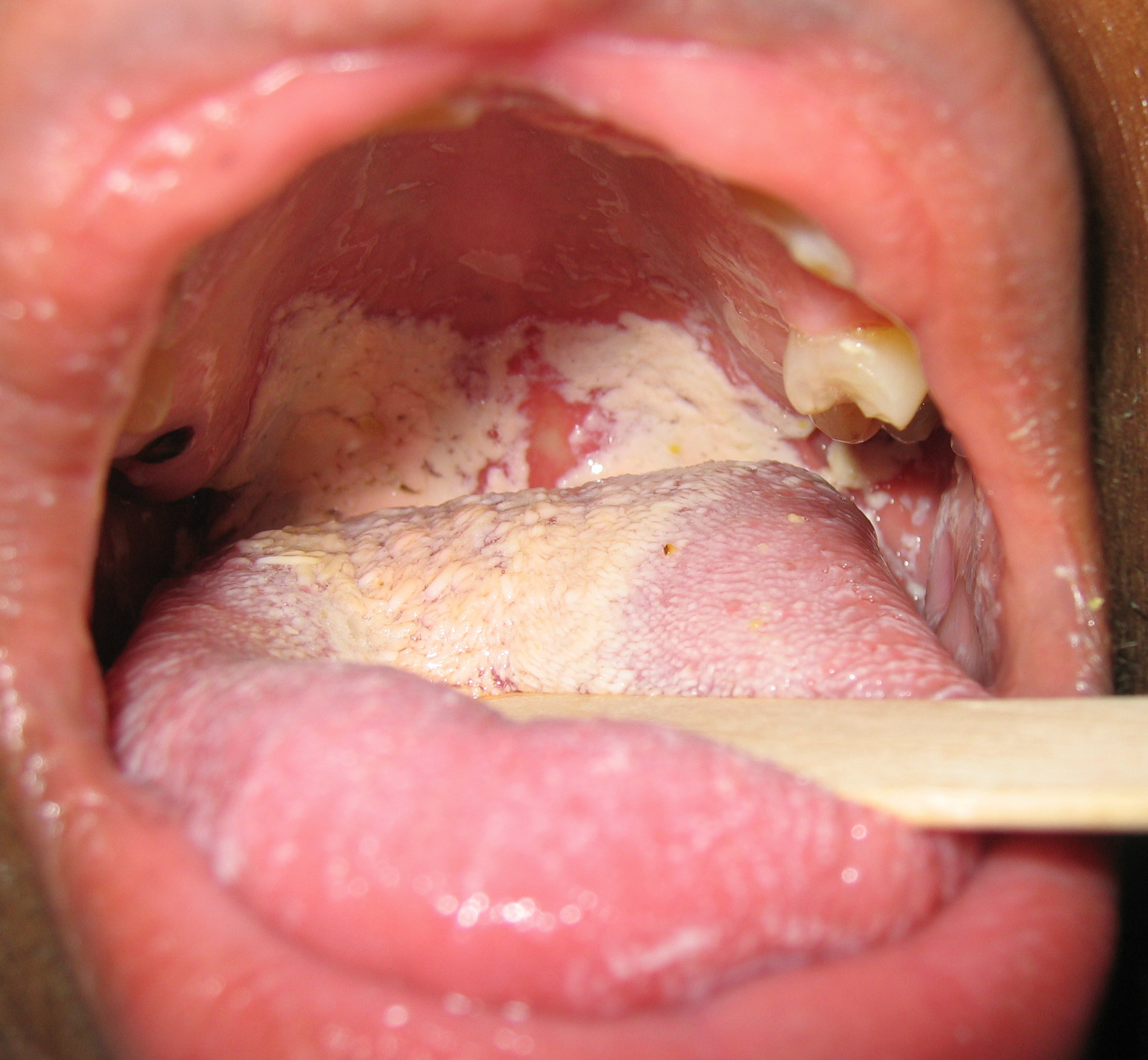
Oral manifestations of HIV infection and AIDS. Chronic oral candidiasis in patient with AIDS. Image courtesy of Professor Peter Anderson DVM PhD and published with permission. © PEIR, University of Alabama at Birmingham, Department of Pathology


-
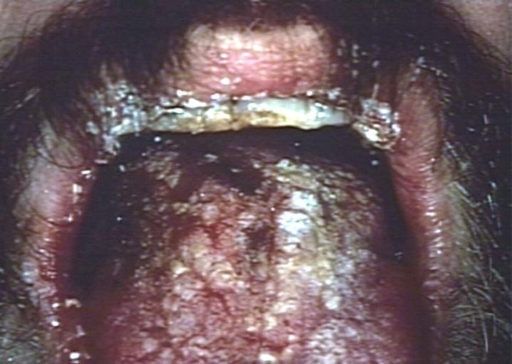
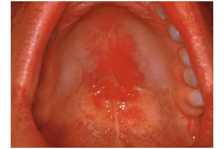
Soft palate showing extensive oral candidiasis in patient with AIDS. Image courtesy of Professor Peter Anderson DVM PhD and published with permission. © PEIR, University of Alabama at Birmingham, Department of Pathology
-
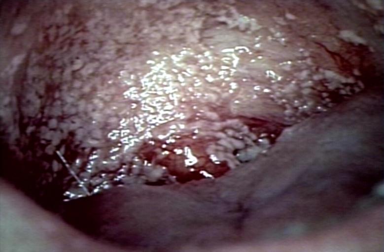
Oral candidiasis Image courtesy of Professor Peter Anderson DVM PhD and published with permission. © PEIR, University of Alabama at Birmingham, Department of Pathology


-
Eczema secondary to candidiasis. Image courtesy of Professor Peter Anderson DVM PhD and published with permission. © PEIR, University of Alabama at Birmingham, Department of Pathology
-
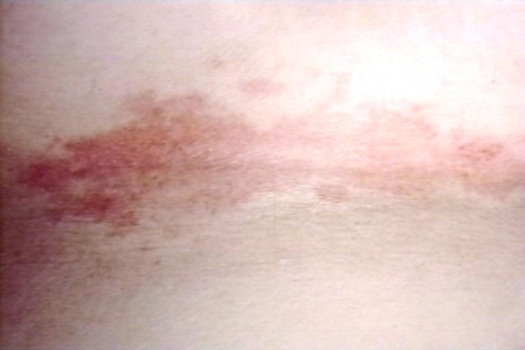
Candidiasis; skinfold. Image courtesy of Professor Peter Anderson DVM PhD and published with permission. © PEIR, University of Alabama at Birmingham, Department of Pathology


-
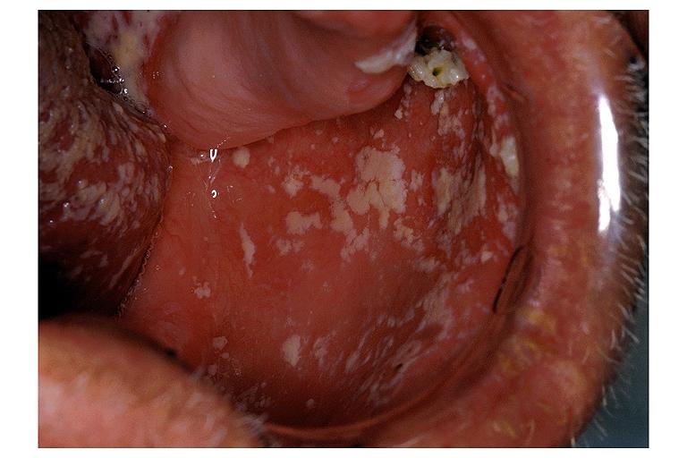
Erythematous candidiasis. Image courtesy of Professor Peter Anderson DVM PhD and published with permission. © PEIR, University of Alabama at Birmingham, Department of Pathology
-
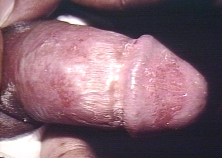
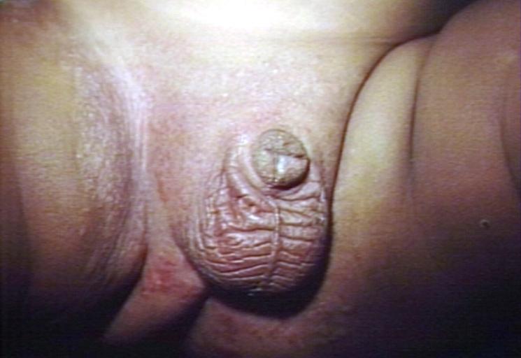
Genital candidiasis. Image courtesy of Professor Peter Anderson DVM PhD and published with permission. © PEIR, University of Alabama at Birmingham, Department of Pathology


-
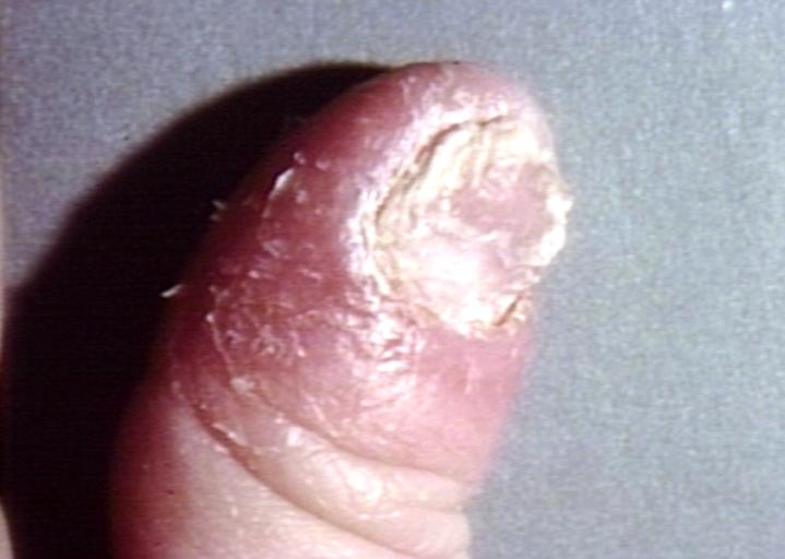
Paronychia: Another manifestation of candidiasis. Image courtesy of Professor Peter Anderson DVM PhD and published with permission. © PEIR, University of Alabama at Birmingham, Department of Pathology
-
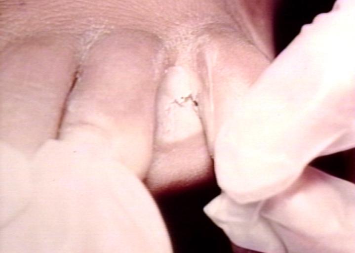
Interdigital candidiasis. Image courtesy of Professor Peter Anderson DVM PhD and published with permission. © PEIR, University of Alabama at Birmingham, Department of Pathology


-
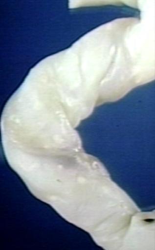
Candidiasis of umblical cord. White spots of colonies are present. Image courtesy of Professor Peter Anderson DVM PhD and published with permission. © PEIR, University of Alabama at Birmingham, Department of Pathology
-
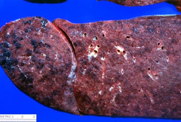
Lung: Candidiasis. Postmortem findings. Image courtesy of Professor Peter Anderson DVM PhD and published with permission. © PEIR, University of Alabama at Birmingham, Department of Pathology


Histopathology
Candidiasis of Esophagus & Colon
{{#ev:youtube|-E-HwjCm2h8}}
Histopathological Findings


Autopsy Findings
At autopsy, there was evidence of disseminated candidiasis.
References
Related Chapters