Candida vulvovaginitis pathophysiology: Difference between revisions
(Created page with "{{Candidiasis}} '''For patient information click here''' {{CMG}} ==Overview== '''Candidiasis''', commonly called '''yeast infection'''...") |
No edit summary |
||
| Line 56: | Line 56: | ||
<youtube v=-E-HwjCm2h8/> | <youtube v=-E-HwjCm2h8/> | ||
===Histopathological Findings=== | |||
[http://www.peir.net Images courtesy of Professor Peter Anderson DVM PhD and published with permission © PEIR, University of Alabama at Birmingham, Department of Pathology] | |||
[[Image:Renal candidiasis 001.jpg|left|thumb|350px|This autopsy photograph of the kidneys demonstrates the multifocal punctate lesions visible on the serosal surface (arrows). Don't confuse these small yellow punctate lesions with the fat that is adherent to the renal capsule. ]] | |||
<br clear="left"/> | |||
[[Image:Renal candidiasis 002.jpeg|left|thumb|350px|This photograph of the cut surface of these kidneys shows that these multifocal punctate lesions are primarily in the cortex (arrows).]] | |||
<br clear="left"/> | |||
[[Image:Renal candidiasis 003.jpeg|left|thumb|350px|This is a low-power photomicrograph of lymph node with three prominent areas of Candida colonies (arrows). Even at this low magnification, the purple-staining yeast and pseudohyphae can be easily seen. This section was stained with Periodic Acid-Schiff Hematoxylin (PASH), which stains the cell wall of fungi to make them more easily visible.]] | |||
<br clear="left"/> | |||
[[Image:Renal candidiasis 004.jpeg|left|thumb|350px|This is a low-power photomicrograph of one of the Candida colonies from this lymph node. The chains of yeast which are termed "pseudohyphae" are apparent at this magnification. ]] | |||
<br clear="left"/> | |||
[[Image:Renal candidiasis 005.jpeg|left|thumb|350px|This higher-power photomicrograph shows the yeasts and pseudohyphae in this focus of Candida organisms. ]] | |||
<br clear="left"/> | |||
[[Image:Renal candidiasis 006.jpeg|left|thumb|350px|This high-power photomicrograph shows the yeasts (1) and pseudohyphae (2). ]] | |||
<br clear="left"/> | |||
[[Image:Renal candidiasis 007.jpeg|left|thumb|350px|This is a low-power photomicrograph of the kidney from this same case. Note the Candida colonies (arrows). The pseudohyphae are evident around the periphery of these colonies even at this low magnification. ]] | |||
<br clear="left"/> | |||
[[Image:Renal candidiasis 008.jpeg|left|thumb|350px|This is a higher-power photomicrograph of a Candida colony in the kidney. Note the pseudohyphae of the Candida organisms. ]] | |||
<br clear="left"/> | |||
==References== | ==References== | ||
Revision as of 20:55, 10 January 2012
|
Candidiasis Main page |
For patient information click here
Editor-In-Chief: C. Michael Gibson, M.S., M.D. [1]
Overview
Candidiasis, commonly called yeast infection or thrush, is a fungal infection (mycosis) of any of the Candida species, of which Candida albicans is the most common.[1][2] Candidiasis thereby encompasses infections that range from superficial, such as oral thrush and vaginitis, to systemic and potentially life-threatening diseases.
Gross Images
-
-
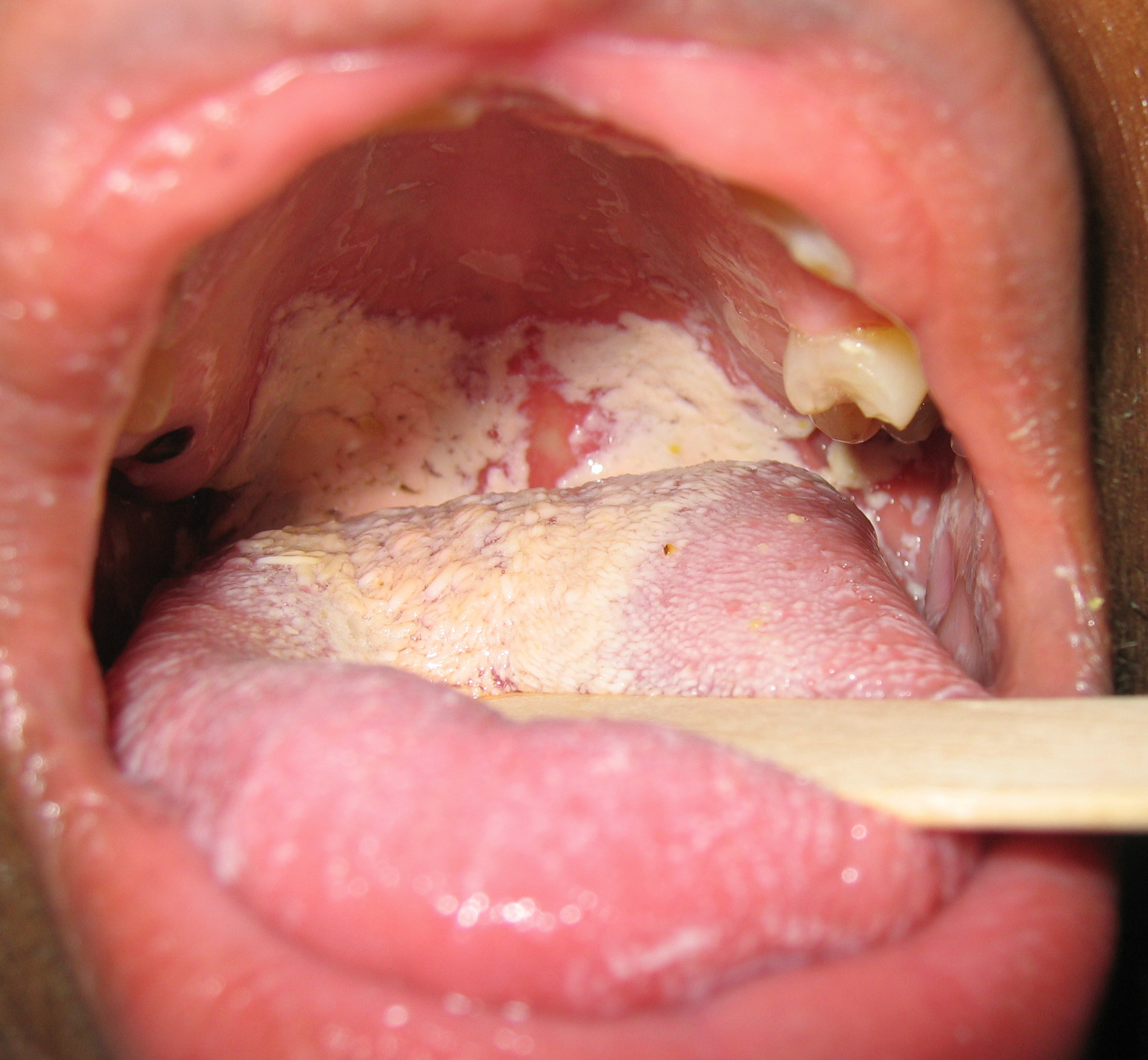
Oral manifestations of HIV infection and AIDS. Chronic oral candidiasis in patient with AIDS. Image courtesy of Professor Peter Anderson DVM PhD and published with permission. © PEIR, University of Alabama at Birmingham, Department of Pathology


-
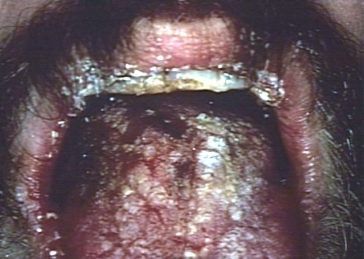
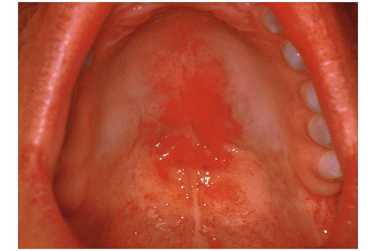
Soft palate showing extensive oral candidiasis in patient with AIDS. Image courtesy of Professor Peter Anderson DVM PhD and published with permission. © PEIR, University of Alabama at Birmingham, Department of Pathology
-
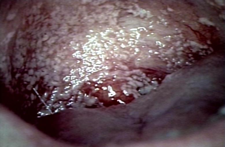
Oral candidiasis Image courtesy of Professor Peter Anderson DVM PhD and published with permission. © PEIR, University of Alabama at Birmingham, Department of Pathology


-
Eczema secondary to candidiasis. Image courtesy of Professor Peter Anderson DVM PhD and published with permission. © PEIR, University of Alabama at Birmingham, Department of Pathology
-
Candidiasis; skinfold. Image courtesy of Professor Peter Anderson DVM PhD and published with permission. © PEIR, University of Alabama at Birmingham, Department of Pathology


-
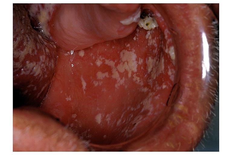
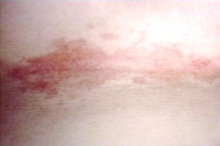
Erythematous candidiasis. Image courtesy of Professor Peter Anderson DVM PhD and published with permission. © PEIR, University of Alabama at Birmingham, Department of Pathology
-
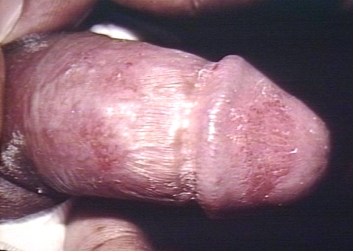
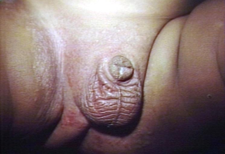
Genital candidiasis. Image courtesy of Professor Peter Anderson DVM PhD and published with permission. © PEIR, University of Alabama at Birmingham, Department of Pathology


-
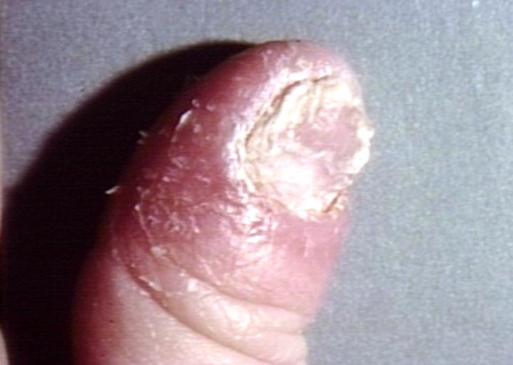
Paronychia: Another manifestation of candidiasis. Image courtesy of Professor Peter Anderson DVM PhD and published with permission. © PEIR, University of Alabama at Birmingham, Department of Pathology
-
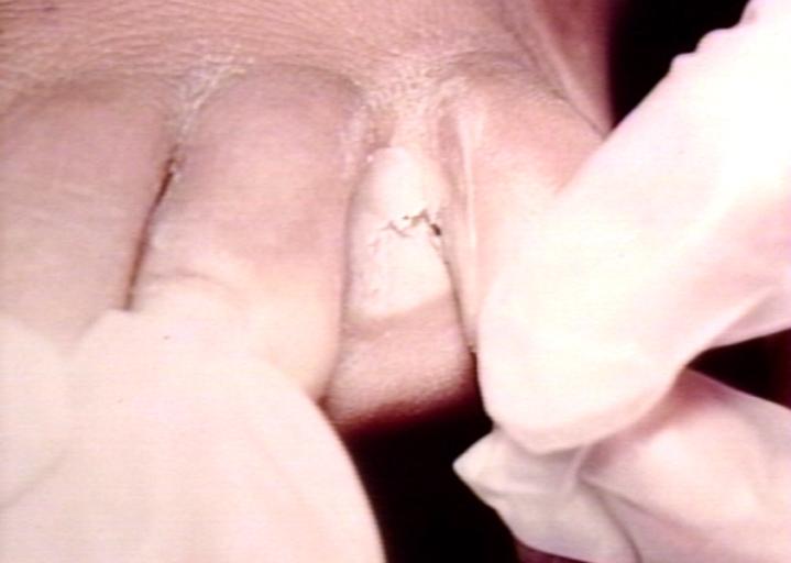
Interdigital candidiasis. Image courtesy of Professor Peter Anderson DVM PhD and published with permission. © PEIR, University of Alabama at Birmingham, Department of Pathology


-
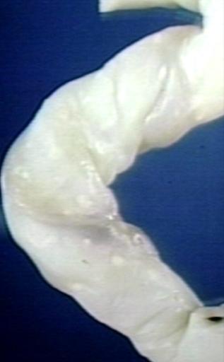
Candidiasis of umblical cord. White spots of colonies are present. Image courtesy of Professor Peter Anderson DVM PhD and published with permission. © PEIR, University of Alabama at Birmingham, Department of Pathology
-
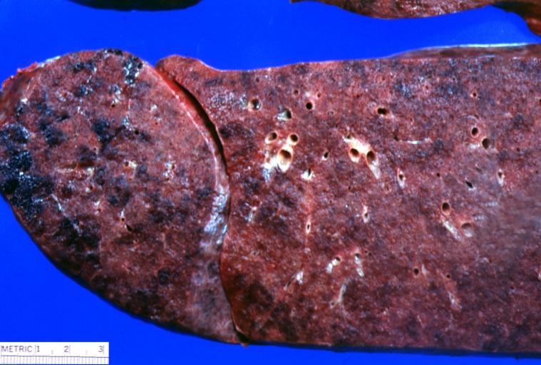
Lung: Candidiasis. Postmortem findings. Image courtesy of Professor Peter Anderson DVM PhD and published with permission. © PEIR, University of Alabama at Birmingham, Department of Pathology


Histopathology
Candidiasis of Esophagus & Colon
<youtube v=-E-HwjCm2h8/>
Histopathological Findings


References
- ↑ Walsh TJ, Dixon DM (1996). "Deep Mycoses". In Baron S et al eds. Baron's Medical Microbiology (via NCBI Bookshelf) (4th ed. ed.). Univ of Texas Medical Branch. ISBN 0-9631172-1-1.
- ↑ MedlinePlus Encyclopedia Vaginal yeast infection
See Also