Brain abscess MRI: Difference between revisions
Jump to navigation
Jump to search
No edit summary |
(→MRI) |
||
| Line 6: | Line 6: | ||
==MRI== | ==MRI== | ||
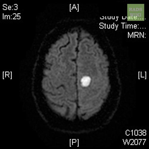
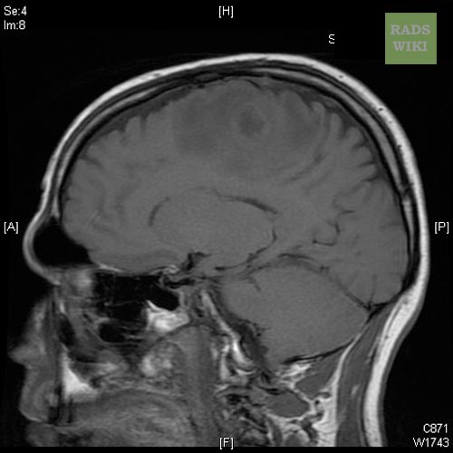
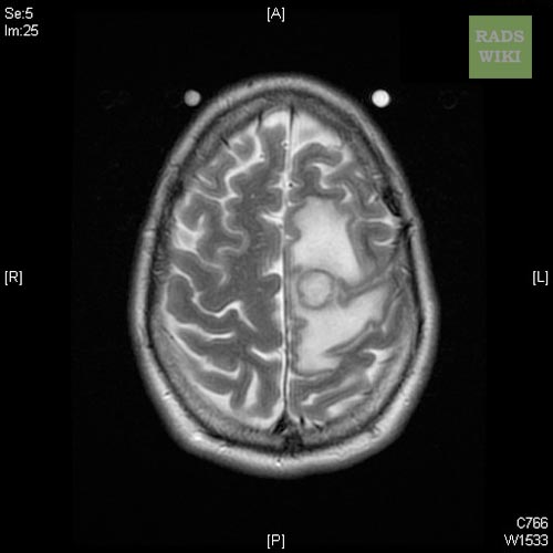
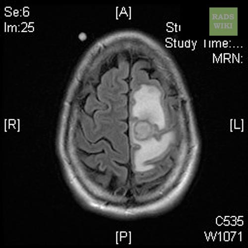
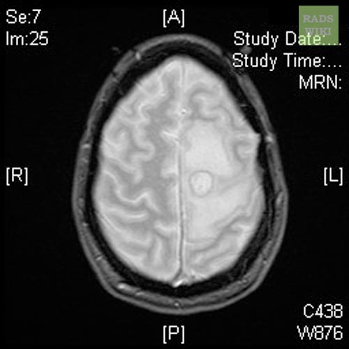
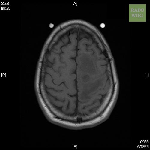
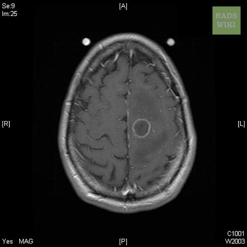
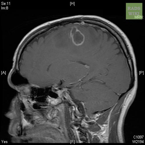
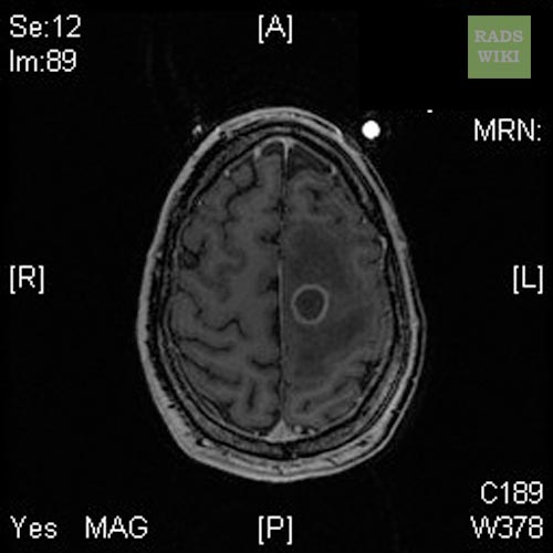
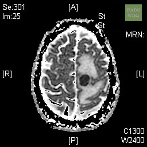
*[[MRI]] is generally thought to be the imaging modality of choice and can more accurately stage the abscess and gauge the response to therapy. | |||
:* T1 images are similar to [[CT]], with a central hypodense signal and surrounding ring-enhancement. | |||
:* T2 images reveal a central hyperintense area surrounded by a well-defined hypointense capsule with surrounding [[edema]]. | |||
([http://www.radswiki.net Images courtesy of RadsWiki]) | ([http://www.radswiki.net Images courtesy of RadsWiki]) | ||
Revision as of 20:17, 4 December 2012
Editor-In-Chief: C. Michael Gibson, M.S., M.D. [1]
|
Brain abscess Microchapters |
|
Diagnosis |
|
Treatment |
|
Case Studies |
|
Brain abscess MRI On the Web |
|
American Roentgen Ray Society Images of Brain abscess MRI |
Please help WikiDoc by adding more content here. It's easy! Click here to learn about editing.
MRI
- MRI is generally thought to be the imaging modality of choice and can more accurately stage the abscess and gauge the response to therapy.
-
Brain abscess
-
Brain abscess
-
Brain abscess
-
Brain abscess
-
Brain abscess
-
Brain abscess
-
Brain abscess
-
Brain abscess
-
Brain abscess
-
Brain abscess


