Atrial septal defect chest x ray
|
Atrial Septal Defect Microchapters | |
|
Treatment | |
|---|---|
|
Surgery | |
|
| |
|
Special Scenarios | |
|
Case Studies | |
|
Atrial septal defect chest x ray On the Web | |
|
American Roentgen Ray Society Images of Atrial septal defect chest x ray | |
|
Risk calculators and risk factors for Atrial septal defect chest x ray | |
Editor-In-Chief: C. Michael Gibson, M.S., M.D. [1]; Associate Editor(s)-In-Chief: Priyamvada Singh, M.B.B.S. [[2]]; Cafer Zorkun, M.D., Ph.D. [3]; Assistant Editor(s)-In-Chief: Kristin Feeney, B.S. [[4]]
Overview
Although not the most preferred methodology, chest x rays may be used as a diagnostic tool in the evaluation of an atrial septal defect. Diagnostic findings may include enlargement of the atrial border or cardiomegaly.
- ==Chest X-ray==
Findings of Chest X-ray seen in atrial septal defects are-
Enlarged right atrial border or cardiomegaly, if significant pulmonary hypertension is present.
- In secundum ASD with large left-to-right shunt - Chest x-ray may show findings suggestive of cardiac enlargement and increased pulmonary vascularity. The increase in pulmonary vascularity typically extends to the periphery of the lung fields, and the pulmonary trunk and central branches appear dilated.
- Triangular appearance of heart- This is seen because the enlarged pulmonary arteries prevents ascending and transverse aorta from forming the normal heart borders.
- Enlargement of the right side of heart, however the left atrium and left ventricle are normal.
- Sinus venosus defect a scimitar sign may be seen. This occurs due to the insertion of the pulmonary vein into the inferior vena cava and can be appreciated as abnormal densities on the chest X-ray. The scimitar sign is a vertical, gently curved, right-sided paracardiac density.
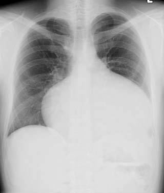
Imaging
-
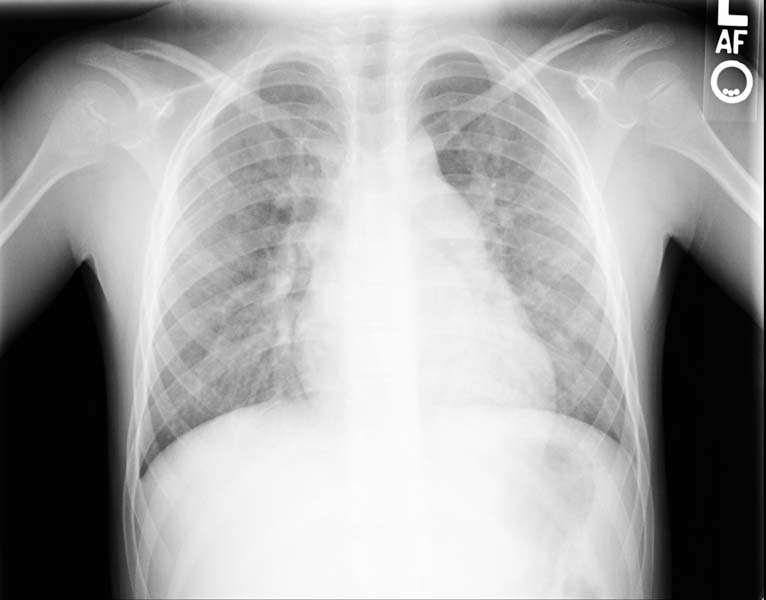
Enlarged right atrial border and mild cardiomegaly.
-
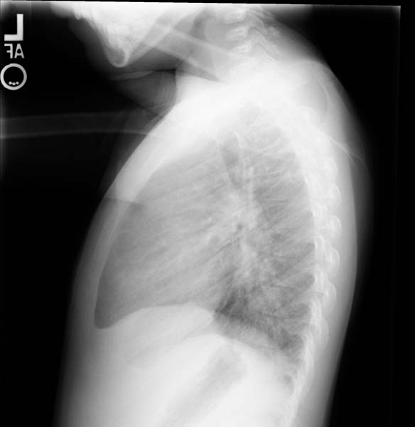
Lateral view
-
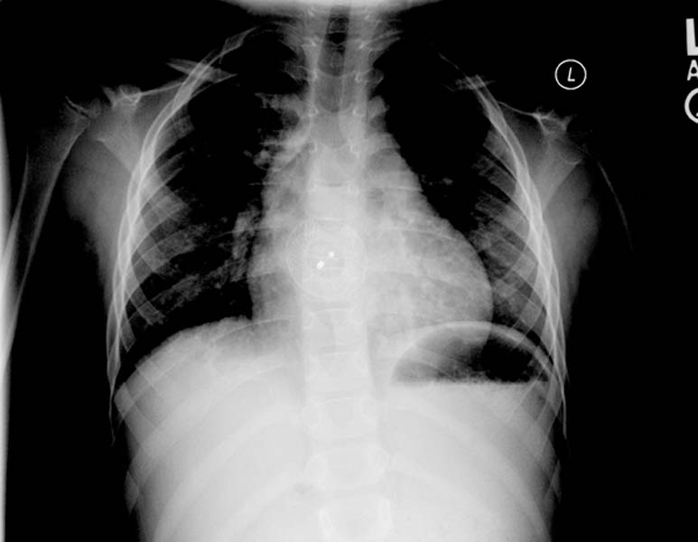
Post repair. Enlarged right atrial border and mild cardiomegaly.
-
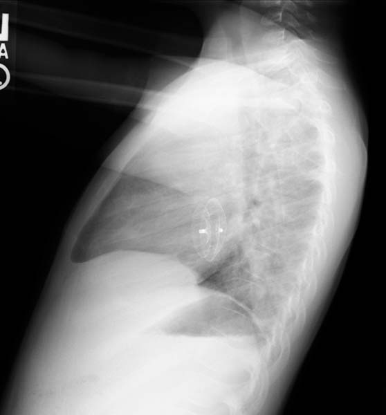
Post repair. Lateral view.
-
ASD. Another patient. Enlarged right atrial border and advanced cardiomegaly.


