Atrial septal defect chest x ray: Difference between revisions
No edit summary |
m (Bot: Adding CME Category::Cardiology) |
||
| (14 intermediate revisions by 5 users not shown) | |||
| Line 1: | Line 1: | ||
__NOTOC__ | |||
{{Atrial septal defect}} | {{Atrial septal defect}} | ||
{{CMG}}; '''Associate Editor(s)-In-Chief:''' [[Priyamvada Singh|Priyamvada Singh, M.B.B.S.]] [mailto: | {{CMG}}; '''Associate Editor(s)-In-Chief:''' [[Priyamvada Singh|Priyamvada Singh, M.B.B.S.]] [mailto:psingh13579@gmail.com]; {{CZ}}; '''Assistant Editor(s)-In-Chief:''' [[Kristin Feeney|Kristin Feeney, B.S.]] [mailto:kfeeney@elon.edu] | ||
==Overview== | ==Overview== | ||
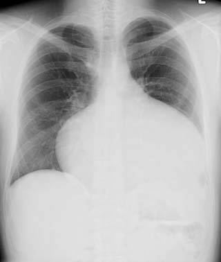
Chest x rays may | Chest x rays may detect an atrial septal defect. Chest x rays can be limited in imaging quality and may only supplement other imaging modalities. The chest x-ray may demonstrate [[cardiomegaly]] (right ventricle and [[right atrial enlargement]]), a prominent [[pulmonary artery]] segment and increased pulmonary vascular markings. | ||
==Chest X Ray== | ==Chest X Ray== | ||
| Line 9: | Line 10: | ||
===Common Findings=== | ===Common Findings=== | ||
CXR findings on an anteroposterior view of the chest x-ray in atrial septal defect may include: <ref name="Abdulla">Abdulla, Ra-id. (2011). Heart Diseases in Children: A Pediatrician's Guide. Springer.</ref> | |||
1) Prominent [[pulmonary artery]], increased pulmonary vascular markings. | |||
2) [[Cardiomegaly]] due to [[right atrial enlargement|right atrial]] and ventricular enlargement. | |||
3)' Triangular appearance of the [[heart]] | |||
* Results from enlargement of pulmonary arteries preventing the ascending and transverse aorta from forming normal heart borders | * Results from enlargement of [[pulmonary arteries]] preventing the ascending and transverse [[aorta]] from forming normal heart borders. | ||
4) [[Scimitar syndrome|Scimitar sign]] | |||
* A vertical, modestly curved, density in the right-side of the pericardium, may be visible | * A vertical, modestly curved, density in the right-side of the [[pericardium]], may be visible. | ||
* Commonly associated with the [[Atrial septal defect sinus venosus|sinus venosus]] atrial septal defect | * Commonly associated with the [[Atrial septal defect sinus venosus|sinus venosus]] atrial septal defect. | ||
* Results from the point of insertion of the pulmonary vein into the [[inferior vena cava]] | * Results from the point of insertion of the [[pulmonary vein]] into the [[inferior vena cava]]. | ||
* May cause abnormal densities within the chest x ray | * May cause abnormal densities within the [[chest x ray]]. | ||
5) Dilatation of the [[superior vena cava]] can be seen in [[Atrial septal defect sinus venosus|sinus venosus]] | |||
===Less Common Findings=== | ===Less Common Findings=== | ||
| Line 32: | Line 32: | ||
* [[Pulmonary edema]] | * [[Pulmonary edema]] | ||
* [[Hypertension|Pulmonary venous hypertension]] | * [[Hypertension|Pulmonary venous hypertension]] | ||
===Imagings=== | |||
=== | |||
<div align="left"> | <div align="left"> | ||
<gallery heights="175" widths="175"> | <gallery heights="175" widths="175"> | ||
| Line 49: | Line 48: | ||
{{WH}} | {{WH}} | ||
{{WS}} | {{WS}} | ||
[[CME Category::Cardiology]] | |||
[[Category:Cardiology]] | |||
[[Category:Congenital heart disease]] | |||
[[Category:Pediatrics]] | |||
[[Category:Embryology]] | |||
[[Category:Disease]] | |||
[[Category:Best pages]] | |||
Latest revision as of 01:44, 15 March 2016
|
Atrial Septal Defect Microchapters | |
|
Treatment | |
|---|---|
|
Surgery | |
|
| |
|
Special Scenarios | |
|
Case Studies | |
|
Atrial septal defect chest x ray On the Web | |
|
American Roentgen Ray Society Images of Atrial septal defect chest x ray | |
|
Risk calculators and risk factors for Atrial septal defect chest x ray | |
Editor-In-Chief: C. Michael Gibson, M.S., M.D. [1]; Associate Editor(s)-In-Chief: Priyamvada Singh, M.B.B.S. [2]; Cafer Zorkun, M.D., Ph.D. [3]; Assistant Editor(s)-In-Chief: Kristin Feeney, B.S. [4]
Overview
Chest x rays may detect an atrial septal defect. Chest x rays can be limited in imaging quality and may only supplement other imaging modalities. The chest x-ray may demonstrate cardiomegaly (right ventricle and right atrial enlargement), a prominent pulmonary artery segment and increased pulmonary vascular markings.
Chest X Ray
Common Findings
CXR findings on an anteroposterior view of the chest x-ray in atrial septal defect may include: [1]
1) Prominent pulmonary artery, increased pulmonary vascular markings.
2) Cardiomegaly due to right atrial and ventricular enlargement.
3)' Triangular appearance of the heart
- Results from enlargement of pulmonary arteries preventing the ascending and transverse aorta from forming normal heart borders.
- A vertical, modestly curved, density in the right-side of the pericardium, may be visible.
- Commonly associated with the sinus venosus atrial septal defect.
- Results from the point of insertion of the pulmonary vein into the inferior vena cava.
- May cause abnormal densities within the chest x ray.
5) Dilatation of the superior vena cava can be seen in sinus venosus
Less Common Findings
- Normal appearance of heart vasculature
- Left heart enlargement/left atrial enlargement
- Pulmonary edema
- Pulmonary venous hypertension
Imagings
-
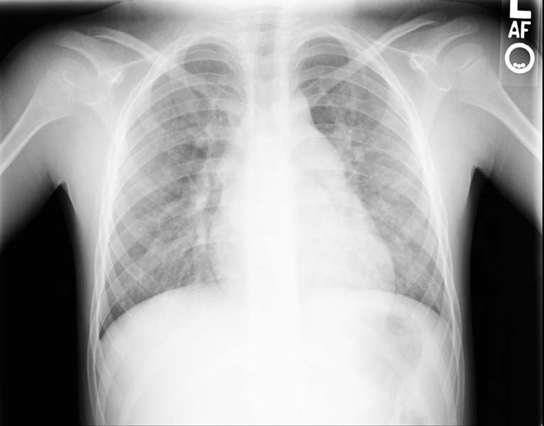
Enlarged right atrial border and mild cardiomegaly.
-
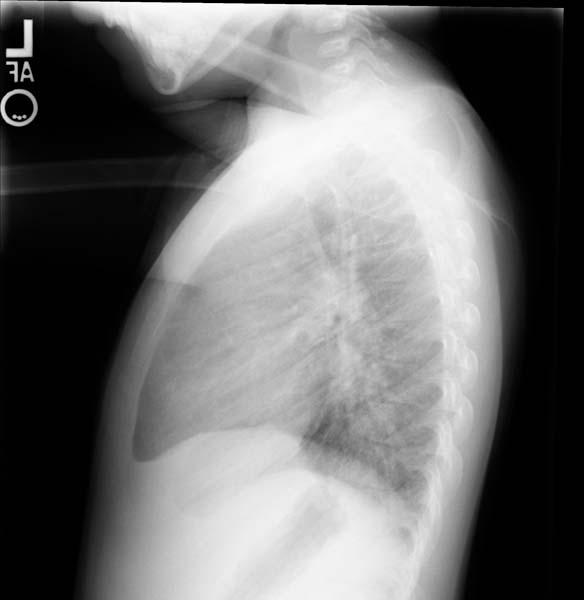
Lateral view
-
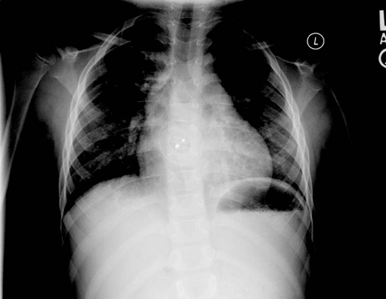
Post repair. Enlarged right atrial border and mild cardiomegaly.
-
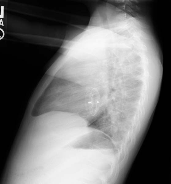
Post repair. Lateral view.
-
ASD. Another patient. Enlarged right atrial border and advanced cardiomegaly.


References
- ↑ Abdulla, Ra-id. (2011). Heart Diseases in Children: A Pediatrician's Guide. Springer.