Pericarditis EKG examples: Difference between revisions
No edit summary |
Esther Lee (talk | contribs) |
||
| Line 1: | Line 1: | ||
__NOTOC__ | __NOTOC__ | ||
{{CMG}} | {{CMG}} | ||
;For | ;For information on electrocardiogram findings in pericarditis, click [[Pericarditis electrocardiogram|here]]. | ||
==Pericarditis EKG Examples== | ==Pericarditis EKG Examples== | ||
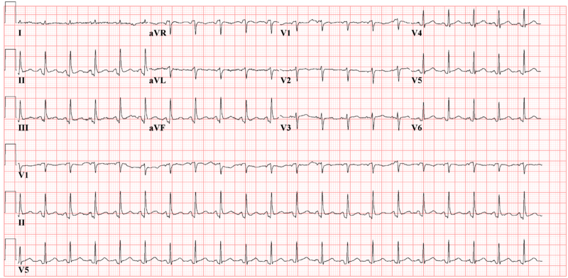
Below is a 12 lead EKG image of acute pericarditis showing PTa depression, but no ST elevation. | |||
[[File:PtaDepressionPericarditis.png|center|800px]] | |||
---- | |||
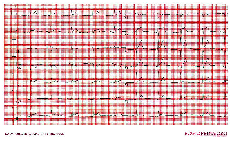
Below is an image of EKG in case of acute pericarditis. It shows clear diffuse ST elevation and some PTa depression. | Below is an image of EKG in case of acute pericarditis. It shows clear diffuse ST elevation and some PTa depression. | ||
[[File:AcutePericarditis.jpg|center|800px]] | [[File:AcutePericarditis.jpg|center|800px]] | ||
| Line 35: | Line 38: | ||
==Sources== | ==Sources== | ||
Copycenter images obtained courtesy of ECGpedia, | Copycenter images obtained courtesy of ECGpedia, http://en.ecgpedia.org/index.php?title=Special:NewFiles&offset=&limit=500 | ||
==References== | ==References== | ||
Revision as of 18:51, 15 October 2012
Editor-In-Chief: C. Michael Gibson, M.S., M.D. [1]
- For information on electrocardiogram findings in pericarditis, click here.
Pericarditis EKG Examples
Below is a 12 lead EKG image of acute pericarditis showing PTa depression, but no ST elevation.
Below is an image of EKG in case of acute pericarditis. It shows clear diffuse ST elevation and some PTa depression.
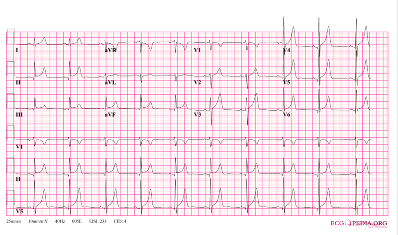
Below is an image of 12 lead EKG in case of pericarditis.
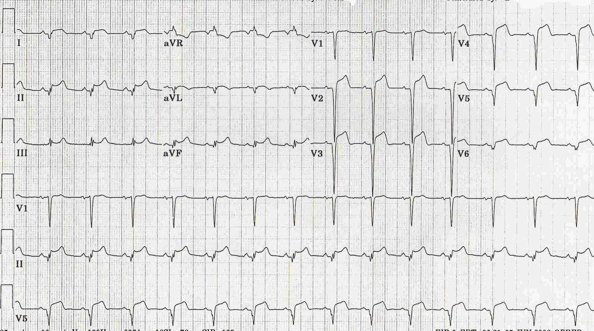
Below is an image of EKG in case of pericarditis.
Below is an image of EKG in case of pericarditis depicting ST segment elevation.

Below is an image of EKG in case of pericarditis depicting ST elevation in most of the leads.
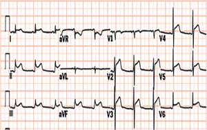
Below is an image of EKG in case of pericarditis PTa depression.(depression between the end of the P-wave and the beginning of the QRS complex)
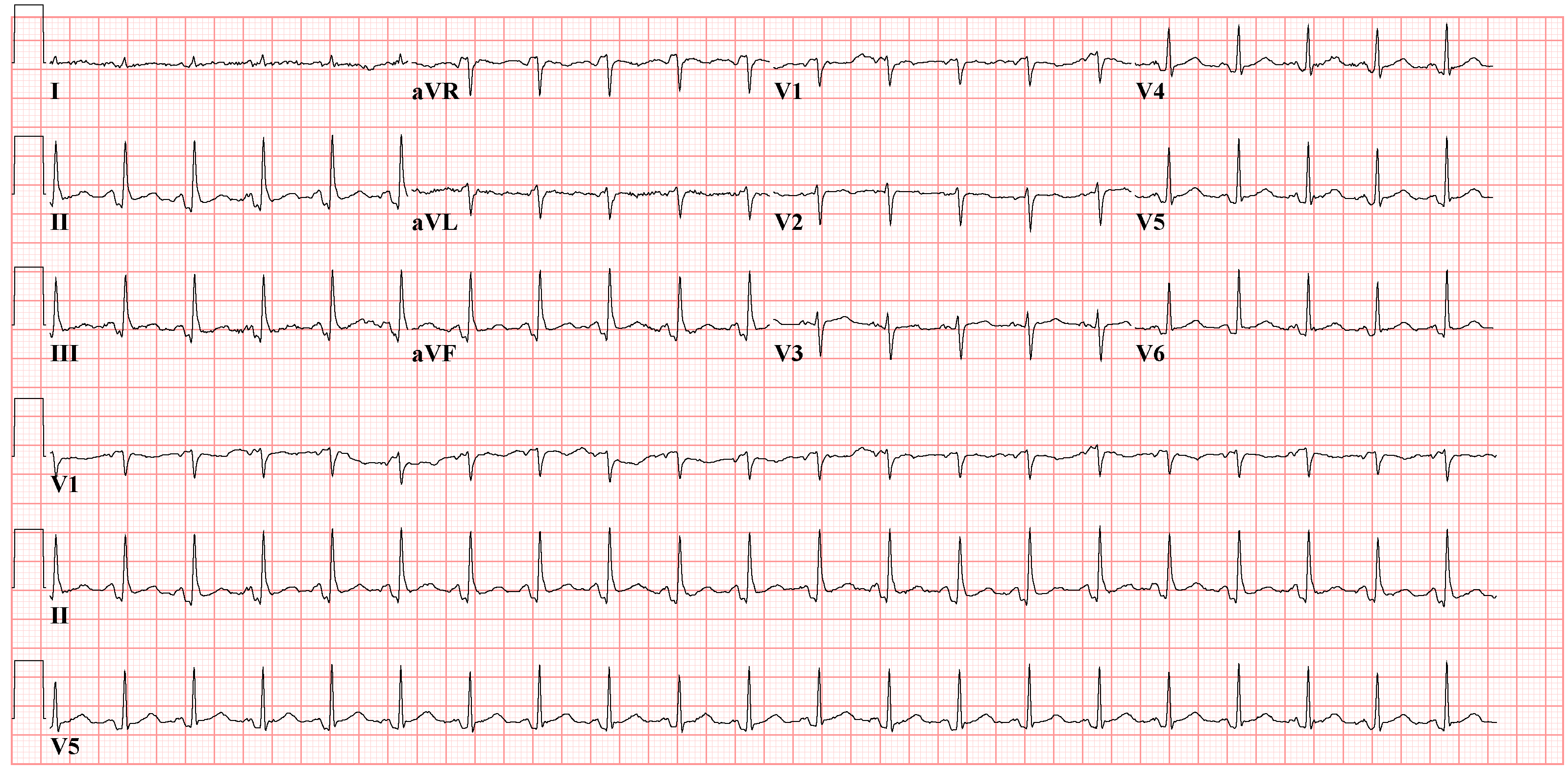
Below is a image of EKG in a case of pericarditis depicting PTa depression.

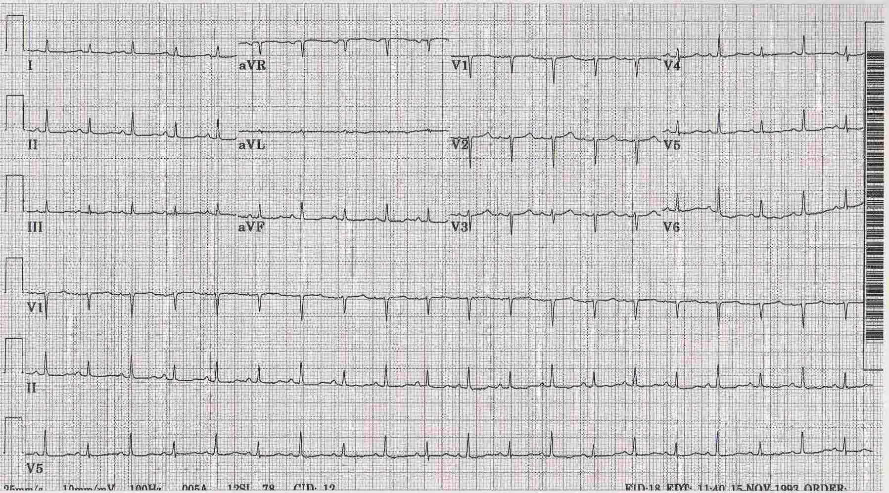
Below is an image of EKG in a case of pericarditis with effusion depicting alterans.
Sources
Copycenter images obtained courtesy of ECGpedia, http://en.ecgpedia.org/index.php?title=Special:NewFiles&offset=&limit=500