Renal artery stenosis ultrasound: Difference between revisions
Jump to navigation
Jump to search
Esther Lee (talk | contribs) No edit summary |
Esther Lee (talk | contribs) |
||
| Line 14: | Line 14: | ||
</gallery> | </gallery> | ||
===Doppler | ===Doppler Ultrasonography=== | ||
[http://www.peir.net Images courtesy of Professor Peter Anderson DVM PhD and published with permission © PEIR, University of Alabama at Birmingham, Department of Pathology] | [http://www.peir.net Images courtesy of Professor Peter Anderson DVM PhD and published with permission © PEIR, University of Alabama at Birmingham, Department of Pathology] | ||
Revision as of 14:51, 28 September 2012
|
Renal artery stenosis Microchapters |
|
Diagnosis |
|---|
|
Treatment |
|
Case Studies |
|
Renal artery stenosis ultrasound On the Web |
|
American Roentgen Ray Society Images of Renal artery stenosis ultrasound |
|
Risk calculators and risk factors for Renal artery stenosis ultrasound |
Editor-In-Chief: C. Michael Gibson, M.S., M.D. [1]
Overview
Ultrasound
-
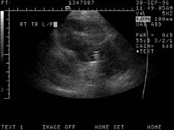
Abdominal pain in a patient with previous renal transplant. 1) Juxtanephric mass surrounding renal vessels, differential includes adenopathy, hematoma and abscess. 2) Renal artery stenosis of CRT.
-

Abdominal pain in a patient with previous renal transplant. 1) Juxtanephric mass surrounding renal vessels, differential includes adenopathy, hematoma and abscess. 2) Renal artery stenosis of CRT

