Acute cholecystitis other diagnostic studies: Difference between revisions
Jump to navigation
Jump to search
No edit summary |
|||
| Line 15: | Line 15: | ||
**[[Bile]] infiltration and [[leucocyte]] margination of blood vessels | **[[Bile]] infiltration and [[leucocyte]] margination of blood vessels | ||
**Lymphatic dilation | **Lymphatic dilation | ||
{| style="border: 0px; font-size: 90%; margin: 3px;" align=center | |||
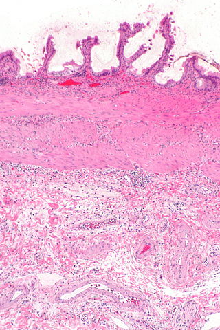
|background: "#FFFFFF;" |[[File:Acute cholecystitis -- low mag.jpg|400px|thumb|center|Histological image of acute cholecystitis; Low magnification. <small> By Nephron - Own work, CC BY-SA 3.0, https://commons.wikimedia.org/w/index.php?curid=30991393 Source:<ref name="urlAcute cholecystitis - Libre Pathology">{{cite web |url=https://librepathology.org/wiki/Acute_cholecystitis |title=Acute cholecystitis - Libre Pathology |format= |work= |accessdate=}}</ref>]] | |||
| | |||
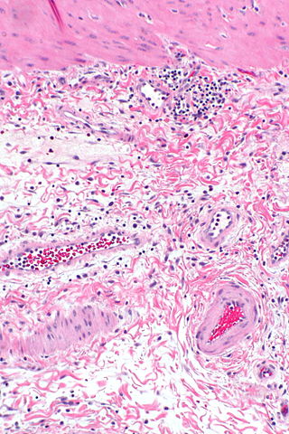
|background: "#FFFFFF;" |[[File:Acute cholecystitis - a -- intermed mag.jpg|400px|thumb|center|Histological image of acute cholecystitis; Intermediate magnification. <small> By Nephron - Own work, CC BY-SA 3.0, https://commons.wikimedia.org/w/index.php?curid=30991393 Source:<ref name="urlAcute cholecystitis - Libre Pathology">{{cite web |url=https://librepathology.org/wiki/Acute_cholecystitis |title=Acute cholecystitis - Libre Pathology |format= |work= |accessdate=}}</ref>]] | |||
<br clear="left" /> | |||
|} | |||
==References== | ==References== | ||
Latest revision as of 19:19, 19 December 2017
|
Acute cholecystitis Microchapters |
|
Diagnosis |
|---|
|
Treatment |
|
Case Studies |
|
Acute cholecystitis other diagnostic studies On the Web |
|
American Roentgen Ray Society Images of Acute cholecystitis other diagnostic studies |
|
Risk calculators and risk factors for Acute cholecystitis other diagnostic studies |
Editor-In-Chief: C. Michael Gibson, M.S., M.D. [1]; Associate Editor(s)-in-Chief: Furqan M M. M.B.B.S[2]
Overview
The histopathological analysis may be helpful in the diagnosis of acute cholecystitis. Findings suggestive of acute cholecystitis include edematous and hemorrhagic gallbladder wall, mucosal necrosis with neutrophil infiltration, eosinophilic infiltration of the gallbladder mucosa, and bile infiltration and leucocyte margination of blood vessels.
Other Diagnostic Studies
- The histopathological analysis may be helpful in the diagnosis of acute cholecystitis. Findings suggestive of acute cholecystitis include:[1][2][3][4]
- Edematous and hemorrhagic gallbladder wall
- Mucosal necrosis with neutrophil infiltration
- Eosinophilic infiltration of the gallblader mucosa
- Specific microscopic features of acalculous cholecystitis:[5]
 |

|
References
- ↑ Owen CC, Bilhartz LE (2003). "Gallbladder polyps, cholesterolosis, adenomyomatosis, and acute acalculous cholecystitis". Semin. Gastrointest. Dis. 14 (4): 178–88. PMID 14719768.
- ↑ McChesney JA, Northup PG, Bickston SJ (2003). "Acute acalculous cholecystitis associated with systemic sepsis and visceral arterial hypoperfusion: a case series and review of pathophysiology". Dig. Dis. Sci. 48 (10): 1960–7. PMID 14627341.
- ↑ Kasprzak A, Malkowski W, Biczysko W, Seraszek A, Sterzyńska K, Zabel M (2011). "Histological alterations of gallbladder mucosa and selected clinical data in young patients with symptomatic gallstones". Pol J Pathol. 62 (1): 41–9. PMID 21574105.
- ↑ Yaylak F, Deger A, Bayhan Z, Kocak C, Zeren S, Kocak FE, Ekici MF, Algın MC (2016). "Histopathological gallbladder morphometric measurements in geriatric patients with symptomatic chronic cholecystitis". Ir J Med Sci. 185 (4): 871–876. doi:10.1007/s11845-015-1385-3. PMID 26602767.
- ↑ Laurila JJ, Ala-Kokko TI, Laurila PA, Saarnio J, Koivukangas V, Syrjälä H, Karttunen TJ (2005). "Histopathology of acute acalculous cholecystitis in critically ill patients". Histopathology. 47 (5): 485–92. doi:10.1111/j.1365-2559.2005.02238.x. PMID 16241996.
- ↑ 6.0 6.1 "Acute cholecystitis - Libre Pathology".