Conjunctivitis physical examination: Difference between revisions
No edit summary |
No edit summary |
||
| Line 4: | Line 4: | ||
==Overview== | ==Overview== | ||
Physical examination of patients with conjunctivitis is usually remarkable for [[swollen]] [[eyelid]], [[epiphora]], [[hyperemia]], and muco-purulent or watery discharge. | |||
==Physical Examination== | ==Physical Examination== | ||
| Line 18: | Line 19: | ||
*[[Corneal]] [[epithelial]] defect (severe cases) | *[[Corneal]] [[epithelial]] defect (severe cases) | ||
===Acute Hemorrhagic Conjunctivitis=== | ===Acute Hemorrhagic Conjunctivitis=== | ||
Ophthalmologic examination of patients with [[acute]] hemorrhagic conjunctivitis is usually remarkable for: | Ophthalmologic examination of patients with [[acute]] hemorrhagic conjunctivitis is usually remarkable for:<ref name="pmid26077630">{{cite journal| author=Jhanji V, Chan TC, Li EY, Agarwal K, Vajpayee RB| title=Adenoviral keratoconjunctivitis. | journal=Surv Ophthalmol | year= 2015 | volume= 60 | issue= 5 | pages= 435-43 | pmid=26077630 | doi=10.1016/j.survophthal.2015.04.001 | pmc= | url=http://www.ncbi.nlm.nih.gov/entrez/eutils/elink.fcgi?dbfrom=pubmed&tool=sumsearch.org/cite&retmode=ref&cmd=prlinks&id=26077630 }} </ref> | ||
*[[Swollen]], | *[[Swollen]], edematous [[eyelid]] | ||
*Eye pain in [[palpation]] | *Eye pain in [[palpation]] | ||
*[[Bulbar]] [[conjunctiva]] [[hemorrhage]] | *[[Bulbar]] [[conjunctiva]] [[hemorrhage]] | ||
| Line 46: | Line 47: | ||
===Allergic Conjunctivitis=== | ===Allergic Conjunctivitis=== | ||
Ophthalmologic examination of patients with allergic conjunctivitis is usually remarkable for: | Ophthalmologic examination of patients with allergic conjunctivitis is usually remarkable for: | ||
*Bilateral conjunctival injection | *[[Bilateral]] conjunctival [[injection]] | ||
*Chemosis | *[[Chemosis]]] | ||
*Watery discharge, or mild mucous discharge | *Watery [[discharge]], or mild mucous discharge | ||
===Keratoconjunctivitis Sicaa=== | ===Keratoconjunctivitis Sicaa=== | ||
Examination should include evaluation of the face, eyelids, blinking patterns, eyelid margins, [[eyelashes]], [[conjunctiva]], [[cornea]], and tear film. | |||
Examination of patients with keratoconjunctivitis sicaa is usually remarkable for: | |||
*More or less pronounced [[conjunctival]] [[redness]] | |||
*Damage to the ocular surface with punctate [[epithelial]] [[erosions]] (superficial punctate [[keratitis]]) | |||
*Thickened eyelid margins and [[telangiectasia]] (signs of meibomian gland dysfunction) | |||
*[[Meibomian gland]] orifices are obstructed with a cloudy, granular or solid [[secretion]] (expressed by exerting considerable pressure on the lower lid) | |||
*[[Blepharitis]] (associated with meibomian gland dysfunction) | |||
*Meibomitis ([[inflammation]] of the meibomian glands) | |||
===Superior Limbic Keratoconjunctivitis=== | ===Superior Limbic Keratoconjunctivitis=== | ||
Ophthalmologic examination of patients with superior limbic keratoconjunctivitis is usually remarkable for: | Ophthalmologic examination of patients with superior limbic keratoconjunctivitis is usually remarkable for: | ||
*Micro-papillary reaction in the upper tarsal conjunctiva, | *Micro-papillary reaction in the upper tarsal conjunctiva, | ||
Hyperemia | *[[Hyperemia]] | ||
Thickening of the superior | *Thickening of the [[superior]] [[bulbar]] [[conjunctiva]] | ||
*Ciliary [[injection]] in the upper bulbar conjunctiva | |||
*[[Corneal]] erosions in the upper quadrants | |||
*[[Diffuse]] [[superficial]] corneal erosions | |||
*Eyelid [[edema]] | |||
[[ | |||
===Images=== | ===Images=== | ||
Revision as of 12:16, 5 July 2016
|
Conjunctivitis Microchapters |
|
Diagnosis |
|---|
|
Treatment |
|
Case Studies |
|
Conjunctivitis physical examination On the Web |
|
American Roentgen Ray Society Images of Conjunctivitis physical examination |
|
Risk calculators and risk factors for Conjunctivitis physical examination |
Editor-In-Chief: C. Michael Gibson, M.S., M.D. [1] Associate Editor(s)-in-Chief: Sara Mehrsefat, M.D. [2]
Overview
Physical examination of patients with conjunctivitis is usually remarkable for swollen eyelid, epiphora, hyperemia, and muco-purulent or watery discharge.
Physical Examination
Viral Conjunctivitis
Patients with viral conjunctivitis usually appear febrile, and they have Preauricular adenopathy. Ophthalmologic examination of patients with viral conjunctivitis is usually remarkable for:
- Epiphora
- Hyperemia
- Chemosis
- Follicular conjunctival reaction
- Pseudomembranous (occasionally)
- Cicatricial conjunctival reaction
- Edematous and ecchymotic eyelids
- Corneal epithelial defect (severe cases)
Acute Hemorrhagic Conjunctivitis
Ophthalmologic examination of patients with acute hemorrhagic conjunctivitis is usually remarkable for:[1]
- Swollen, edematous eyelid
- Eye pain in palpation
- Bulbar conjunctiva hemorrhage
Bacterial Conjunctivitis
Ophthalmologic examination of patients with bacterial conjunctivitis is usually remarkable for:
- Bulbar conjunctival injection
- Palpebral conjunctival papillary reaction
- Muco-purulent or watery discharge
- Chemosis
- Lid erythema
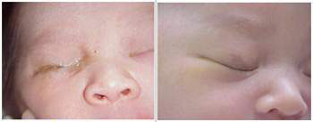
Neonatal Conjunctivitis
Ophthalmologic examination of patients with neonatal conjunctivitis is usually remarkable for:
- Neisseria gonorrhea
- Chemosis
- Severe lid edema and
- Mucopurulent discharge
- Corneal involvement (diffuse epithelial edema, ulceration, corneal perforation, and endophthalmitis
- Chlamydia trachomatis
- Chemical
- Mild conjunctival injection
- Tearing
Allergic Conjunctivitis
Ophthalmologic examination of patients with allergic conjunctivitis is usually remarkable for:
Keratoconjunctivitis Sicaa
Examination should include evaluation of the face, eyelids, blinking patterns, eyelid margins, eyelashes, conjunctiva, cornea, and tear film. Examination of patients with keratoconjunctivitis sicaa is usually remarkable for:
- More or less pronounced conjunctival redness
- Damage to the ocular surface with punctate epithelial erosions (superficial punctate keratitis)
- Thickened eyelid margins and telangiectasia (signs of meibomian gland dysfunction)
- Meibomian gland orifices are obstructed with a cloudy, granular or solid secretion (expressed by exerting considerable pressure on the lower lid)
- Blepharitis (associated with meibomian gland dysfunction)
- Meibomitis (inflammation of the meibomian glands)
Superior Limbic Keratoconjunctivitis
Ophthalmologic examination of patients with superior limbic keratoconjunctivitis is usually remarkable for:
- Micro-papillary reaction in the upper tarsal conjunctiva,
- Hyperemia
- Thickening of the superior bulbar conjunctiva
- Ciliary injection in the upper bulbar conjunctiva
- Corneal erosions in the upper quadrants
- Diffuse superficial corneal erosions
- Eyelid edema
Images
The following are gross images associated with rheumatic fever.[2]
-
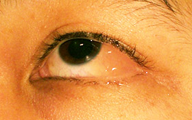
One eye with conjunctivitis.
-
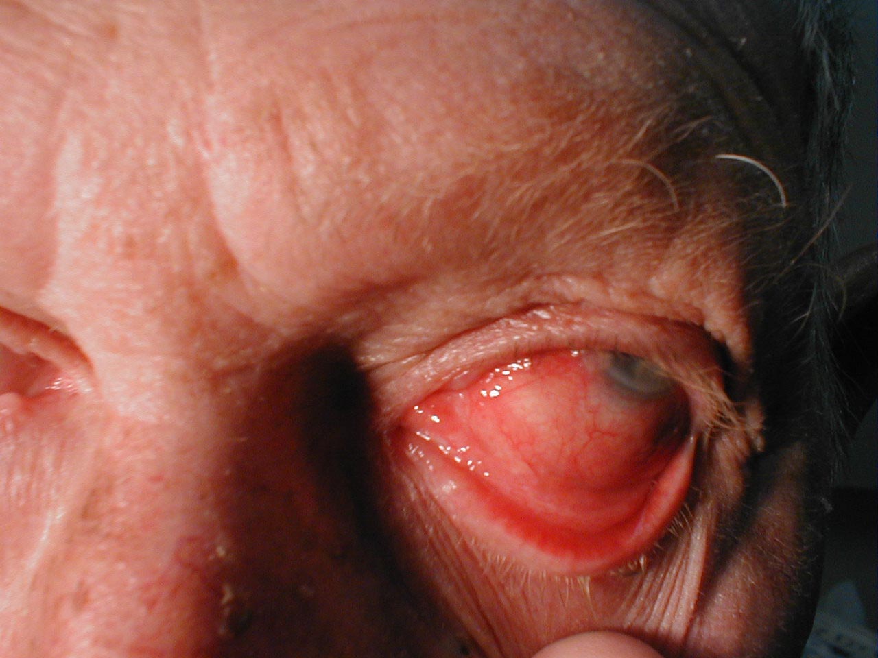
Bacterial Conjunctivitis
-
Chemical Conjunctivitis


.jpg)
(Images shown below are courtesy of Charlie Goldberg, M.D., UCSD School of Medicine and VA Medical Center, San Diego, CA)
-
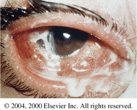
Conjunctivitis: Note inflamed conjunctiva of sclera and reflection onto underside of eyelid.
-
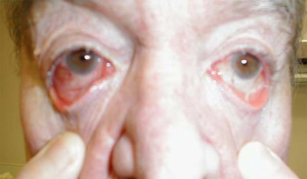
Conjunctivitis: Marked bilateral inflammation involving conjunctiva that covers sclera and under surface of eyelid. Thick exudate can also be seen.
-
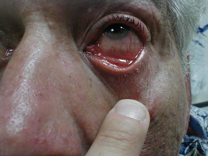
Conjunctivitis: Inflammation of conjunctiva covering sclera and under surface of eyelid.


References
- ↑ Jhanji V, Chan TC, Li EY, Agarwal K, Vajpayee RB (2015). "Adenoviral keratoconjunctivitis". Surv Ophthalmol. 60 (5): 435–43. doi:10.1016/j.survophthal.2015.04.001. PMID 26077630.
- ↑ http://picasaweb.google.com/mcmumbi/USMLEIIImages/