Squamous cell carcinoma of the skin history and symptoms
|
Squamous cell carcinoma of the skin Microchapters |
|
Differentiating Squamous cell carcinoma of the skin from other Diseases |
|---|
|
Diagnosis |
|
Treatment |
|
Case Studies |
|
Squamous cell carcinoma of the skin history and symptoms On the Web |
|
American Roentgen Ray Society Images of Squamous cell carcinoma of the skin history and symptoms |
|
FDA on Squamous cell carcinoma of the skin history and symptoms |
|
CDC on Squamous cell carcinoma of the skin history and symptoms |
|
Squamous cell carcinoma of the skin history and symptoms in the news |
|
Blogs on Squamous cell carcinoma of the skin history and symptoms |
|
Directions to Hospitals Treating Squamous cell carcinoma of the skin |
|
Risk calculators and risk factors for Squamous cell carcinoma of the skin history and symptoms |
Editor-In-Chief: C. Michael Gibson, M.S., M.D. [1]; Associate Editor(s)-in-Chief: Aditya Govindavarjhulla, M.B.B.S. [2],Raviteja Guddeti, M.B.B.S. [3]
Overview
Most Squamous cell carcinomas (SCC) arise on the sun-exposed skin of the head and neck, with fewer lesions arising on the extremities and occasional tumors occurring on the trunk. Early lesions frequently present as a red, scaly spots. Later lesions may form nodules or firm plaques, either of which can ulcerate ( http://tray.dermatology.uiowa.edu). Diagnosis is established by biopsy and histopathological confirmation. Complete excision is curative in the vast majority of cases. Occasionally squamous cell carcinoma will invade along the perineural layer of peripheral nerves and will extend well beyond the clinically apparent mass. Local recurrence is more common in these instances and when present on the head, direct intracranial extension may occur. Metastases to draining lymph nodes occurs in a minority of cases and disseminated disease is the cause of most squamous cell carcinoma-related deaths. Higher rates of metastasis (~15%) are observed with primary lesions of the lips or ears (Rowe et al., 1992). Radiation therapy is helpful in some cases of locally recurrent disease in which complete resection is difficult to achieve and in cases of limited metastatic disease.
History and Symptoms
- Lesions of invasive SCC are often asymptomatic but may be painful or pruritic.
- Local neurologic symptoms (eg, numbness, stinging, burning, paresthesias, paralysis, or visual changes) occur in approximately one-third of patients with histologic perineural invasion by the tumor.[1]
It is easy to find it as it appears mostly in the noticeable regions of the body like face, ears, neck, arms, etc. It grows slowly and presentation depends on the part of the body that is involved. It may vary from simple growing lump , plaque or a bleeding ulcer.[2]
Systemic symptoms of this carcinoma are seen in the advanced stages where the cancer disseminates causing easy fatigability. But few symptoms like dysphagia, odynophagia are seen when tongue, lips or esophagus are involved.
Skin
Presentation : They usually notice plaque. Ulcers are commonly seen over the face causing disfiguration. At times they can be exophytic over the lips etc.
Tongue and Esophagus
-
 Squamous cell carcinoma in oral cavity.
Image courtesy of Professor Peter Anderson DVM PhD and published with permission © PEIR, University of Alabama at Birmingham, Department of Pathology -
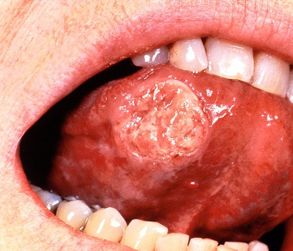
![A large squamous cell carcinoma of the tongue[3]](/images/e/e9/Squamous_cell_carcinoma.jpg) A large squamous cell carcinoma of the tongue[3]
A large squamous cell carcinoma of the tongue[3]

![A large squamous cell carcinoma of the tongue[3]](/index.php/File:Squamous_cell_carcinoma.jpg)
Presentation : Its usually ulcerated in the lateral part of the tongue, pink - red color. Patient finds tough to eat or swallow. As it bleeds on touch or biting. They have dysphagia/ odynophagia when esophagus is involved. In later stages even liquids are tough to go thru. Swelling of lymph nodes is seen in advanced stages where we can find lumps in the neck
Nasopharynx
Presentation : Patient presents with symptoms from the mass effect of the tumour. They include nasal discharge, bleeding, obstruction; ear infection, deafness , tinnitus are complained. Other important complaints include headache and neck swelling due to lymph nodal spread.[4]
Lungs
Presentation : Persons who present with SCC of lungs doesn't present usually with any typical symptoms of pulmonary. But in few we may see persistent cough, hemoptysis when it is exophytic and occupies whole of the bronchi which can even lead to recurrent infections.
Penis
Presentation : Unhealed lesions, subtle indurations may be the intial presentation. Often presentation to the clinic is delayed due to embarrassment most of the times. Warts can be a pre-disposing factor.Large warts lead to infections and necrosis leading to hemorrhages at times.
References
- ↑ Reule RB, Golda NJ, Wheeland RG (October 2009). "Treatment of cutaneous squamous cell carcinoma with perineural invasion using Mohs micrographic surgery: report of two cases and review of the literature". Dermatol Surg. 35 (10): 1559–66. doi:10.1111/j.1524-4725.2009.01276.x. PMID 19681994.
- ↑ www.ncbi.nlm.nih.gov/pubmedhealth/PMH0001832/
- ↑ http://picasaweb.google.com/mcmumbi/USMLEIIImages
- ↑ Sham JS, Poon YF, Wei WI, Choy D. Nasopharyngeal carcinoma in young patients. Cancer. Jun 1 1990;65(11):2606-10.