Sartorius muscle
|
WikiDoc Resources for Sartorius muscle |
|
Articles |
|---|
|
Most recent articles on Sartorius muscle Most cited articles on Sartorius muscle |
|
Media |
|
Powerpoint slides on Sartorius muscle |
|
Evidence Based Medicine |
|
Clinical Trials |
|
Ongoing Trials on Sartorius muscle at Clinical Trials.gov Trial results on Sartorius muscle Clinical Trials on Sartorius muscle at Google
|
|
Guidelines / Policies / Govt |
|
US National Guidelines Clearinghouse on Sartorius muscle NICE Guidance on Sartorius muscle
|
|
Books |
|
News |
|
Commentary |
|
Definitions |
|
Patient Resources / Community |
|
Patient resources on Sartorius muscle Discussion groups on Sartorius muscle Patient Handouts on Sartorius muscle Directions to Hospitals Treating Sartorius muscle Risk calculators and risk factors for Sartorius muscle
|
|
Healthcare Provider Resources |
|
Causes & Risk Factors for Sartorius muscle |
|
Continuing Medical Education (CME) |
|
International |
|
|
|
Business |
|
Experimental / Informatics |
Overview
The sartorius muscle is a long thin muscle that runs down the length of the thigh. It is the longest muscle in the human body. Its upper portion forms the lateral border of the femoral triangle.
Origin and insertion
The sartorius muscle arises by tendinous fibres from the anterior superior iliac spine, running obliquely across the upper and anterior part of the thigh in an inferomedial direction.
It descends as far as the medial side of the knee, passing behind the medial condyle of the femur to end in a tendon.
This tendon curves anteriorly to join the tendons of the gracilis and semitendinous muscles which together form the pes anserinus, finally inserting into the proximal part of the tibia on the medial surface of its body.
Etymology
The name sartorius is the Latin word for "sartorial" (i.e. "to do with tailoring", in turn from sartor i.e. "tailor", in turn from sartus i.e. "patched" or "repaired", in turn from sarcio i.e. "to patch", "to repair").
This name was chosen in reference to the cross-legged position in which tailors once sat.
Actions
Assists in flexion, abduction and lateral rotation of hip, and flexion and medial rotation of knee. Looking at the bottom of your foot, like you are checking to see if you stepped in dog poop, demonstrates all 5 actions of sartorius.
Innervation
Situated in the anterior fascial compartment of the thigh, sartorius is innervated via branches of the femoral nerve.
Variations
Slips of origin from the outer end of the inguinal ligament, the notch of the ilium, the ilio-pectineal line or the pubis occur.
The muscle may be split into two parts, and one part may be inserted into the fascia lata, the femur, the ligament of the patella or the tendon of the Semitendinosus.
The tendon of insertion may end in the fascia lata, the capsule of the knee-joint, or the fascia of the leg.
The muscle may be absent.
Common Injuries of the Sartorius Muscle
Overextension of the hip may cause a strain of the muscle at its attachment point (the iliac crest).
Additional images
-
Bones of the right leg. Anterior surface.
-
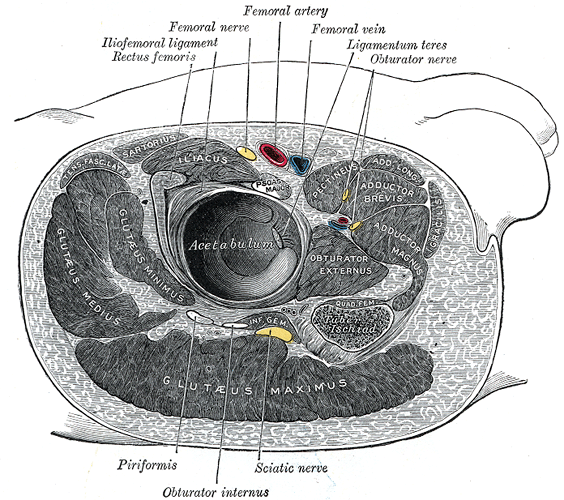
Structures surrounding right hip-joint.
-
Muscles of the iliac and anterior femoral regions.
-
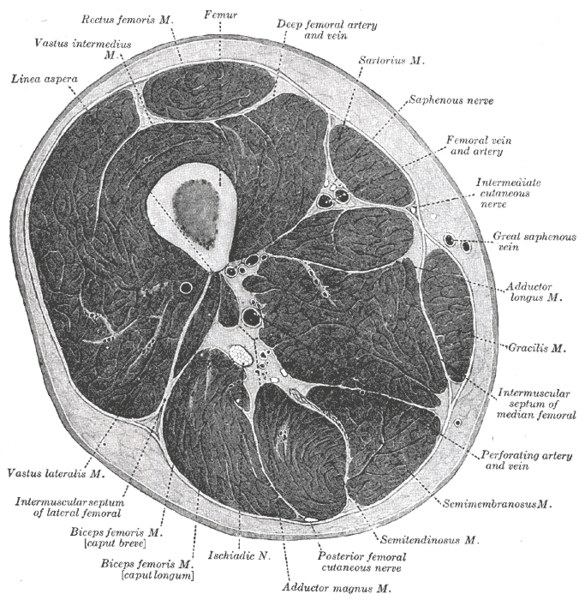
Cross-section through the middle of the thigh.
-
Muscles of the gluteal and posterior femoral regions.
-
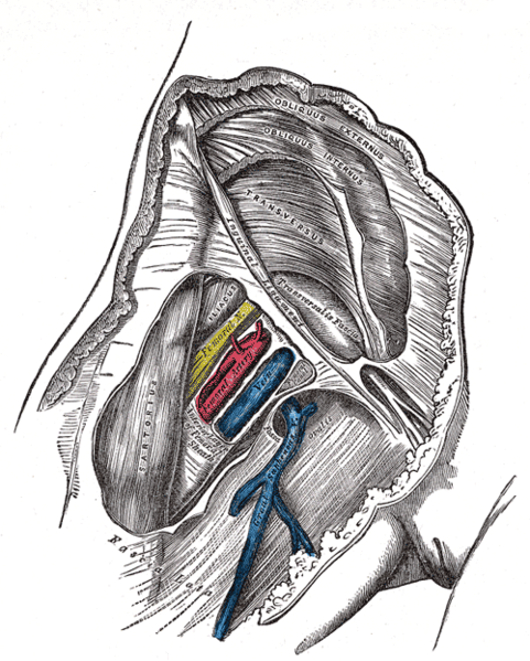
Femoral sheath laid open to show its three compartments.
-
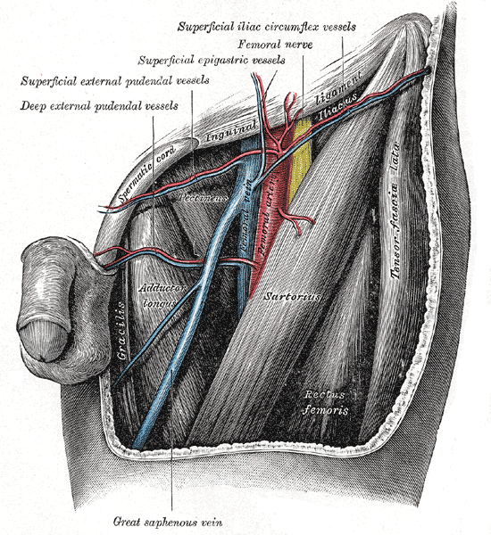
The left femoral triangle.
-
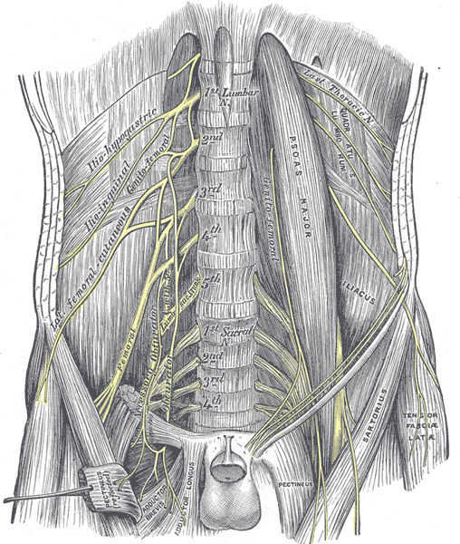
The lumbar plexus and its branches.
-
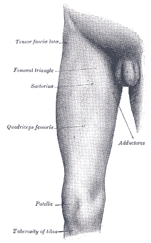
Front and medial aspect of right thigh.
External links
- Template:GPnotebook
- Template:MuscleLoyola
- Template:SUNYAnatomyLabs
- Template:ViennaCrossSection
- Template:ViennaCrossSection
