Renal ectopia ultrasound
|
Renal ectopia Microchapters |
|
Diagnosis |
|---|
|
Treatment |
|
Case Studies |
|
Renal ectopia ultrasound On the Web |
|
American Roentgen Ray Society Images of Renal ectopia ultrasound |
|
Risk calculators and risk factors for Renal ectopia ultrasound |
Editor-In-Chief: C. Michael Gibson, M.S., M.D. [1]; Associate Editor(s)-in-Chief:
Overview
There are no echocardiography/ultrasound findings associated with [disease name].
OR
Echocardiography/ultrasound may be helpful in the diagnosis of [disease name]. Findings on an echocardiography/ultrasound suggestive of/diagnostic of [disease name] include [finding 1], [finding 2], and [finding 3].
OR
There are no echocardiography/ultrasound findings associated with [disease name]. However, an echocardiography/ultrasound may be helpful in the diagnosis of complications of [disease name], which include [complication 1], [complication 2], and [complication 3].
Echocardiography/Ultrasound
There are no echocardiography/ultrasound findings associated with [disease name].
OR
Echocardiography/ultrasound may be helpful in the diagnosis of [disease name]. Findings on an echocardiography/ultrasound suggestive of/diagnostic of [disease name] include:
- [Finding 1]
- [Finding 2]
- [Finding 3]
OR
There are no echocardiography/ultrasound findings associated with [disease name]. However, an echocardiography/ultrasound may be helpful in the diagnosis of complications of [disease name], which include:
- [Complication 1]
- [Complication 2]
- [Complication 3]
CT Scan
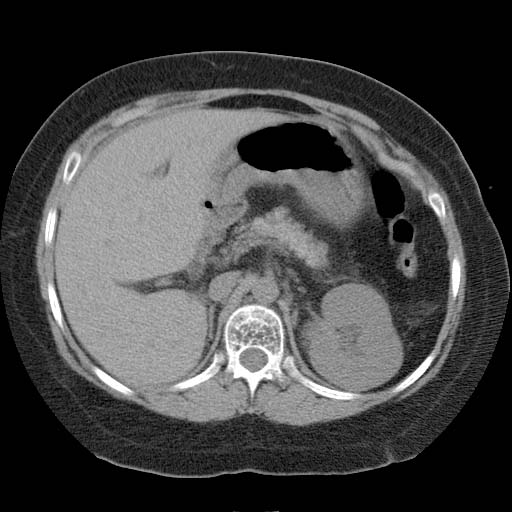
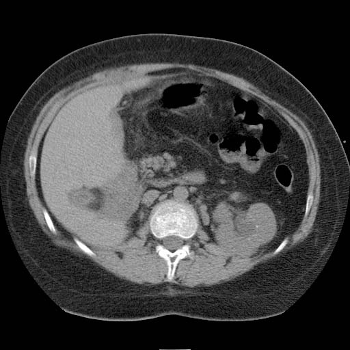
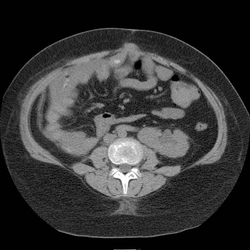
Findings on CT should be interpreted in light of bowel laxity in the region of the empty renal fossa. In particular, distinction must be made from internal hernia. Patient #1: CT images demonstrate a pelvic kidney


Patient #1: CT images demonstrate cross-fused renal ectopia
-
 Kidney 1
 -
-
 Kidney 2


Patient #2: Renal scan images demonstrate cross-fused renal ectopia


