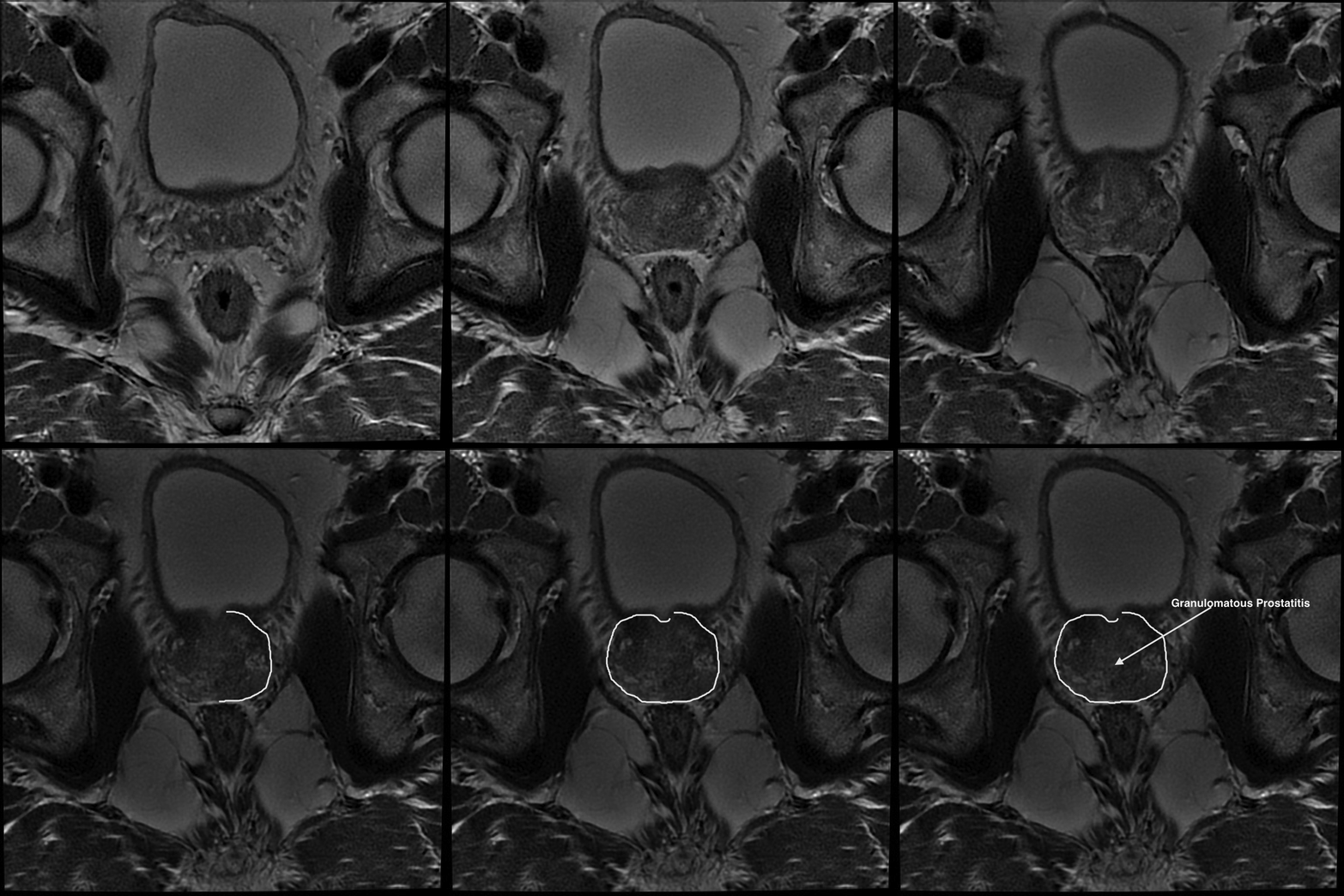
Prostatitis imaging findings
|
Prostatitis Microchapters |
|
Diagnosis |
|
Treatment |
|
Case Studies |
|
Prostatitis imaging findings On the Web |
|
American Roentgen Ray Society Images of Prostatitis imaging findings |
|
Risk calculators and risk factors for Prostatitis imaging findings |
Editor-In-Chief: C. Michael Gibson, M.S., M.D. [1] Associate Editor(s)-in-Chief: Maliha Shakil, M.D. [2]
Overview
CT scan in a patient with prostatitis shows edema of the prostate gland with diffuse enlargement, mostly in the peripheral zone. An abscess may be seen as a rim enhancing hypodensity which can either have single or multiple loculi. On ultrasonography, focal hypoechoic area in the periphery of the prostate represents prostatitis. Fluid collection can show abscess formation. Colour Doppler ultrasound may also prove to be very effective. MRI is also an important investigation and depicts diffuse enlargement of the gland.[1][2][3]
Imaging findings
CT
When an abscess is included in the differential diagnosis of prostatitis, a contrast CT scan is preferred. CT scan in a patient with prostatitis shows edema of the prostate gland with diffuse enlargement, mostly in the peripheral zone. An abscess can also be identified as a rim enhancing hypodensity which can either be unilocular or multilocular. Central zone can be involved rarely such as after the transurethral resection of the prostate (TURP).[1][2][4]
Ultrasound
On ultrasonography, focal hypoechoic region located in the peripheral part of the prostate represents prostatitis. Fluid collection can show abscess formation. Colour Doppler ultrasound may also prove to be very effective.[1][5]
MRI
MRI depicts diffuse enlargement of the gland. Inflammatory changes of the fat around the prostate and the seminal vesicles. It has been shown that combining the morphology with the conventional MRI investigation can be more effective in differentiating prostatitis from early peripheral prostatic carcinoma.[1][6][7]
Findings of acute prostatitis on MRI include:
- T1: peripheral zone iso- or hypo-intense to transitional zone
- T2: hyperintense
- Gd (C+): diffusely enhancing
Prostate Imaging Reporting and Data System
Prostate Imaging reporting and Data System or PIRADS score is very helpful in diagnosing prostatitis and differentiating it from prostatic cancer.[8][9]
- A higher PIRADS score can differentiate prostatic inflammation from carcinoma more reliably
- Decreased diffusion favours the diagnosis of prostatitis
Images

References
- ↑ 1.0 1.1 1.2 1.3 Prostatitis. Radiopaedia 2016. http://radiopaedia.org/articles/prostatitis. Accessed on Feb 09, 2017
- ↑ 2.0 2.1 Sharp VJ, Takacs EB, Powell CR (2010). "Prostatitis: diagnosis and treatment". Am Fam Physician. 82 (4): 397–406. PMID 20704171.
- ↑ Choon-Young Kim, Sang-Woo Lee, Seock Hwan Choi, Seung Hyun Son, Ji-Hoon Jung, Chang-Hee Lee, Shin Young Jeong, Byeong-Cheol Ahn & Jaetae Lee (2016). "Granulomatous Prostatitis After Intravesical Bacillus Calmette-Guerin Instillation Therapy: A Potential Cause of Incidental F-18 FDG Uptake in the Prostate Gland on F-18 FDG PET/CT in Patients with Bladder Cancer". Nuclear medicine and molecular imaging. 50 (1): 31–37. doi:10.1007/s13139-015-0364-y. PMID 26941857. Unknown parameter
|month=ignored (help) - ↑ Robert Lucaj & Dwight M. Achong (2017). "Concurrent Diffuse Pyelonephritis and Prostatitis: Discordant Findings on Sequential FDG PET/CT and 67Ga SPECT/CT Imaging". Clinical nuclear medicine. 42 (1): 73–75. doi:10.1097/RLU.0000000000001415. PMID 27824318. Unknown parameter
|month=ignored (help) - ↑ Dong Sup Lee, Hyun-Sop Choe, Hee Youn Kim, Sun Wook Kim, Sang Rak Bae, Byung Il Yoon & Seung-Ju Lee (2016). "Acute bacterial prostatitis and abscess formation". BMC urology. 16 (1): 38. doi:10.1186/s12894-016-0153-7. PMID 27388006. Unknown parameter
|month=ignored (help) - ↑ Rajeev Jyoti, Noel Hamesh Jina & Hodo Z. Haxhimolla (2016). "In-gantry MRI guided prostate biopsy diagnosis of prostatitis and its relationship with PIRADS V.2 based score". Journal of medical imaging and radiation oncology. doi:10.1111/1754-9485.12555. PMID 27987276. Unknown parameter
|month=ignored (help) - ↑ P. Li, Y. Huang, Y. Li, L. Cai, G. H. Ji, Y. Zheng & Z. Q. Chen (2016). "[Application evaluation of multi-parametric MRI in the diagnosis and differential diagnosis of early prostate cancer and prostatitis]". Zhonghua yi xue za zhi. 96 (37): 2973–2977. PMID 27760657. Unknown parameter
|month=ignored (help) - ↑ Rajeev Jyoti, Noel Hamesh Jina & Hodo Z. Haxhimolla (2016). "In-gantry MRI guided prostate biopsy diagnosis of prostatitis and its relationship with PIRADS V.2 based score". Journal of medical imaging and radiation oncology. doi:10.1111/1754-9485.12555. PMID 27987276. Unknown parameter
|month=ignored (help) - ↑ Michael Meier-Schroers, Guido Kukuk, Karsten Wolter, Georges Decker, Stefan Fischer, Christian Marx, Frank Traeber, Alois Martin Sprinkart, Wolfgang Block, Hans Heinz Schild & Winfried Willinek (2016). "Differentiation of prostatitis and prostate cancer using the Prostate Imaging-Reporting and Data System (PI-RADS)". European journal of radiology. 85 (7): 1304–1311. doi:10.1016/j.ejrad.2016.04.014. PMID 27235878. Unknown parameter
|month=ignored (help) - ↑ Libre Pathology https://radiopaedia.org/cases/granulomatous-prostatitis-1 Accessed on Feb 13, 2017