Mast cell tumor physical examination
|
Mast cell tumor Microchapters |
|
Diagnosis |
|---|
|
Treatment |
|
Case Studies |
|
Mast cell tumor physical examination On the Web |
|
American Roentgen Ray Society Images of Mast cell tumor physical examination |
|
Risk calculators and risk factors for Mast cell tumor physical examination |
Editor-In-Chief: C. Michael Gibson, M.S., M.D. [1];Associate Editor(s)-in-Chief: Jesus Rosario Hernandez, M.D. [2], Suveenkrishna Pothuru, M.B,B.S. [3]
Overview
Physical examination for mast cell tumor include inspection for a large assortment of types of skin lesions, testing for dermatographism (Darier's sign), and palpating for hepatosplenomegaly and lymphadenopathy.
Physical Examination
Vital signs
Skin
- Urticaria pigmentosa:[1]
- Fixed, reddish brown lesions appears as maculo-papules, plaques, nodules, or blisters.[2]
- Urticaria Pigmentosa (UP) lesions tend to be larger, better delineated, and more hyperpigmented in children, as compared to adults, who tend to have numerous small lesions that coalesce to form mottled areas.
- The trunk and thigh are more commonly involved with sparing of face, palms and soles.
- Darier’s sign:[3]
- Lesions urticate in response to physical irritation.
- Localized erythema and urticaria erupts within short period of time (minutes) in response to physical irritation.
- Diffuse Cutaneous Mastocytosis
- Diffuse infiltrative yellow-orange xanthogranuloma-like subcutaneous nodules, or as a widespread urticarial eruption with bullae and redness.[2]
- Telangiectasia macularis eruptiva perstans[4]
- It is a rare form of mastocytosis, and presents as brownish macules and telangiectasia.
- Not associated with pruritus and blistering.
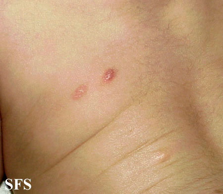
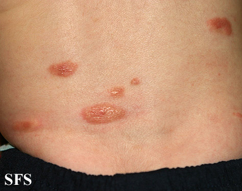
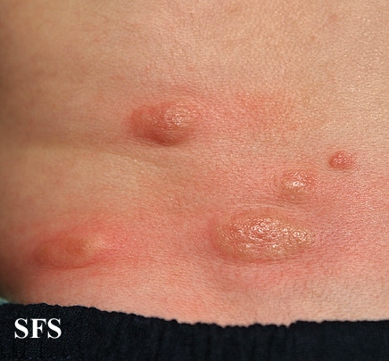
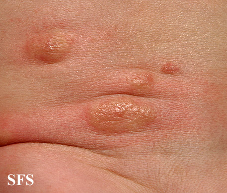
Abdomen
-
Mastocytoma.
Adapted from Atlas<ref name="www.atlasdermatologico.com.br">"Dermatology Atlas". -
Mastocytoma.
Adapted from Atlas<ref name="www.atlasdermatologico.com.br">"Dermatology Atlas". -
Mastocytoma.
Adapted from Atlas<ref name="www.atlasdermatologico.com.br">"Dermatology Atlas". -
Mastocytoma.
Adapted from Atlas<ref name="www.atlasdermatologico.com.br">"Dermatology Atlas".
References
- ↑ CAPLAN RM (February 1963). "The natural course of urticaria pigmentosa. Analysis and follow-up of 112 cases". Arch Dermatol. 87: 146–57. PMID 14018418.
- ↑ 2.0 2.1 Ferrante, Giuliana; Scavone, Valeria; Muscia, Maria; Adrignola, Emilia; Corsello, Giovanni; Passalacqua, Giovanni; La Grutta, Stefania (2015). "The care pathway for children with urticaria, angioedema, mastocytosis". World Allergy Organization Journal. 8 (1): 5. doi:10.1186/s40413-014-0052-x. ISSN 1939-4551.
- ↑ Soter NA (June 2000). "Mastocytosis and the skin". Hematol. Oncol. Clin. North Am. 14 (3): 537–55, vi. PMID 10909039.
- ↑ Williams KW, Metcalfe DD, Prussin C, Carter MC, Komarow HD (2014). "Telangiectasia macularis eruptiva perstans or highly vascularized urticaria pigmentosa?". J Allergy Clin Immunol Pract. 2 (6): 813–5. doi:10.1016/j.jaip.2014.07.002. PMC 4254445. PMID 25439382.


