Internuclear ophthalmoplegia
| Internuclear ophthalmoplegia | |
 | |
|---|---|
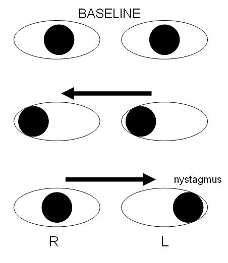
| Schematic demonstrating right internuclear ophthalmoplegia, caused by injury of the right medial longitudinal fasciculus. | |
| ICD-10 | H51.2 |
| ICD-9 | 378.86 |
| DiseasesDB | 6853 |
Editor-In-Chief: C. Michael Gibson, M.S., M.D. [1]
Internuclear ophthalmoplegia or INO is a physical finding, or sign, that is a particular form of eye muscle weakness or ophthalmoparesis. It can affect either the right or left eye. It is a disorder of conjugate lateral gaze. The affected eye shows impairment of adduction. The partner eye diverges from the affected eye during abduction, producing horizontal diplopia, in other words, if the affected eye is on the right, the patient will see "double" when looking to the left and the images will be side by side. During extreme abduction, compensatory nystagmus can be seen in the partner eye.
Causes
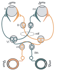
The disorder is caused by injury or dysfunction in the medial longitudinal fasciculus (MLF), a heavily-myelinated tract that allows conjugate eye movement by connecting the paramedian pontine reticular formation (PPRF) -abducens nucleus complex of one side to the oculomotor nucleus of the opposite side.
In young patients with bilateral INO, multiple sclerosis is nearly always the cause. In older patients with one-sided lesions a stroke is a distinct possibility.
Variants
A rostral lesion within the midbrain may affect the convergence center thus causing bilateral divergence of the eyes which is known as the WEBINO syndrome ( Wall Eyed Bilateral INO) as each eye looks at the opposite "wall".
If the lesion affects the PPRF and the MLF on the same side (the MLF having crossed from the opposite side) then the "one and a half syndrome" occurs which, simply put, involves paralysis of all conjugate horizontal eye movements other than abduction of the eye on the opposite side to the lesion.
External links
- Template:GPnotebook
- Animation at mrcophth.com
