Epilepsy eeg
|
Epilepsy Microchapters |
|
Diagnosis |
|---|
|
Treatment |
|
Case Studies |
|
Epilepsy eeg On the Web |
|
American Roentgen Ray Society Images of Epilepsy eeg |
Editor-In-Chief: C. Michael Gibson, M.S., M.D. [1]; Associate Editor(s)-in-Chief: Fahimeh Shojaei, M.D.
Overview
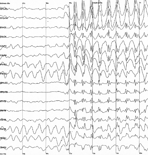
An EEG may be helpful in the diagnosis of epilepsy. Findings on an EEG suggestive of epilepsy include synchronous generalized spikes and waves in all leads in tonic-clonic seizures, spike and wave activity at a frequency of approximately 3 HZ in absence seizures, localized epileptic activity over the seizure focus in focal seizures with intact consciousness and temporal slow waves or spikes in focal seizures with impaired consciousness.
Electroencephalogram
An EEG may be helpful in the diagnosis of epilepsy. Findings on an EEG suggestive of epilepsy include:[1]
| Epilepsy | |||||||||||||||||||||||||||||||||||||||||||||||
| Generalized | Focal | ||||||||||||||||||||||||||||||||||||||||||||||
| Tonic–clonic Seizure | Absences | Focal seizures without altered consciousness | Focal seizures with altered consciousness | ||||||||||||||||||||||||||||||||||||||||||||
| Synchronous generalized spikes and waves in all leads | Bursts of synchronous, generalized
spike-and-wave activity at a frequency of approximately 3 Hz | Localized epileptic activity over the seizure focus | Temporal slow waves or spikes. In the interictal period/Normal EEG | ||||||||||||||||||||||||||||||||||||||||||||
NOTE: Video-EEG monitoring is a combination of recording EEG and clinical behavior of the patient. Although, it's it's more expensive, it is more effective in differentiating different type if seizures.[2]
 |
 |
References
- ↑ Mattle, Heinrich (2017). Fundamentals of neurology : an illustrated guide. Stuttgart New York: Thieme. ISBN 9783131364524.
- ↑ Worrell GA, Lagerlund TD, Buchhalter JR (September 2002). "Role and limitations of routine and ambulatory scalp electroencephalography in diagnosing and managing seizures". Mayo Clin. Proc. 77 (9): 991–8. doi:10.4065/77.9.991. PMID 12233935.