Elbow
|
WikiDoc Resources for Elbow |
|
Articles |
|---|
|
Media |
|
Evidence Based Medicine |
|
Clinical Trials |
|
Ongoing Trials on Elbow at Clinical Trials.gov Clinical Trials on Elbow at Google
|
|
Guidelines / Policies / Govt |
|
US National Guidelines Clearinghouse on Elbow
|
|
Books |
|
News |
|
Commentary |
|
Definitions |
|
Patient Resources / Community |
|
Directions to Hospitals Treating Elbow Risk calculators and risk factors for Elbow
|
|
Healthcare Provider Resources |
|
Continuing Medical Education (CME) |
|
International |
|
|
|
Business |
|
Experimental / Informatics |
Editor-In-Chief: C. Michael Gibson, M.S., M.D. [1]
The elbow-joint is a ginglymus or hinge joint. Three bones form the elbow joint: the humerus of the upper arm, and the paired radius and ulna of the forearm.
The bony prominence at the very tip of the elbow is the olecranon process of the ulna.
Movements
Two main movements are possible at the elbow:
- The hinge-like bending and straightening of the elbow (flexion and extension) happens at the articulation ("joint") between the humerus and the ulna.
- The complex action of turning the forearm over (pronation or supination) happens at the articulation between the radius and the ulna (this movement also occurs at the wrist joint).
In the anatomical position (with the forearm supine), the radius and ulna lie parallel to each other. During pronation, the ulna remains fixed, and the radius rolls around it at both the wrist and the elbow joints. In the prone position, the radius and ulna appear crossed.
Most of the force through the elbow joint is transferred between the humerus and the ulna. Very little force is transmitted between the humerus and the radius. (By contrast, at the wrist joint, most of the force is transferred between the radius and the carpus, with the ulna taking very little part in the wrist joint).
Muscles, arteries, and nerves
The muscles in relation with the joint are:
- in front, the Brachialis
- behind, the Triceps brachii and Anconæus
- laterally, the Supinator, and the common tendon of origin of the Extensor muscles
- medially, the common tendon of origin of the Flexor muscles, and the Flexor carpi ulnaris
The arteries supplying the joint are derived from the anastomosis between the profunda and the superior and inferior ulnar collateral branches of the brachial, with the anterior, posterior, and interosseous recurrent branches of the ulnar, and the recurrent branch of the radial. These vessels form a complete anastomotic network around the joint.
The nerves of the joint are a twig from the ulnar, as it passes between the medial condyle and the olecranon; a filament from the musculocutaneous, and two from the median.
Portions of joint
The elbow-joint comprises three different portions. All these articular surfaces are enveloped by a common synovial membrane, and the movements of the whole joint should be studied together.
| Joint | From | To | Description |
| humeroulnar joint | ulna | humerus | Is a simple hinge-joint, and allows of movements of flexion and extension only. |
| humeroradial joint | head of the radius | capitulum of the humerus | Is an arthrodial joint. |
| proximal radioulnar joint | radius | ulna | In any position of flexion or extension, the radius, carrying the hand with it, can be rotated in it. This movement includes pronation and supination. |
The combination of the movements of flexion and extension of the forearm with those of pronation and supination of the hand, which is ensured by the two being performed at the same joint, is essential to the accuracy of the various minute movements of the hand.
The hand is only directly articulated to the distal surface of the radius, and the ulnar notch on the lower end of the radius travels around the lower end of the ulna. The ulna is excluded from the wrist-joint by the articular disk.
Thus, rotation of the head of the radius around an axis passing through the center of the radial head of the humerus imparts circular movement to the hand through a very considerable arc.
Ligaments
The trochlea of the humerus is received into the semilunar notch of the ulna, and the capitulum of the humerus articulates with the fovea on the head of the radius. The articular surfaces are connected together by a capsule, which is thickened medially and laterally, and, to a less extent, in front and behind. These thickened portions are usually described as distinct ligaments.
The major ligaments are the ulnar collateral ligament, radial collateral ligament, and annular ligament.
Synovial membrane
The synovial membrane is very extensive. It extends from the margin of the articular surface of the humerus, and lines the coronoid, radial and olecranon fossæ on that bone; it is reflected over the deep surface of the capsule and forms a pouch between the radial notch, the deep surface of the annular ligament, and the circumference of the head of the radius. Projecting between the radius and ulna into the cavity is a crescentic fold of synovial membrane, suggesting the division of the joint into two; one the humeroradial, the other the humeroulnar.
Between the capsule and the synovial membrane are three masses of fat:
- the largest, over the olecranon fossa, is pressed into the fossa by the Triceps brachii during the flexion;
- the second, over the coronoid fossa,
- and the third, over the radial fossa, are pressed by the Brachialis into their respective fossæ during extension.
Terminology: "Elbow" and "Ell"
The now obsolete length unit ell relates closely to the elbow. This becomes especially visible when considering the Germanic origins of both words, Elle (ell, defined as the length of an arm from shoulder to fingertips) and Ellbogen (elbow).
It is unknown when or why the second "l" was dropped from English usage of the word, but a more precise suggested spelling would be "ellbow" for the joint and "ellbone" for the ulna, the etymological originator of both unit and joint.
Carrying angle

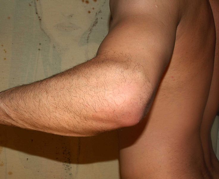
When the arm is extended, with the palm facing forward or up, the bones of the humerus and forearm are not perfectly aligned. The deviation from a straight line (generally on the order of 5-10°) occurs in the direction of the thumb, and is referred to as the carrying angle (visible in the right half of the picture, right). In females the carrying angle is greater than in males.[1]
The carrying angle can influence how objects are held by individuals - those with a more extreme carrying angle may be more likely to supinate the forearm when holding objects in the hand to keep the elbow closer to the body.
Diagnostic Findings
MRI
-
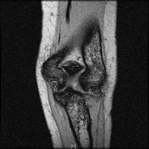 Normal elbow: Cor MPGR
 -
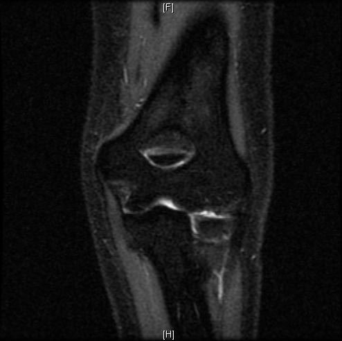 Normal elbow: Cor IR
 -
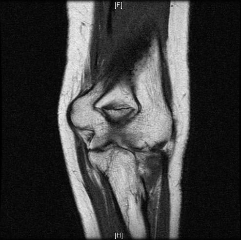 Normal elbow: Cor PD


-
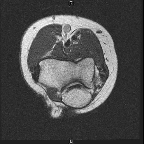 Normal elbow: Axl PD
 -
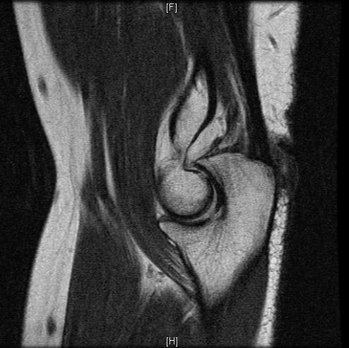 Normal elbow: Sag PD
 -
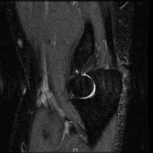 Normal elbow: Sag IR


See also
Additional images
-
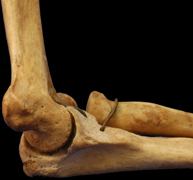 Medial Humerus Radius Ulna Articulated
 -
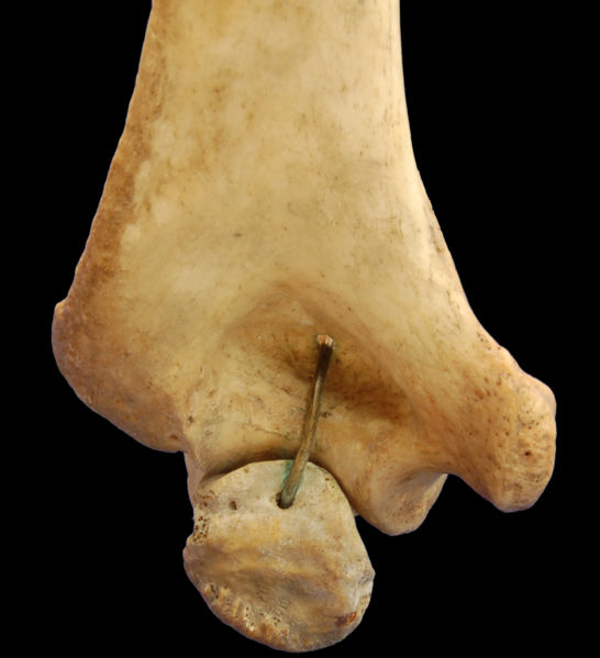
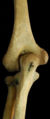 Left Human Posterior Distal Humerus Extended
 -
 Left Human Posterior Distal Humerus Flexed
 -
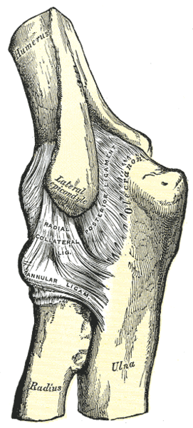 Left elbow-joint, showing posterior and radial collateral ligaments.
 -
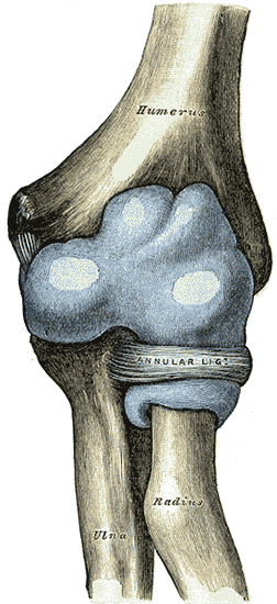 Capsule of elbow-joint (distended). Anterior aspect.
 -
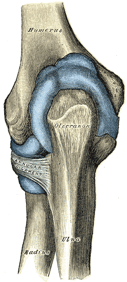 Capsule of elbow-joint (distended). Posterior aspect.
 -
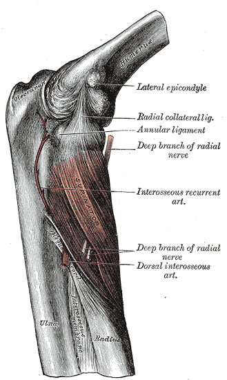 The Supinator. Posterior view.
 -
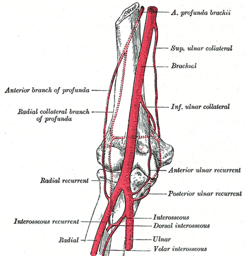 Diagram of the anastomosis around the elbow-joint.
 -
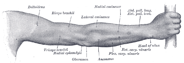 Back of right upper extremity.
 -
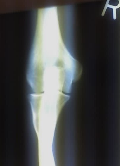 Close-up radiograph, right elbow-joint
 -
 Left male elbow


References
- ↑ Steel, F (1958). "The carrying angle in man". Journal of Anatomy. 92 (2): 315–7. PMID 13525245. Unknown parameter
|coauthors=ignored (help)
| File:Wiktionary-logo-en-v2.svg | Look up Elbow in Wiktionary, the free dictionary. |
Template:Gray's Template:Human anatomical features Template:Joints of upper limbs
ar:مرفق ca:Colze da:Albue de:Ellbogengelenk eo:Kubuto it:Gomito he:מרפק la:Cubitum (anatomia) hu:Könyök nl:Elleboog no:Albue simple:Elbow fi:Kyynärnivel sv:Armbågsled yi:עלנבויגן sl:Komolec tl:Siko