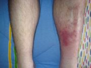
Cellulitis physical examination
|
Cellulitis Microchapters |
|
Diagnosis |
|---|
|
Treatment |
|
Case Studies |
|
Cellulitis physical examination On the Web |
|
American Roentgen Ray Society Images of Cellulitis physical examination |
|
Risk calculators and risk factors for Cellulitis physical examination |
Editor-In-Chief: C. Michael Gibson, M.S., M.D. [1]; Associate Editor(s)-in-Chief: Aditya Govindavarjhulla, M.B.B.S.
Overview
Cellulitis is an acute, spreading infection of the deeper dermis and subcutaneous tissue, usually complicating a wound, ulcer, or dermatosis. It is characterized by rapidly expanding areas of edema, erythema, and warmth, sometimes accompanied by lymphangitis and inflammation of the regional lymph nodes. Unlike erysipelas which affects the upper dermis and the superficial lymphatics, cellulitis lacks sharp demarcation from uninvolved skin and usually does not present with an indurated, "peau d'orange" surface with a raised border. The diagnosis of cellulitis is based on the morphology of lesion and the clinical setting.
Physical Examination
Diagnoses by primary care physicians[1] and hospitalists[2] may be inaccurate and can be improved with consultation by a dermatologist[3]. Common causes of pseudocellulitis are eczematous dermatitis, stasis dermatitis, and erythema chronicum migrans.[3] [4]
Skin
- Redness, warmth, and swelling of the skin may be present
- Possible drainage, if there is an infection
- Swollen glands (lymph nodes) near the affected area
- A health care provider may mark the edges of the redness with a pen, to see if the redness goes past the marked border over the next several days. [5]

Gallery
-
Positive amidase test for Nocardia asteroids, one of the etiologic agents for Nocardiosis. From Public Health Image Library (PHIL). [6]
-
Child developed a secondary staphylococcal infection at the smallpox vaccination site. From Public Health Image Library (PHIL). [6]
-
Child developed a secondary infection subsequent to having received a smallpox vaccination. From Public Health Image Library (PHIL). [6]
-
Left upper arm of a middle-aged woman who’d received a primary smallpox vaccination, and thereafter, developed local erythema, and a “bull’s eye” surrounding the site. From Public Health Image Library (PHIL). [6]
-
Patient with nocardiosis infection of his right upper arm due to Gram-positive Nocardia brasiliensis bacteria, which manifested into a cellulitic inflammation known as an actinomycotic mycetoma. From Public Health Image Library (PHIL). [6]
-
Posterior perspective of patient with nocardiosis infection of his right upper arm due to Gram-positive Nocardia brasiliensis bacteria. From Public Health Image Library (PHIL). [6]
-
Posterior perspective of patient with nocardiosis infection of his right upper arm due to Gram-positive Nocardia brasiliensis bacteria. From Public Health Image Library (PHIL). [6]
-
Anterior perspective of patient with nocardiosis infection of his right upper arm due to Gram-positive Nocardia brasiliensis bacteria. From Public Health Image Library (PHIL). [6]
-
Close view of the lips and nose of a male patient infected with the dermatophytic fungus, Trichophyton mentagrophytes. From Public Health Image Library (PHIL). [6]
-
Lesions that were diagnosed as ringworm, attributed to a dermatophytic fungal organism, Trichophyton verrucosum. From Public Health Image Library (PHIL). [6]
-
Lesions that were diagnosed as ringworm, attributed to a dermatophytic fungal organism, Trichophyton verrucosum. From Public Health Image Library (PHIL). [6]
-
Patient’s right foot displayed a rash that had been diagnosed as tinea pedis, caused by the dermatophytic fungal organism, Trichophyton mentagrophytes. From Public Health Image Library (PHIL). [6]
References
- ↑ Weng QY, Raff AB, Cohen JM, Gunasekera N, Okhovat JP, Vedak P; et al. (2016). "Costs and Consequences Associated With Misdiagnosed Lower Extremity Cellulitis". JAMA Dermatol. doi:10.1001/jamadermatol.2016.3816. PMID 27806170.
- ↑ Cutler TS, Jannat-Khah DP, Kam B, Mages KC, Evans AT (2023). "Prevalence of misdiagnosis of cellulitis: A systematic review and meta-analysis". J Hosp Med. 18 (3): 254–261. doi:10.1002/jhm.12977. PMID 36189619 Check
|pmid=value (help). - ↑ 3.0 3.1 Arakaki RY, Strazzula L, Woo E, Kroshinsky D (2014). "The impact of dermatology consultation on diagnostic accuracy and antibiotic use among patients with suspected cellulitis seen at outpatient internal medicine offices: a randomized clinical trial". JAMA Dermatol. 150 (10): 1056–61. doi:10.1001/jamadermatol.2014.1085. PMID 25143179.
- ↑ Keller, Emily C.; Tomecki, Kenneth J.; Alraies, M. Chadi (2012). "Distinguishing cellulitis from its mimics". Cleveland Clinic Journal of Medicine. 79 (8): 547–552. doi:10.3949/ccjm.79a.11121. ISSN 0891-1150.
- ↑ Raff, Adam B.; Kroshinsky, Daniela (2016). "Cellulitis". JAMA. 316 (3): 325. doi:10.1001/jama.2016.8825. ISSN 0098-7484.
- ↑ 6.00 6.01 6.02 6.03 6.04 6.05 6.06 6.07 6.08 6.09 6.10 6.11 "Public Health Image Library (PHIL)".
![Positive amidase test for Nocardia asteroids, one of the etiologic agents for Nocardiosis. From Public Health Image Library (PHIL). [6]](/images/d/df/Cellulitis28.jpeg)
![Child developed a secondary staphylococcal infection at the smallpox vaccination site. From Public Health Image Library (PHIL). [6]](/images/5/5d/Cellulitis21.jpeg)
![Child developed a secondary infection subsequent to having received a smallpox vaccination. From Public Health Image Library (PHIL). [6]](/images/e/e6/Cellulitis20.jpeg)
![Left upper arm of a middle-aged woman who’d received a primary smallpox vaccination, and thereafter, developed local erythema, and a “bull’s eye” surrounding the site. From Public Health Image Library (PHIL). [6]](/images/2/27/Cellulitis19.jpeg)
![Patient with nocardiosis infection of his right upper arm due to Gram-positive Nocardia brasiliensis bacteria, which manifested into a cellulitic inflammation known as an actinomycotic mycetoma. From Public Health Image Library (PHIL). [6]](/images/3/3d/Cellulitis17.jpeg)
![Posterior perspective of patient with nocardiosis infection of his right upper arm due to Gram-positive Nocardia brasiliensis bacteria. From Public Health Image Library (PHIL). [6]](/images/f/fd/Cellulitis15.jpeg)
![Posterior perspective of patient with nocardiosis infection of his right upper arm due to Gram-positive Nocardia brasiliensis bacteria. From Public Health Image Library (PHIL). [6]](/images/c/c2/Cellulitis14.jpeg)
![Anterior perspective of patient with nocardiosis infection of his right upper arm due to Gram-positive Nocardia brasiliensis bacteria. From Public Health Image Library (PHIL). [6]](/images/2/2a/Cellulitis12.jpeg)
![Close view of the lips and nose of a male patient infected with the dermatophytic fungus, Trichophyton mentagrophytes. From Public Health Image Library (PHIL). [6]](/images/a/a7/Cellulitis09.jpeg)
![Lesions that were diagnosed as ringworm, attributed to a dermatophytic fungal organism, Trichophyton verrucosum. From Public Health Image Library (PHIL). [6]](/images/4/4a/Cellulitis06.jpeg)
![Lesions that were diagnosed as ringworm, attributed to a dermatophytic fungal organism, Trichophyton verrucosum. From Public Health Image Library (PHIL). [6]](/images/6/67/Cellulitis05.jpeg)
![Patient’s right foot displayed a rash that had been diagnosed as tinea pedis, caused by the dermatophytic fungal organism, Trichophyton mentagrophytes. From Public Health Image Library (PHIL). [6]](/images/3/33/Cellulitis04.jpeg)