Breast lumps ultrasound
Jump to navigation
Jump to search
|
Breast lumps Microchapters |
|
Diagnosis |
|---|
|
Treatment |
|
Case Studies |
|
Breast lumps ultrasound On the Web |
|
American Roentgen Ray Society Images of Breast lumps ultrasound |
|
Risk calculators and risk factors for Breast lumps ultrasound |
Editor-In-Chief: C. Michael Gibson, M.S., M.D. [1]; Associate Editor(s)-in-Chief: Shadan Mehraban, M.D.[2]
Overview
Breast ultrasound is the first imaging modality in patients with palpable masses under age 40 years old and is adjunctive modality to mammography for patients older than 40 years. Breast sonography is a type of imaging used to confirm abnormal findings on mammography or MRI. Breast ultrasound improves breast cancer detection rate.
Ultrasound
- Indications of breast ultrasonography:[1][2]
- The first imaging modality in patients with palpable masses under age 40 years old
- Adjunctive modality to mammography for patients older than 40 years
- Abnormal findings on mammography or MRI
- Breast implants issues
- Determination of masses with microcalcification and architectural distortion findings on mammography
- Screening method for high risk individuals for breast cancer who have contraindications for breast MRI
- Evaluating axillary lymphadenopathy
- Breast ultrasound improves breast cancer detection rate.
- According to the fact that 11% of palpable breast cancers were detected by ultrasound while these lesions were occult on mammography features.
- Combination of mammography and ultrasound increase cancer detection rate to 14%.[3]
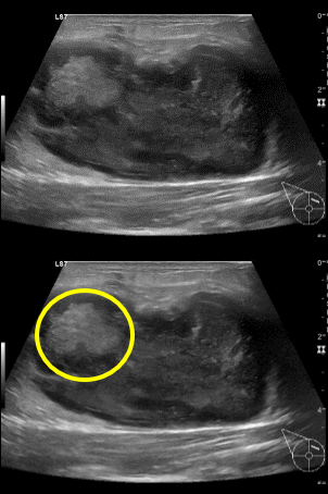
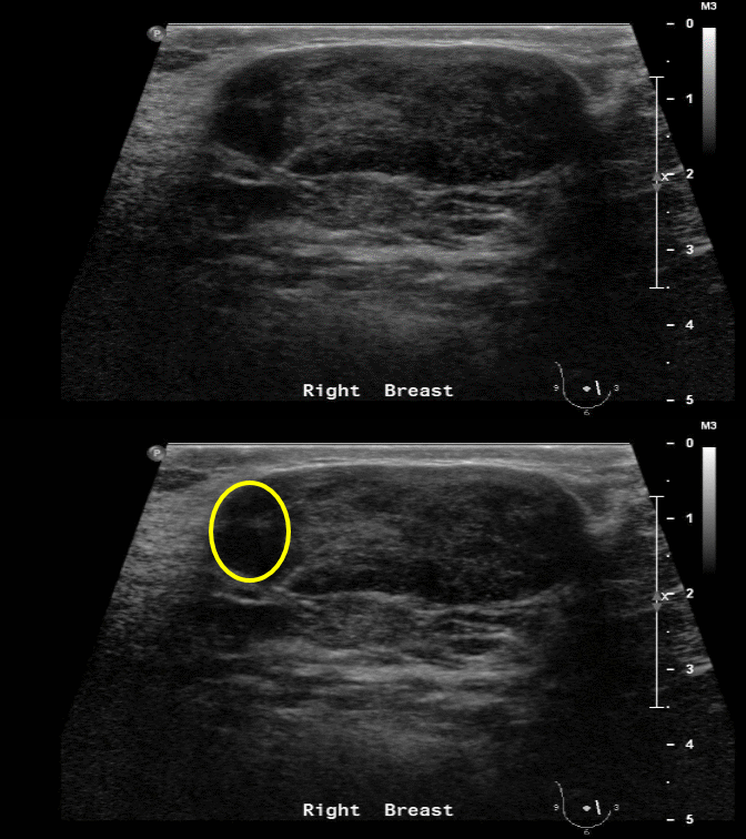
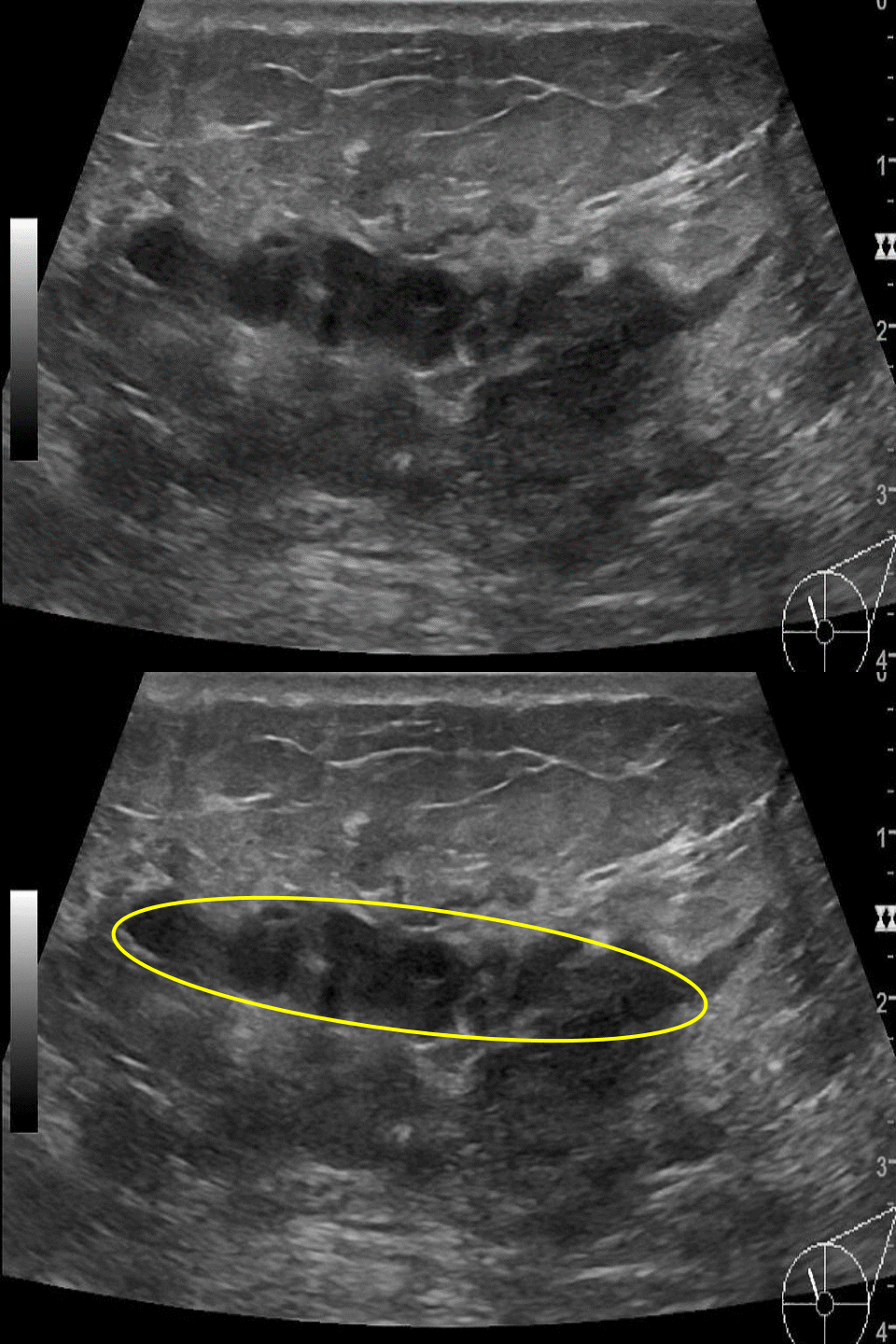
| Types of breast lumps | Characteristic findings |
|---|---|
| Cyst |
|
| Abscess |
|
| Mastitis |
|
| Galactocele |
|
| Seroma |
|
| Liponecrosis |
|
| Hemangioma |
|
| Fibroadenoma |
|
| Phyllodes tumor |
|
| Hamartoma |
|
| Papilloma |
|
 |
 |
 |
References
- ↑ Shah R, Rosso K, Nathanson SD (2014). "Pathogenesis, prevention, diagnosis and treatment of breast cancer". World J Clin Oncol. 5 (3): 283–98. doi:10.5306/wjco.v5.i3.283. PMC 4127601. PMID 25114845.
- ↑ Lehman CD, Lee AY, Lee CI (2014). "Imaging management of palpable breast abnormalities". AJR Am J Roentgenol. 203 (5): 1142–53. doi:10.2214/AJR.14.12725. PMID 25341156.
- ↑ Moss HA, Britton PD, Flower CD, Freeman AH, Lomas DJ, Warren RM (1999). "How reliable is modern breast imaging in differentiating benign from malignant breast lesions in the symptomatic population?". Clin Radiol. 54 (10): 676–82. PMID 10541394.
- ↑ Masciadri N, Ferranti C (2011). "Benign breast lesions: Ultrasound". J Ultrasound. 14 (2): 55–65. doi:10.1016/j.jus.2011.03.002. PMC 3558101. PMID 23396888.