Atrial septal defect ostium secundum anatomy
|
Atrial septal defect ostium secundum Microchapters |
|
Diagnosis |
|---|
|
Treatment |
|
Case Studies |
|
Atrial septal defect ostium secundum anatomy On the Web |
|
American Roentgen Ray Society Images of Atrial septal defect ostium secundum anatomy |
|
Directions to Hospitals Treating Atrial septal defect ostium secundum |
|
Risk calculators and risk factors for Atrial septal defect ostium secundum anatomy |
Editor-In-Chief: C. Michael Gibson, M.S., M.D. [1]; Associate Editors-In-Chief: Priyamvada Singh, M.B.B.S. [2]; Cafer Zorkun, M.D., Ph.D. [3]; Assistant Editor-In-Chief: Kristin Feeney, B.S. [4]
Overview
During fetal development, the septal wall may fail to fuse causing an atrial septal defect to arise. An ostium secundum atrial septal defect is one such type of malformation arising from the irregular development of the foramen ovale, septum secundum or septum primum. It is the most common type of atrial septal defect.
Anatomy


Embryogenesis
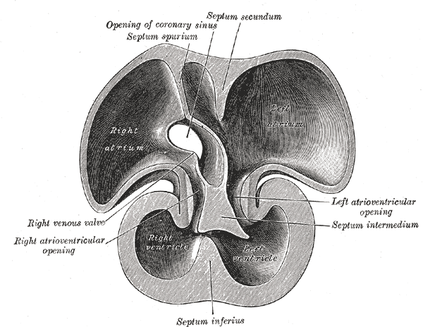
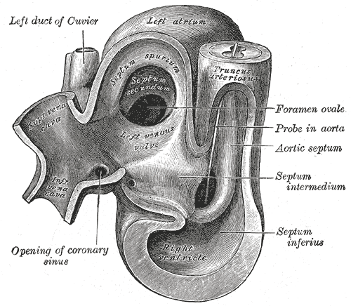
- The septum secundum is semilunar in shape. It grows downward from the upper wall of the atrium immediately to the right of the primary septum and foramen ovale. Shortly after birth the septum secundum fuses with the primary septum and through this means the foramen ovale is closed. Sometimes the septal fusion is incomplete and the upper part of the foramen remains patent.
- The limbus fossae ovalis denotes the free margin of the septum secundum. The ostium secundum (or foramen secundum) is a foramen in the septum primum. It should not be confused with the foramen ovale, which is a foramen in the septum secundum. It can arises from an enlarged foramen ovale, inadequate growth of the septum secundum, or excessive absorption of the septum.