Sarcoidosis: Difference between revisions
| Line 25: | Line 25: | ||
==Causes and pathophysiology== | ==Causes and pathophysiology== | ||
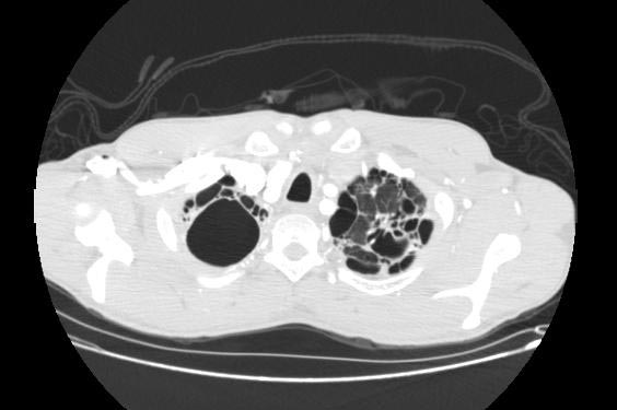
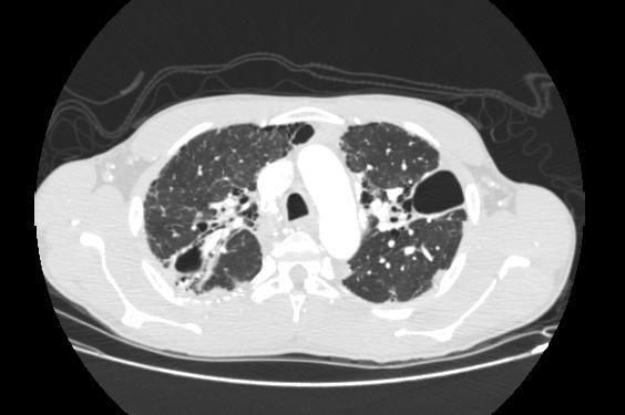
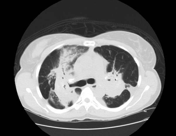
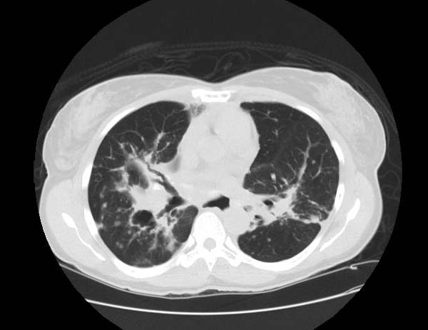
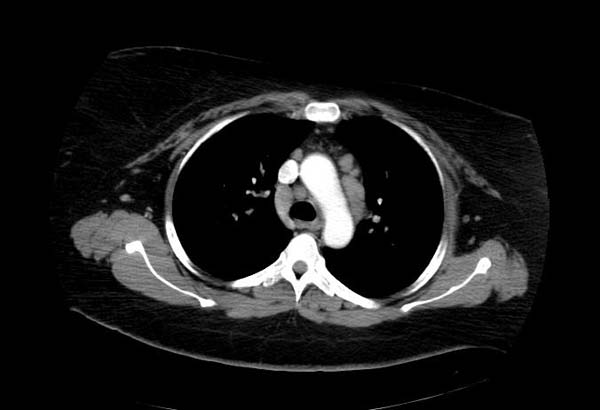
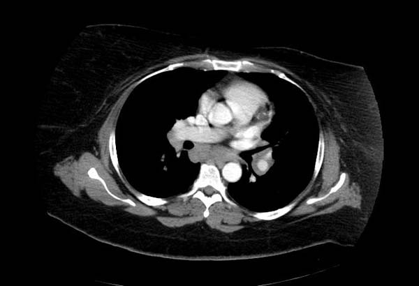
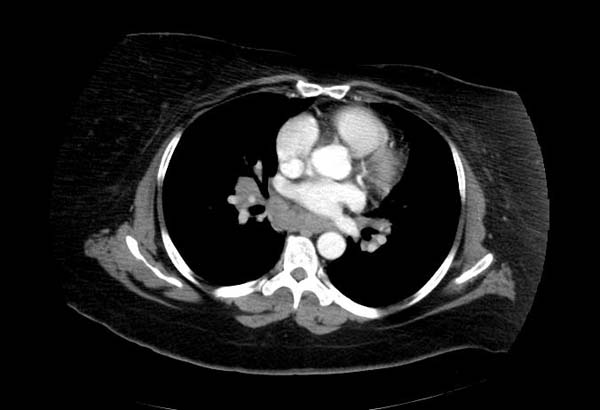
==Image Examples== | ==Image Examples== | ||
Revision as of 14:24, 26 September 2012
For patient information click here
| Sarcoidosis | |
_lymph_node_biopsy.jpg) | |
|---|---|
| Sarcoidosis in a Lymph Node. | |
| ICD-10 | D86 |
| ICD-9 | 135 |
| OMIM | 181000 |
| DiseasesDB | 11797 |
| MedlinePlus | 000076 |
| MeSH | D012507 |
|
Sarcoidosis Microchapters |
|
Diagnosis |
|---|
|
Treatment |
|
Case Studies |
|
Sarcoidosis On the Web |
|
American Roentgen Ray Society Images of Sarcoidosis |
Editor-in-Chief: Philip Marcus, M.D., M.P.H. [1], Division of Pulmonary Medicine St. Francis Hospital-The Heart Center, Roslyn, NY
Associate Editor-In-Chief: Cafer Zorkun, M.D., Ph.D. [2] and Hilary Womble, M. D.[[3]]
Click Here For The Chapter On The Heart In Sarcoidosis
Causes and pathophysiology
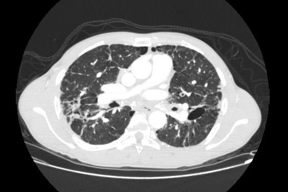
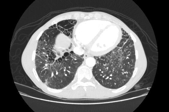
Image Examples
-
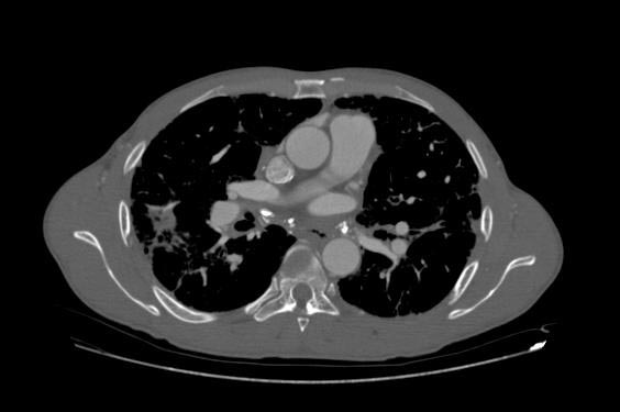
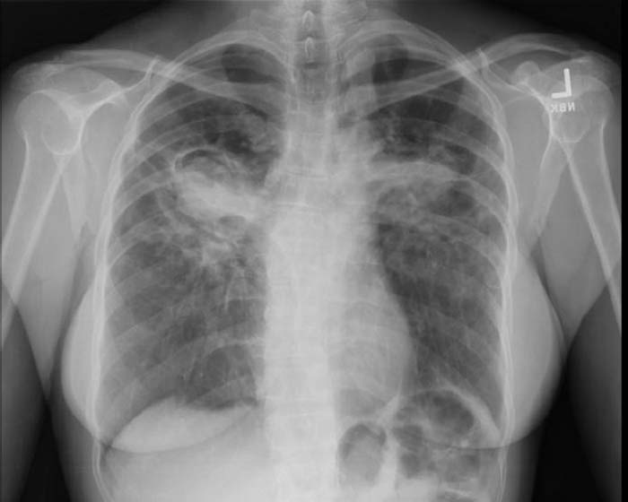
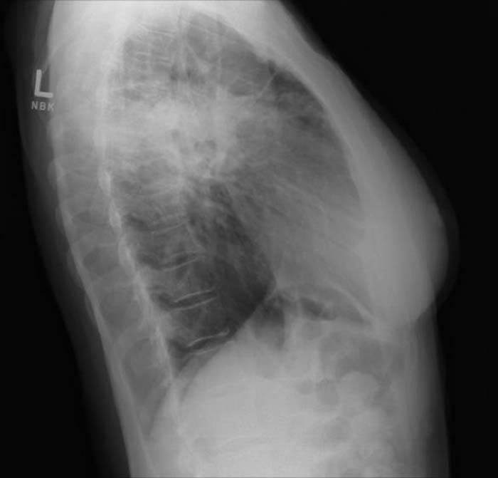
Sarcoidosis
-
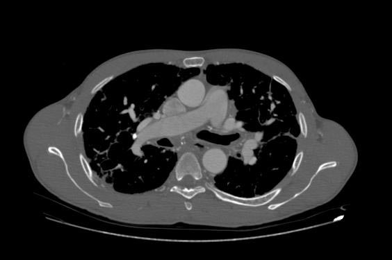
Sarcoidosis
-
Sarcoidosis


-
Sarcoidosis
-
Sarcoidosis
-
Sarcoidosis


-
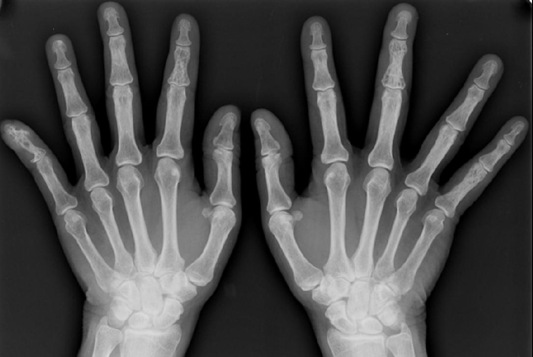
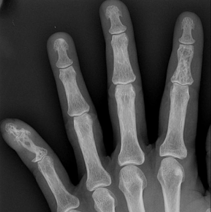
Osseous sarcoidosis
-
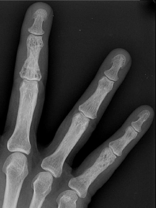
Osseous sarcoidosis
-
Osseous sarcoidosis


-
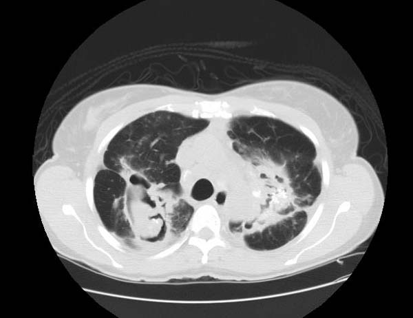
Complicated sarcoidosis
-
Complicated sarcoidosis
-
Complicated sarcoidosis


-
Complicated sarcoidosis
-
Complicated sarcoidosis


-
Sarcoidosis
-
Sarcoidosis
-
Sarcoidosis


Treatment
Corticosteroids, most commonly prednisone, have been the standard treatment for many years. In some patients, this treatment can slow or reverse the course of the disease, but other patients unfortunately do not respond to steroid therapy. The use of corticosteroids in mild disease is controversial because in many cases the disease remits spontaneously. Additionally, corticosteroids have many recognized dose- and duration-related side effects (which can be reduced through the use of alternate-day dosing for those on chronic prednisone therapy [1]), and their use is generally limited to severe, progressive, or organ-threatening disease. The influence of corticosteroids or other immunosuppressants on the natural history is unclear.
Severe symptoms are generally treated with steroids, and steroid-sparing agents such as azathioprine and methotrexate are often used. Rarely, cyclophosphamide has also been used. As the granulomas are caused by collections of immune system cells, particularly T cells, there has been some early indications of success using immunosuppressants, interleukin-2 inhibitors or anti-tumor necrosis factor-alpha treatment (such as infliximab). Unfortunately, none of these have provided reliable treatment and there can be significant side effects such as an increased risk of reactivating latent tuberculosis.
Avoidance of sunlight and Vitamin D foods may be helpful in patients who are susceptible to developing hypercalcemia.
Case Examples
Case #1
Clinical Summary
This 33-year-old white female was admitted for evaluation of abnormal findings on a chest x-ray. She was asymptomatic and a physical examination revealed no significant abnormalities. Laboratory results indicated hypercalcemia and elevated gamma globulin. Radiographic examination showed enlarged subcarinal, hilar, and right paratracheal lymph nodes. A right paratracheal lymph node was biopsied. Special stains for acid-fast bacilli and fungi were negative and a diagnosis of sarcoidosis was made.
Histopathological Findings


See also
References
External links
- The Foundation for Sarcoidosis Research
- Pathology Images of Sarcoidosis and Other Granulomatous Diseases
- Microscopy of granulomas in sarcoidosis
- Template:GPnotebook
- The Sarcoidosis Community is The Foundation for Sarcoidosis Research's online community
- Dorothy P. and Richard P. Simmons Center for Interstitial Lung Disease
- MedPix Pulmonary Sarcoid
da:Sarkoidose de:Sarkoidose eo:Sarkoidozo eu:Sarkoidosia he:סרקואידוזיס lb:Sarkoidos nl:Sarcoïdose no:Sarkoidose nn:sarkoidose sq:Sarkoidoza fi:Sarkoidoosi sv:Sarkoidos