Sandbox:iqra: Difference between revisions
No edit summary |
No edit summary |
||
| Line 29: | Line 29: | ||
! style="background:#4479BA; color: #FFFFFF;" align="center" |Imaging | ! style="background:#4479BA; color: #FFFFFF;" align="center" |Imaging | ||
|- | |- | ||
! rowspan=" | ! rowspan="54" |Abdominal causes | ||
! rowspan=" | ! rowspan="39" |Inflammatory causes | ||
! rowspan="9" |Pancreato-biliary disorders | ! rowspan="9" |Pancreato-biliary disorders | ||
| colspan="1" rowspan="1" style="padding: 5px 5px; background: #DCDCDC;" align="center" | [[Cholangitis|Acute cholangitis]] | | colspan="1" rowspan="1" style="padding: 5px 5px; background: #DCDCDC;" align="center" | [[Cholangitis|Acute cholangitis]] | ||
| Line 153: | Line 153: | ||
| style="padding: 5px 5px; background: #F5F5F5;" align="center" | + | | style="padding: 5px 5px; background: #F5F5F5;" align="center" | + | ||
| style="padding: 5px 5px; background: #F5F5F5;" align="center" | − | | style="padding: 5px 5px; background: #F5F5F5;" align="center" | − | ||
| style="padding: 5px 5px; background: #F5F5F5;" align="center" | | | style="padding: 5px 5px; background: #F5F5F5;" align="center" | - | ||
| style="padding: 5px 5px; background: #F5F5F5;" align="center" | − | | style="padding: 5px 5px; background: #F5F5F5;" align="center" | − | ||
| style="padding: 5px 5px; background: #F5F5F5;" align="center" | − | | style="padding: 5px 5px; background: #F5F5F5;" align="center" | − | ||
| Line 160: | Line 160: | ||
| style="padding: 5px 5px; background: #F5F5F5;" align="left" | | | style="padding: 5px 5px; background: #F5F5F5;" align="left" | | ||
* Increased AMA level, abnormal [[LFTs]] | * Increased AMA level, abnormal [[LFTs]] | ||
| style="padding: 5px 5px; background: #F5F5F5;" align="left" | | | style="padding: 5px 5px; background: #F5F5F5;" align="left" |ERCP | ||
| style="padding: 5px 5px; background: #F5F5F5;" align="left" | | | style="padding: 5px 5px; background: #F5F5F5;" align="left" | | ||
|- | |- | ||
| Line 199: | Line 199: | ||
| style="padding: 5px 5px; background: #F5F5F5;" align="center" | − | | style="padding: 5px 5px; background: #F5F5F5;" align="center" | − | ||
| style="padding: 5px 5px; background: #F5F5F5;" align="center" | − | | style="padding: 5px 5px; background: #F5F5F5;" align="center" | − | ||
| style="padding: 5px 5px; background: #F5F5F5;" align="left" | | | style="padding: 5px 5px; background: #F5F5F5;" align="left" |Normal to hyperactive for dislodged stone | ||
| style="padding: 5px 5px; background: #F5F5F5;" align="left" | | | style="padding: 5px 5px; background: #F5F5F5;" align="left" | | ||
* [[Leukocytosis]] | * [[Leukocytosis]] | ||
| Line 205: | Line 205: | ||
| style="padding: 5px 5px; background: #F5F5F5;" align="left" |Fatty food intolerance | | style="padding: 5px 5px; background: #F5F5F5;" align="left" |Fatty food intolerance | ||
|- | |- | ||
! colspan="1" rowspan=" | ! colspan="1" rowspan="8" style="padding: 5px 5px; background: #DCDCDC;" align="center" | Gastric causes | ||
| colspan="1" rowspan="1" style="padding: 5px 5px; background: #DCDCDC;" align="center" | [[Peptic Ulcer Disease|Peptic ulcer disease]] | | colspan="1" rowspan="1" style="padding: 5px 5px; background: #DCDCDC;" align="center" | [[Peptic Ulcer Disease|Peptic ulcer disease]] | ||
| style="padding: 5px 5px; background: #F5F5F5;" align="center" |Diffuse | | style="padding: 5px 5px; background: #F5F5F5;" align="center" |Diffuse | ||
| Line 215: | Line 215: | ||
* Gastric ulcer- [[melena]] and [[hematemesis]] | * Gastric ulcer- [[melena]] and [[hematemesis]] | ||
* Duodenal ulcer- [[melena]] and [[hematochezia]] | * Duodenal ulcer- [[melena]] and [[hematochezia]] | ||
| style="padding: 5px 5px; background: #F5F5F5;" align="center" | | | style="padding: 5px 5px; background: #F5F5F5;" align="center" | Positive if perforated | ||
| style="padding: 5px 5px; background: #F5F5F5;" align="center" | | | style="padding: 5px 5px; background: #F5F5F5;" align="center" | Positive if perforated | ||
| style="padding: 5px 5px; background: #F5F5F5;" align="center" | | | style="padding: 5px 5px; background: #F5F5F5;" align="center" | Positive if perforated | ||
| style="padding: 5px 5px; background: #F5F5F5;" align="left" |N | | style="padding: 5px 5px; background: #F5F5F5;" align="left" |N | ||
| style="padding: 5px 5px; background: #F5F5F5;" align="left" | | | style="padding: 5px 5px; background: #F5F5F5;" align="left" | | ||
| Line 226: | Line 226: | ||
| style="padding: 5px 5px; background: #F5F5F5;" align="left" |Air under [[diaphragm]] in upright [[CXR]] | | style="padding: 5px 5px; background: #F5F5F5;" align="left" |Air under [[diaphragm]] in upright [[CXR]] | ||
| style="padding: 5px 5px; background: #F5F5F5;" align="left" |Upper GI [[endoscopy]] for diagnosis | | style="padding: 5px 5px; background: #F5F5F5;" align="left" |Upper GI [[endoscopy]] for diagnosis | ||
|- | |||
!Disease | |||
!Abdominal Pain | |||
! colspan="1" rowspan="1" |Fever | |||
!Rigors and chills | |||
!Jaundice | |||
!Diarrhea | |||
!GI Bleed | |||
!Hypo- | |||
tension | |||
! colspan="1" rowspan="1" |Guarding | |||
!Rebound Tenderness | |||
!Bowel sounds | |||
! colspan="1" rowspan="1" |Lab Findings | |||
!Imaging | |||
!Comments | |||
|- | |- | ||
| style="padding: 5px 5px; background: #DCDCDC;" align="center" |[[Gastritis|Gastritis]] | | style="padding: 5px 5px; background: #DCDCDC;" align="center" |[[Gastritis|Gastritis]] | ||
| Line 233: | Line 249: | ||
| style="padding: 5px 5px; background: #F5F5F5;" align="center" |− | | style="padding: 5px 5px; background: #F5F5F5;" align="center" |− | ||
| style="padding: 5px 5px; background: #F5F5F5;" align="center" | − | | style="padding: 5px 5px; background: #F5F5F5;" align="center" | − | ||
| style="padding: 5px 5px; background: #F5F5F5;" align="center" | | | style="padding: 5px 5px; background: #F5F5F5;" align="center" | Positive in chronic gastritis | ||
| style="padding: 5px 5px; background: #F5F5F5;" align="center" |− | | style="padding: 5px 5px; background: #F5F5F5;" align="center" |− | ||
| style="padding: 5px 5px; background: #F5F5F5;" align="center" |− | | style="padding: 5px 5px; background: #F5F5F5;" align="center" |− | ||
| Line 253: | Line 269: | ||
| style="padding: 5px 5px; background: #F5F5F5;" align="center" |− | | style="padding: 5px 5px; background: #F5F5F5;" align="center" |− | ||
| style="padding: 5px 5px; background: #F5F5F5;" align="left" |N | | style="padding: 5px 5px; background: #F5F5F5;" align="left" |N | ||
| style="padding: 5px 5px; background: #F5F5F5;" align="left" | | | style="padding: 5px 5px; background: #F5F5F5;" align="left" |N | ||
| style="padding: 5px 5px; background: #F5F5F5;" align="left" |Gastric emptying studies | | style="padding: 5px 5px; background: #F5F5F5;" align="left" |Gastric emptying studies | ||
| style="padding: 5px 5px; background: #F5F5F5;" align="left" |[[Endoscopy]] for alarm signs | | style="padding: 5px 5px; background: #F5F5F5;" align="left" |[[Esophageal]] [[manometry]], [[Endoscopy]] for alarm signs | ||
|- | |- | ||
| style="padding: 5px 5px; background: #DCDCDC;" align="center" |[[Gastric outlet obstruction|Gastric outlet obstruction]] | | style="padding: 5px 5px; background: #DCDCDC;" align="center" |[[Gastric outlet obstruction|Gastric outlet obstruction]] | ||
| Line 305: | Line 321: | ||
| style="padding: 5px 5px; background: #F5F5F5;" align="center" | ± | | style="padding: 5px 5px; background: #F5F5F5;" align="center" | ± | ||
| style="padding: 5px 5px; background: #F5F5F5;" align="center" | − | | style="padding: 5px 5px; background: #F5F5F5;" align="center" | − | ||
| style="padding: 5px 5px; background: #F5F5F5;" align="center" | | | style="padding: 5px 5px; background: #F5F5F5;" align="center" | Positive, depends on site | ||
| style="padding: 5px 5px; background: #F5F5F5;" align="center" | + | | style="padding: 5px 5px; background: #F5F5F5;" align="center" | + | ||
| style="padding: 5px 5px; background: #F5F5F5;" align="center" | + | | style="padding: 5px 5px; background: #F5F5F5;" align="center" | + | ||
| Line 334: | Line 350: | ||
| style="padding: 5px 5px; background: #F5F5F5;" align="left" |Postgastrectomy | | style="padding: 5px 5px; background: #F5F5F5;" align="left" |Postgastrectomy | ||
|- | |- | ||
! rowspan=" | ! | ||
!Disease | |||
!Abdominal Pain | |||
! colspan="1" rowspan="1" |Fever | |||
!Rigors and chills | |||
!Jaundice | |||
!Diarrhea | |||
!GI Bleed | |||
!Hypo- | |||
tension | |||
! colspan="1" rowspan="1" |Guarding | |||
!Rebound Tenderness | |||
!Bowel sounds | |||
! colspan="1" rowspan="1" |Lab Findings | |||
!Imaging | |||
!Comments | |||
|- | |||
! rowspan="12" style="padding: 5px 5px; background: #DCDCDC;" align="center" |Intestinal causes | |||
| colspan="1" rowspan="1" style="padding: 5px 5px; background: #DCDCDC;" align="center" |[[Acute appendicitis]] | | colspan="1" rowspan="1" style="padding: 5px 5px; background: #DCDCDC;" align="center" |[[Acute appendicitis]] | ||
| style="padding: 5px 5px; background: #F5F5F5;" align="center" |Starts in [[epigastrium]], migrates to RLQ | | style="padding: 5px 5px; background: #F5F5F5;" align="center" |Starts in [[epigastrium]], migrates to RLQ | ||
| style="padding: 5px 5px; background: #F5F5F5;" align="center" | + | | style="padding: 5px 5px; background: #F5F5F5;" align="center" | + | ||
| style="padding: 5px 5px; background: #F5F5F5;" align="center" | | | style="padding: 5px 5px; background: #F5F5F5;" align="center" | Positive in pyogenic appendicitis | ||
| style="padding: 5px 5px; background: #F5F5F5;" align="center" | − | | style="padding: 5px 5px; background: #F5F5F5;" align="center" | − | ||
| style="padding: 5px 5px; background: #F5F5F5;" align="center" | − | | style="padding: 5px 5px; background: #F5F5F5;" align="center" | − | ||
| style="padding: 5px 5px; background: #F5F5F5;" align="center" | − | | style="padding: 5px 5px; background: #F5F5F5;" align="center" | − | ||
| style="padding: 5px 5px; background: #F5F5F5;" align="center" | | | style="padding: 5px 5px; background: #F5F5F5;" align="center" | Positive in perforated appendicitis | ||
| style="padding: 5px 5px; background: #F5F5F5;" align="center" | + | | style="padding: 5px 5px; background: #F5F5F5;" align="center" | + | ||
| style="padding: 5px 5px; background: #F5F5F5;" align="center" | + | | style="padding: 5px 5px; background: #F5F5F5;" align="center" | + | ||
| Line 348: | Line 381: | ||
| style="padding: 5px 5px; background: #F5F5F5;" align="left" | | | style="padding: 5px 5px; background: #F5F5F5;" align="left" | | ||
* [[Leukocytosis]] | * [[Leukocytosis]] | ||
| style="padding: 5px 5px; background: #F5F5F5;" align="left" |Ultrasound | | style="padding: 5px 5px; background: #F5F5F5;" align="left" |Ct scan and | ||
Ultrasound | |||
| style="padding: 5px 5px; background: #F5F5F5;" align="left" |[[Nausea and vomiting|Nausea & vomiting]], [[decreased appetite]] | | style="padding: 5px 5px; background: #F5F5F5;" align="left" |[[Nausea and vomiting|Nausea & vomiting]], [[decreased appetite]] | ||
|- | |- | ||
| Line 358: | Line 392: | ||
| style="padding: 5px 5px; background: #F5F5F5;" align="center" | ± | | style="padding: 5px 5px; background: #F5F5F5;" align="center" | ± | ||
| style="padding: 5px 5px; background: #F5F5F5;" align="center" | [[Hematochezia]] | | style="padding: 5px 5px; background: #F5F5F5;" align="center" | [[Hematochezia]] | ||
| style="padding: 5px 5px; background: #F5F5F5;" align="center" | | | style="padding: 5px 5px; background: #F5F5F5;" align="center" | Positive in perforated diverticulitis | ||
| style="padding: 5px 5px; background: #F5F5F5;" align="center" | + | | style="padding: 5px 5px; background: #F5F5F5;" align="center" | + | ||
| style="padding: 5px 5px; background: #F5F5F5;" align="center" | + | | style="padding: 5px 5px; background: #F5F5F5;" align="center" | + | ||
| Line 372: | Line 406: | ||
| style="padding: 5px 5px; background: #F5F5F5;" align="center" | − | | style="padding: 5px 5px; background: #F5F5F5;" align="center" | − | ||
| style="padding: 5px 5px; background: #F5F5F5;" align="center" | ± | | style="padding: 5px 5px; background: #F5F5F5;" align="center" | ± | ||
| style="padding: 5px 5px; background: #F5F5F5;" align="center" | | | style="padding: 5px 5px; background: #F5F5F5;" align="center" | + | ||
| style="padding: 5px 5px; background: #F5F5F5;" align="center" | [[Hematochezia]] | | style="padding: 5px 5px; background: #F5F5F5;" align="center" | [[Hematochezia]] | ||
| style="padding: 5px 5px; background: #F5F5F5;" align="center" |− | | style="padding: 5px 5px; background: #F5F5F5;" align="center" |− | ||
| style="padding: 5px 5px; background: #F5F5F5;" align="center" |− | | style="padding: 5px 5px; background: #F5F5F5;" align="center" |− | ||
| style="padding: 5px 5px; background: #F5F5F5;" align="center" |− | | style="padding: 5px 5px; background: #F5F5F5;" align="center" |− | ||
| style="padding: 5px 5px; background: #F5F5F5;" align="left" | | | style="padding: 5px 5px; background: #F5F5F5;" align="left" |Normal or hyperactive | ||
| style="padding: 5px 5px; background: #F5F5F5;" align="left" | | | style="padding: 5px 5px; background: #F5F5F5;" align="left" | | ||
* [[Anti-neutrophil cytoplasmic antibody]] ([[P-ANCA]]) in [[Ulcerative colitis]] | * [[Anti-neutrophil cytoplasmic antibody]] ([[P-ANCA]]) in [[Ulcerative colitis]] | ||
| Line 389: | Line 423: | ||
| style="padding: 5px 5px; background: #DCDCDC;" align="center" |[[Irritable bowel syndrome]] | | style="padding: 5px 5px; background: #DCDCDC;" align="center" |[[Irritable bowel syndrome]] | ||
| style="padding: 5px 5px; background: #F5F5F5;" align="center" |Diffuse | | style="padding: 5px 5px; background: #F5F5F5;" align="center" |Diffuse | ||
| style="padding: 5px 5px; background: #F5F5F5;" align="center" | | | style="padding: 5px 5px; background: #F5F5F5;" align="center" | - | ||
| style="padding: 5px 5px; background: #F5F5F5;" align="center" | − | | style="padding: 5px 5px; background: #F5F5F5;" align="center" | − | ||
| style="padding: 5px 5px; background: #F5F5F5;" align="center" | − | | style="padding: 5px 5px; background: #F5F5F5;" align="center" | − | ||
| style="padding: 5px 5px; background: #F5F5F5;" align="center" | | | style="padding: 5px 5px; background: #F5F5F5;" align="center" | ± | ||
| style="padding: 5px 5px; background: #F5F5F5;" align="center" | − | | style="padding: 5px 5px; background: #F5F5F5;" align="center" | − | ||
| style="padding: 5px 5px; background: #F5F5F5;" align="center" | − | | style="padding: 5px 5px; background: #F5F5F5;" align="center" | − | ||
| Line 398: | Line 432: | ||
| style="padding: 5px 5px; background: #F5F5F5;" align="center" | − | | style="padding: 5px 5px; background: #F5F5F5;" align="center" | − | ||
| style="padding: 5px 5px; background: #F5F5F5;" align="left" |N | | style="padding: 5px 5px; background: #F5F5F5;" align="left" |N | ||
| style="padding: 5px 5px; background: #F5F5F5;" align="left" | | | style="padding: 5px 5px; background: #F5F5F5;" align="left" |Normal | ||
| style="padding: 5px 5px; background: #F5F5F5;" align="left" | | | style="padding: 5px 5px; background: #F5F5F5;" align="left" |Normal | ||
| style="padding: 5px 5px; background: #F5F5F5;" align="left" |Symptomatic treatment | | style="padding: 5px 5px; background: #F5F5F5;" align="left" |Symptomatic treatment | ||
* High [[dietary fiber]] | * High [[dietary fiber]] | ||
| Line 435: | Line 469: | ||
* [[Arthritis]] | * [[Arthritis]] | ||
* [[Ascites]] | * [[Ascites]] | ||
|- | |||
!Disease | |||
!Abdominal Pain | |||
! colspan="1" rowspan="1" |Fever | |||
!Rigors and chills | |||
!Jaundice | |||
!Diarrhea | |||
!GI Bleed | |||
!Hypo- | |||
tension | |||
! colspan="1" rowspan="1" |Guarding | |||
!Rebound Tenderness | |||
!Bowel sounds | |||
! colspan="1" rowspan="1" |Lab Findings | |||
!Imaging | |||
!Comments | |||
|- | |- | ||
| style="padding: 5px 5px; background: #DCDCDC;" align="center" |[[Toxic megacolon]] | | style="padding: 5px 5px; background: #DCDCDC;" align="center" |[[Toxic megacolon]] | ||
| Line 445: | Line 495: | ||
| style="padding: 5px 5px; background: #F5F5F5;" align="center" | + | | style="padding: 5px 5px; background: #F5F5F5;" align="center" | + | ||
| style="padding: 5px 5px; background: #F5F5F5;" align="center" | ± | | style="padding: 5px 5px; background: #F5F5F5;" align="center" | ± | ||
| style="padding: 5px 5px; background: #F5F5F5;" align="center" | | | style="padding: 5px 5px; background: #F5F5F5;" align="center" | + | ||
| style="padding: 5px 5px; background: #F5F5F5;" align="left" |Hypoactive | | style="padding: 5px 5px; background: #F5F5F5;" align="left" |Hypoactive | ||
| style="padding: 5px 5px; background: #F5F5F5;" align="left" | | | style="padding: 5px 5px; background: #F5F5F5;" align="left" | | ||
| Line 453: | Line 503: | ||
*[[Metabolic alkalosis]] associated with a poor [[prognosis]] | *[[Metabolic alkalosis]] associated with a poor [[prognosis]] | ||
*[[Metabolic acidosis]] secondary to [[ischemic colitis]] | *[[Metabolic acidosis]] secondary to [[ischemic colitis]] | ||
| style="padding: 5px 5px; background: #F5F5F5;" align="left" |CT | | style="padding: 5px 5px; background: #F5F5F5;" align="left" |CT and [[Ultrasound]] shows: | ||
*Loss of colonic haustration | |||
[[Ultrasound]] shows: | |||
*Loss of | |||
*Hypoechoic and thickened bowel walls with irregular internal margins in the [[sigmoid]] and descending colon | *Hypoechoic and thickened bowel walls with irregular internal margins in the [[sigmoid]] and descending colon | ||
*Prominent dilation of the transverse colon (>6 cm) | *Prominent dilation of the transverse colon (>6 cm) | ||
| Line 512: | Line 557: | ||
| style="padding: 5px 5px; background: #F5F5F5;" align="center" | + | | style="padding: 5px 5px; background: #F5F5F5;" align="center" | + | ||
| style="padding: 5px 5px; background: #F5F5F5;" align="center" |[[Hematochezia]] | | style="padding: 5px 5px; background: #F5F5F5;" align="center" |[[Hematochezia]] | ||
| style="padding: 5px 5px; background: #F5F5F5;" align="center" | | | style="padding: 5px 5px; background: #F5F5F5;" align="center" | Positive in fulminant colitis | ||
| style="padding: 5px 5px; background: #F5F5F5;" align="center" |± | | style="padding: 5px 5px; background: #F5F5F5;" align="center" |± | ||
| style="padding: 5px 5px; background: #F5F5F5;" align="center" |± | | style="padding: 5px 5px; background: #F5F5F5;" align="center" |± | ||
| Line 524: | Line 569: | ||
* Edema | * Edema | ||
| style="padding: 5px 5px; background: #F5F5F5;" align="left" | | | style="padding: 5px 5px; background: #F5F5F5;" align="left" | | ||
|- | |||
!Disease | |||
!Abdominal Pain | |||
! colspan="1" rowspan="1" |Fever | |||
!Rigors and chills | |||
!Jaundice | |||
!Diarrhea | |||
!GI Bleed | |||
!Hypo- | |||
tension | |||
! colspan="1" rowspan="1" |Guarding | |||
!Rebound Tenderness | |||
!Bowel sounds | |||
! colspan="1" rowspan="1" |Lab Findings | |||
!Imaging | |||
!Comments | |||
|- | |- | ||
| style="padding: 5px 5px; background: #DCDCDC;" align="center" |Colon carcinoma | | style="padding: 5px 5px; background: #DCDCDC;" align="center" |Colon carcinoma | ||
| Line 554: | Line 615: | ||
| style="padding: 5px 5px; background: #F5F5F5;" align="center" |− | | style="padding: 5px 5px; background: #F5F5F5;" align="center" |− | ||
| style="padding: 5px 5px; background: #F5F5F5;" align="center" | + | | style="padding: 5px 5px; background: #F5F5F5;" align="center" | + | ||
| style="padding: 5px 5px; background: #F5F5F5;" align="center" | | | style="padding: 5px 5px; background: #F5F5F5;" align="center" | Positive in Hep A and E | ||
| style="padding: 5px 5px; background: #F5F5F5;" align="center" | − | | style="padding: 5px 5px; background: #F5F5F5;" align="center" | − | ||
| style="padding: 5px 5px; background: #F5F5F5;" align="center" | | | style="padding: 5px 5px; background: #F5F5F5;" align="center" | Positive in fulminant hepatitis | ||
| style="padding: 5px 5px; background: #F5F5F5;" align="center" | | | style="padding: 5px 5px; background: #F5F5F5;" align="center" | Positive in acute | ||
| style="padding: 5px 5px; background: #F5F5F5;" align="center" | + | | style="padding: 5px 5px; background: #F5F5F5;" align="center" | + | ||
| style="padding: 5px 5px; background: #F5F5F5;" align="left" |N | | style="padding: 5px 5px; background: #F5F5F5;" align="left" |N | ||
| Line 563: | Line 624: | ||
* Abnormal LFTs | * Abnormal LFTs | ||
* Viral serology | * Viral serology | ||
| style="padding: 5px 5px; background: #F5F5F5;" align="left" | | | style="padding: 5px 5px; background: #F5F5F5;" align="left" |US | ||
| style="padding: 5px 5px; background: #F5F5F5;" align="left" |Hep A and E have fecoral route of transmission and Hep B and C transmits via blood transfusion and sexual contact. | | style="padding: 5px 5px; background: #F5F5F5;" align="left" |Hep A and E have fecoral route of transmission and Hep B and C transmits via blood transfusion and sexual contact. | ||
|- | |- | ||
| style="padding: 5px 5px; background: #DCDCDC;" align="center" |[[Liver abscess]] | | style="padding: 5px 5px; background: #DCDCDC;" align="center" |[[Liver abscess]] | ||
| Line 593: | Line 637: | ||
| style="padding: 5px 5px; background: #F5F5F5;" align="center" | + | | style="padding: 5px 5px; background: #F5F5F5;" align="center" | + | ||
| style="padding: 5px 5px; background: #F5F5F5;" align="center" |± | | style="padding: 5px 5px; background: #F5F5F5;" align="center" |± | ||
| style="padding: 5px 5px; background: #F5F5F5;" align="center" |Normal | | style="padding: 5px 5px; background: #F5F5F5;" align="center" |Normal or hypoactive | ||
| style="padding: 5px 5px; background: #F5F5F5;" align="left" | | | style="padding: 5px 5px; background: #F5F5F5;" align="left" | | ||
* CBC | * CBC | ||
| Line 630: | Line 674: | ||
* [[Spider nevi]] | * [[Spider nevi]] | ||
* [[Asterixis]] | * [[Asterixis]] | ||
|- | |||
!Disease | |||
!Abdominal Pain | |||
! colspan="1" rowspan="1" |Fever | |||
!Rigors and chills | |||
!Jaundice | |||
!Diarrhea | |||
!GI Bleed | |||
!Hypo- | |||
tension | |||
! colspan="1" rowspan="1" |Guarding | |||
!Rebound Tenderness | |||
!Bowel sounds | |||
! colspan="1" rowspan="1" |Lab Findings | |||
!Imaging | |||
!Comments | |||
|- | |- | ||
| style="padding: 5px 5px; background: #DCDCDC;" align="center" |[[Budd-Chiari syndrome|Budd-Chiari syndrome]] | | style="padding: 5px 5px; background: #DCDCDC;" align="center" |[[Budd-Chiari syndrome|Budd-Chiari syndrome]] | ||
| Line 692: | Line 752: | ||
| style="padding: 5px 5px; background: #F5F5F5;" align="center" | + | | style="padding: 5px 5px; background: #F5F5F5;" align="center" | + | ||
| style="padding: 5px 5px; background: #F5F5F5;" align="center" | − | | style="padding: 5px 5px; background: #F5F5F5;" align="center" | − | ||
| style="padding: 5px 5px; background: #F5F5F5;" align="center" | | | style="padding: 5px 5px; background: #F5F5F5;" align="center" | + | ||
| style="padding: 5px 5px; background: #F5F5F5;" align="center" | + | | style="padding: 5px 5px; background: #F5F5F5;" align="center" | + | ||
| style="padding: 5px 5px; background: #F5F5F5;" align="center" |− | | style="padding: 5px 5px; background: #F5F5F5;" align="center" |− | ||
| Line 709: | Line 769: | ||
* Stigmata of liver disease | * Stigmata of liver disease | ||
* Cruveilhier- Baumgarten murmur | * Cruveilhier- Baumgarten murmur | ||
|- | |||
! | |||
!Disease | |||
!Abdominal Pain | |||
! colspan="1" rowspan="1" |Fever | |||
!Rigors and chills | |||
!Jaundice | |||
!Diarrhea | |||
!GI Bleed | |||
!Hypo- | |||
tension | |||
! colspan="1" rowspan="1" |Guarding | |||
!Rebound Tenderness | |||
!Bowel sounds | |||
! colspan="1" rowspan="1" |Lab Findings | |||
!Imaging | |||
!Comments | |||
|- | |- | ||
! style="padding: 5px 5px; background: #DCDCDC;" align="center" | Peritoneal causes | ! style="padding: 5px 5px; background: #DCDCDC;" align="center" | Peritoneal causes | ||
| Line 731: | Line 808: | ||
! colspan="2" rowspan="2" style="padding: 5px 5px; background: #DCDCDC;" align="center" |Renal causes | ! colspan="2" rowspan="2" style="padding: 5px 5px; background: #DCDCDC;" align="center" |Renal causes | ||
| style="padding: 5px 5px; background: #DCDCDC;" align="center" |Pyelonephritis | | style="padding: 5px 5px; background: #DCDCDC;" align="center" |Pyelonephritis | ||
| style="padding: 5px 5px; background: #F5F5F5;" align="center" | | | style="padding: 5px 5px; background: #F5F5F5;" align="center" |Unilateral | ||
| style="padding: 5px 5px; background: #F5F5F5;" align="center" | + | | style="padding: 5px 5px; background: #F5F5F5;" align="center" | + | ||
| style="padding: 5px 5px; background: #F5F5F5;" align="center" |± | | style="padding: 5px 5px; background: #F5F5F5;" align="center" |± | ||
| Line 738: | Line 815: | ||
| style="padding: 5px 5px; background: #F5F5F5;" align="center" | - | | style="padding: 5px 5px; background: #F5F5F5;" align="center" | - | ||
| style="padding: 5px 5px; background: #F5F5F5;" align="center" | + | | style="padding: 5px 5px; background: #F5F5F5;" align="center" | + | ||
| style="padding: 5px 5px; background: #F5F5F5;" align="center" | | | style="padding: 5px 5px; background: #F5F5F5;" align="center" | - | ||
| style="padding: 5px 5px; background: #F5F5F5;" align="center" | | | style="padding: 5px 5px; background: #F5F5F5;" align="center" | - | ||
| style="padding: 5px 5px; background: #F5F5F5;" align="center" |Hypoactive | | style="padding: 5px 5px; background: #F5F5F5;" align="center" |Hypoactive | ||
| style="padding: 5px 5px; background: #F5F5F5;" align="left" | | | style="padding: 5px 5px; background: #F5F5F5;" align="left" | | ||
| Line 749: | Line 826: | ||
* MRI | * MRI | ||
| style="padding: 5px 5px; background: #F5F5F5;" align="left" | | | style="padding: 5px 5px; background: #F5F5F5;" align="left" | | ||
* | *CVA tenderness | ||
|- | |- | ||
| style="padding: 5px 5px; background: #DCDCDC;" align="center" |[[Renal colic]] | | style="padding: 5px 5px; background: #DCDCDC;" align="center" |[[Renal colic]] | ||
| Line 782: | Line 859: | ||
| style="padding: 5px 5px; background: #F5F5F5;" align="left" |[[Leukocytosis]] with left shift indicates complications | | style="padding: 5px 5px; background: #F5F5F5;" align="left" |[[Leukocytosis]] with left shift indicates complications | ||
| style="padding: 5px 5px; background: #F5F5F5;" align="left" |[[Abdominal X-ray|Abdominal X ray]] | | style="padding: 5px 5px; background: #F5F5F5;" align="left" |[[Abdominal X-ray|Abdominal X ray]] | ||
* | * Dilated loops of bowel with air fluid levels | ||
* | * Gasless abdomen | ||
| style="padding: 5px 5px; background: #F5F5F5;" align="left" | | | style="padding: 5px 5px; background: #F5F5F5;" align="left" | | ||
* "Target sign"– , indicative of intussusception | * "Target sign"– , indicative of intussusception | ||
| Line 818: | Line 895: | ||
| style="padding: 5px 5px; background: #F5F5F5;" align="left" |Ultrasound | | style="padding: 5px 5px; background: #F5F5F5;" align="left" |Ultrasound | ||
| style="padding: 5px 5px; background: #F5F5F5;" align="left" |[[Nausea and vomiting|Nausea & vomiting]] | | style="padding: 5px 5px; background: #F5F5F5;" align="left" |[[Nausea and vomiting|Nausea & vomiting]] | ||
|- | |||
! | |||
! | |||
!Disease | |||
!Abdominal Pain | |||
! colspan="1" rowspan="1" |Fever | |||
!Rigors and chills | |||
!Jaundice | |||
!Diarrhea | |||
!GI Bleed | |||
!Hypo- | |||
tension | |||
! colspan="1" rowspan="1" |Guarding | |||
!Rebound Tenderness | |||
!Bowel sounds | |||
! colspan="1" rowspan="1" |Lab Findings | |||
!Imaging | |||
!Comments | |||
|- | |- | ||
! rowspan="4" style="padding: 5px 5px; background: #DCDCDC;" align="center" |Vascular Disorders | ! rowspan="4" style="padding: 5px 5px; background: #DCDCDC;" align="center" |Vascular Disorders | ||
| Line 827: | Line 922: | ||
| style="padding: 5px 5px; background: #F5F5F5;" align="center" | − | | style="padding: 5px 5px; background: #F5F5F5;" align="center" | − | ||
| style="padding: 5px 5px; background: #F5F5F5;" align="center" | + | | style="padding: 5px 5px; background: #F5F5F5;" align="center" | + | ||
| style="padding: 5px 5px; background: #F5F5F5;" align="center" | | | style="padding: 5px 5px; background: #F5F5F5;" align="center" | + | ||
| style="padding: 5px 5px; background: #F5F5F5;" align="center" | + if bowel becomes gangrenous | | style="padding: 5px 5px; background: #F5F5F5;" align="center" | + if bowel becomes gangrenous | ||
| style="padding: 5px 5px; background: #F5F5F5;" align="center" | + if bowel becomes gangrenous | | style="padding: 5px 5px; background: #F5F5F5;" align="center" | + if bowel becomes gangrenous | ||
| Line 846: | Line 941: | ||
| style="padding: 5px 5px; background: #F5F5F5;" align="center" | − | | style="padding: 5px 5px; background: #F5F5F5;" align="center" | − | ||
| style="padding: 5px 5px; background: #F5F5F5;" align="center" | + | | style="padding: 5px 5px; background: #F5F5F5;" align="center" | + | ||
| style="padding: 5px 5px; background: #F5F5F5;" align="center" | | | style="padding: 5px 5px; background: #F5F5F5;" align="center" | + | ||
| style="padding: 5px 5px; background: #F5F5F5;" align="center" | + | | style="padding: 5px 5px; background: #F5F5F5;" align="center" | + | ||
| style="padding: 5px 5px; background: #F5F5F5;" align="center" |<nowiki>+</nowiki> | | style="padding: 5px 5px; background: #F5F5F5;" align="center" |<nowiki>+</nowiki> | ||
| Line 857: | Line 952: | ||
* Double halo appearance, thumbprinting | * Double halo appearance, thumbprinting | ||
* Thickening of bowel | * Thickening of bowel | ||
| style="padding: 5px 5px; background: #F5F5F5;" align="left" | | | style="padding: 5px 5px; background: #F5F5F5;" align="left" |May lead to shock | ||
|- | |- | ||
! rowspan="2" style="padding: 5px 5px; background: #DCDCDC;" align="center" |Hemorrhagic causes | ! rowspan="2" style="padding: 5px 5px; background: #DCDCDC;" align="center" |Hemorrhagic causes | ||
| Line 866: | Line 961: | ||
| style="padding: 5px 5px; background: #F5F5F5;" align="center" | − | | style="padding: 5px 5px; background: #F5F5F5;" align="center" | − | ||
| style="padding: 5px 5px; background: #F5F5F5;" align="center" | − | | style="padding: 5px 5px; background: #F5F5F5;" align="center" | − | ||
| style="padding: 5px 5px; background: #F5F5F5;" align="center" | | | style="padding: 5px 5px; background: #F5F5F5;" align="center" |+ | ||
| style="padding: 5px 5px; background: #F5F5F5;" align="center" | + | | style="padding: 5px 5px; background: #F5F5F5;" align="center" | + | ||
| style="padding: 5px 5px; background: #F5F5F5;" align="center" | − | | style="padding: 5px 5px; background: #F5F5F5;" align="center" | − | ||
| Line 883: | Line 978: | ||
| style="padding: 5px 5px; background: #F5F5F5;" align="center" | − | | style="padding: 5px 5px; background: #F5F5F5;" align="center" | − | ||
| style="padding: 5px 5px; background: #F5F5F5;" align="center" | − | | style="padding: 5px 5px; background: #F5F5F5;" align="center" | − | ||
| style="padding: 5px 5px; background: #F5F5F5;" align="center" | | | style="padding: 5px 5px; background: #F5F5F5;" align="center" | + | ||
| style="padding: 5px 5px; background: #F5F5F5;" align="center" | + | | style="padding: 5px 5px; background: #F5F5F5;" align="center" | + | ||
| style="padding: 5px 5px; background: #F5F5F5;" align="center" | − | | style="padding: 5px 5px; background: #F5F5F5;" align="center" | − | ||
| Line 891: | Line 986: | ||
| style="padding: 5px 5px; background: #F5F5F5;" align="left" |CT scan | | style="padding: 5px 5px; background: #F5F5F5;" align="left" |CT scan | ||
| style="padding: 5px 5px; background: #F5F5F5;" align="left" |History of [[trauma]] | | style="padding: 5px 5px; background: #F5F5F5;" align="left" |History of [[trauma]] | ||
|- | |||
! | |||
! | |||
!Disease | |||
!Abdominal Pain | |||
! colspan="1" rowspan="1" |Fever | |||
!Rigors and chills | |||
!Jaundice | |||
!Diarrhea | |||
!GI Bleed | |||
!Hypo- | |||
tension | |||
! colspan="1" rowspan="1" |Guarding | |||
!Rebound Tenderness | |||
!Bowel sounds | |||
! colspan="1" rowspan="1" |Lab Findings | |||
!Imaging | |||
!Comments | |||
|- | |- | ||
! rowspan="4" style="padding: 5px 5px; background: #DCDCDC;" align="center" |Gynaecological Causes | ! rowspan="4" style="padding: 5px 5px; background: #DCDCDC;" align="center" |Gynaecological Causes | ||
| Line 997: | Line 1,110: | ||
* Spiral [[CT pulmonary angiogram]] | * Spiral [[CT pulmonary angiogram]] | ||
| style="padding: 5px 5px; background: #F5F5F5;" align="left" | | | style="padding: 5px 5px; background: #F5F5F5;" align="left" | | ||
* Dyspnea | |||
* Tachycardia | |||
* Pleuretic chest pain | |||
|- | |- | ||
! colspan="2" style="padding: 5px 5px; background: #DCDCDC;" align="center" |Pneumonia | ! colspan="2" style="padding: 5px 5px; background: #DCDCDC;" align="center" |Pneumonia | ||
| Line 1,008: | Line 1,124: | ||
| style="padding: 5px 5px; background: #F5F5F5;" align="center" | - | | style="padding: 5px 5px; background: #F5F5F5;" align="center" | - | ||
| style="padding: 5px 5px; background: #F5F5F5;" align="center" | - | | style="padding: 5px 5px; background: #F5F5F5;" align="center" | - | ||
| style="padding: 5px 5px; background: #F5F5F5;" align="center" | | | style="padding: 5px 5px; background: #F5F5F5;" align="center" |Normal or hypoactive | ||
| style="padding: 5px 5px; background: #F5F5F5;" align="left" | | | style="padding: 5px 5px; background: #F5F5F5;" align="left" | | ||
* ABGs | * ABGs | ||
| Line 1,022: | Line 1,138: | ||
| colspan="2" style="padding: 5px 5px; background: #DCDCDC;" align="center" |[[Myocardial Infarction]] | | colspan="2" style="padding: 5px 5px; background: #DCDCDC;" align="center" |[[Myocardial Infarction]] | ||
| style="padding: 5px 5px; background: #F5F5F5;" align="center" |[[Epigastric]] | | style="padding: 5px 5px; background: #F5F5F5;" align="center" |[[Epigastric]] | ||
| style="padding: 5px 5px; background: #F5F5F5;" align="center" | | | style="padding: 5px 5px; background: #F5F5F5;" align="center" |± | ||
| style="padding: 5px 5px; background: #F5F5F5;" align="center" | − | | style="padding: 5px 5px; background: #F5F5F5;" align="center" | − | ||
| style="padding: 5px 5px; background: #F5F5F5;" align="center" | − | | style="padding: 5px 5px; background: #F5F5F5;" align="center" | − | ||
| Line 1,032: | Line 1,148: | ||
| style="padding: 5px 5px; background: #F5F5F5;" align="left" |N | | style="padding: 5px 5px; background: #F5F5F5;" align="left" |N | ||
| style="padding: 5px 5px; background: #F5F5F5;" align="left" | | | style="padding: 5px 5px; background: #F5F5F5;" align="left" | | ||
* [[Cardiac enzymes]] | * [[Cardiac enzymes]] | ||
| style="padding: 5px 5px; background: #F5F5F5;" align="left" |[[Echocardiogram]] | | style="padding: 5px 5px; background: #F5F5F5;" align="left" |[[ECG]] | ||
[[Echocardiogram]] | |||
* Wall motion abnormality | * Wall motion abnormality | ||
* Wall rupture | * Wall rupture | ||
| Line 1,050: | Line 1,166: | ||
|- | |- | ||
| | | | ||
<figure-inline><figure-inline>[[Image:Right_upper_quadrant.PNG|link=Right upper quadrant abdominal pain resident survival guide|339x339px]]</figure-inline></figure-inline> | <figure-inline><figure-inline><figure-inline>[[Image:Right_upper_quadrant.PNG|link=Right upper quadrant abdominal pain resident survival guide|339x339px]]</figure-inline></figure-inline></figure-inline> | ||
| | | | ||
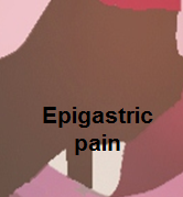
<figure-inline><figure-inline>[[Image:Epigastric_quadrant_pain.PNG|link=Epigastric pain resident survival guide|179x179px]]</figure-inline></figure-inline> | <figure-inline><figure-inline><figure-inline>[[Image:Epigastric_quadrant_pain.PNG|link=Epigastric pain resident survival guide|179x179px]]</figure-inline></figure-inline></figure-inline> | ||
| | | | ||
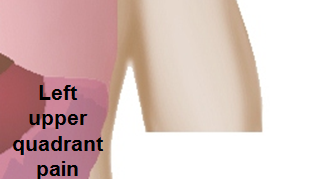
<figure-inline><figure-inline>[[Image:Left_upper_quadrant.PNG|link=Left upper quadrant abdominal pain resident survival guide|329x329px]]</figure-inline></figure-inline> | <figure-inline><figure-inline><figure-inline>[[Image:Left_upper_quadrant.PNG|link=Left upper quadrant abdominal pain resident survival guide|329x329px]]</figure-inline></figure-inline></figure-inline> | ||
|- | |- | ||
| | | | ||
<figure-inline><figure-inline>[[Image:Right_flank_quadrant.PNG|link=Right flank pain resident survival guide|338x338px]]</figure-inline></figure-inline> | <figure-inline><figure-inline><figure-inline>[[Image:Right_flank_quadrant.PNG|link=Right flank pain resident survival guide|338x338px]]</figure-inline></figure-inline></figure-inline> | ||
| | | | ||
<figure-inline><figure-inline>[[Image:Umbilical_pain.PNG|link=Umbilical region pain resident survival guide|165x165px]]</figure-inline></figure-inline> | <figure-inline><figure-inline><figure-inline>[[Image:Umbilical_pain.PNG|link=Umbilical region pain resident survival guide|165x165px]]</figure-inline></figure-inline></figure-inline> | ||
| | | | ||
<figure-inline><figure-inline>[[Image:Left_flank_quadrant.PNG|link=Left flank quadrant abdominal pain resident survival guide|335x335px]]</figure-inline></figure-inline> | <figure-inline><figure-inline><figure-inline>[[Image:Left_flank_quadrant.PNG|link=Left flank quadrant abdominal pain resident survival guide|335x335px]]</figure-inline></figure-inline></figure-inline> | ||
|- | |- | ||
| | | | ||
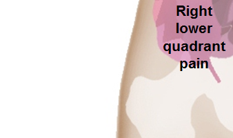
<figure-inline><figure-inline>[[Image:Right_lower_quadrant.PNG|link=Right lower quadrant abdominal pain resident survival guide|338x338px]]</figure-inline></figure-inline> | <figure-inline><figure-inline><figure-inline>[[Image:Right_lower_quadrant.PNG|link=Right lower quadrant abdominal pain resident survival guide|338x338px]]</figure-inline></figure-inline></figure-inline> | ||
| | | | ||
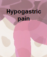
<figure-inline><figure-inline>[[Image:Hypogastric.PNG|link=Hypogastric pain resident survival guide|199x199px]]</figure-inline></figure-inline> | <figure-inline><figure-inline><figure-inline>[[Image:Hypogastric.PNG|link=Hypogastric pain resident survival guide|199x199px]]</figure-inline></figure-inline></figure-inline> | ||
| | | | ||
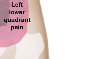
<figure-inline><figure-inline>[[Image:Left_lower_quadrant.PNG|link=Left lower quadrant abdominal pain resident survival guide|335x335px]]</figure-inline></figure-inline> | <figure-inline><figure-inline><figure-inline>[[Image:Left_lower_quadrant.PNG|link=Left lower quadrant abdominal pain resident survival guide|335x335px]]</figure-inline></figure-inline></figure-inline> | ||
|} | |} | ||
Revision as of 15:19, 29 November 2017
Abbreviations: RUQ= Right upper quadrant of the abdomen, LUQ= Left upper quadrant, LLQ= Left lower quadrant, RLQ= Right lower quadrant, LFT= Liver function test, SIRS= Systemic inflammatory response syndrome, ERCP= Endoscopic retrograde cholangiopancreatography, IV= Intravenous, N= Normal, AMA= Anti mitochondrial antibodies, LDH= Lactate dehydrogenase, GI= Gastrointestinal, CXR= Chest X ray, IgA= Immunoglobulin A, IgG= Immunoglobulin G, IgM= Immunoglobulin M, CT= Computed tomography, PMN= Polymorphonuclear cells, ESR= Erythrocyte sedimentation rate, CRP= C-reactive protein, TS= Transferrin saturation, SF= Serum Ferritin, SMA= Superior mesenteric artery, SMV= Superior mesenteric vein, ECG= Electrocardiogram
| ||||||||||||||||||||||||||||||||||||||||||||||||||||||||||||||||||||||||||||||||||||||||||||||||||||||||||||||||||||||||||||||||||||||||||||||||||||||||||||||||||||||||||||||||||||||||||||||||||||||||||||||||||||||||||||||||||||||||||||||||||||||||||||||||||||||||||||||||||||||||||||||||||||||||||||||||||||||||||||||||||||||||||||||||||||||||||||||||||||||||||||||||||||||||||||||||||||||||||||||||||||||||||||||||||||||||||||||||||||||||||||||||||||||||||||||||||||||||||||||||||||||||||||||||||||||||||||||||||||||||||||||||||||||||||||||||||||||||||||||||||||||||||||||||||||||||||||||||||||||||||||||||||||||||||||||||||||||||||||||||||||||||||||||||||||||||||||||||||||||||||||||||||||||||||||||||||||||||||||||||||||||||||||||||||||||||||||||||||||||||||||||||||||||||||||||||||||||||||||||||||||||||||||||||||||||||||||||||||||||||||||||||||||||||||||||||||||||||||||||||||
|
<figure-inline><figure-inline><figure-inline> |
<figure-inline><figure-inline><figure-inline> |
<figure-inline><figure-inline><figure-inline> |
|
<figure-inline><figure-inline><figure-inline> |
<figure-inline><figure-inline><figure-inline> |
<figure-inline><figure-inline><figure-inline> |
|
<figure-inline><figure-inline><figure-inline> |
<figure-inline><figure-inline><figure-inline> |
<figure-inline><figure-inline><figure-inline> |