Sandbox:iqra: Difference between revisions
No edit summary |
No edit summary |
||
| Line 29: | Line 29: | ||
! style="background:#4479BA; color: #FFFFFF;" align="center" |Imaging | ! style="background:#4479BA; color: #FFFFFF;" align="center" |Imaging | ||
|- | |- | ||
! rowspan=" | ! rowspan="46" |Abdominal causes | ||
! | ! rowspan="33" |Inflammatory causes | ||
! rowspan=" | ! rowspan="8" |Pancreato-biliary disorders | ||
| colspan="1" rowspan="1" style="padding: 5px 5px; background: #DCDCDC;" align="center" | [[Cholangitis|Acute cholangitis]] | | colspan="1" rowspan="1" style="padding: 5px 5px; background: #DCDCDC;" align="center" | [[Cholangitis|Acute cholangitis]] | ||
| style="padding: 5px 5px; background: #F5F5F5;" align="center" | [[RUQ]] | | style="padding: 5px 5px; background: #F5F5F5;" align="center" | [[RUQ]] | ||
| Line 90: | Line 73: | ||
| style="padding: 5px 5px; background: #F5F5F5;" align="center" | − | | style="padding: 5px 5px; background: #F5F5F5;" align="center" | − | ||
| style="padding: 5px 5px; background: #F5F5F5;" align="center" | ± | | style="padding: 5px 5px; background: #F5F5F5;" align="center" | ± | ||
| style="padding: 5px 5px; background: #F5F5F5;" align="center" | | | style="padding: 5px 5px; background: #F5F5F5;" align="center" | - | ||
| style="padding: 5px 5px; background: #F5F5F5;" align="center" | | | style="padding: 5px 5px; background: #F5F5F5;" align="center" | - | ||
| style="padding: 5px 5px; background: #F5F5F5;" align="left" |N | | style="padding: 5px 5px; background: #F5F5F5;" align="left" |N | ||
| style="padding: 5px 5px; background: #F5F5F5;" align="left" | | | style="padding: 5px 5px; background: #F5F5F5;" align="left" | | ||
| Line 113: | Line 96: | ||
| style="padding: 5px 5px; background: #F5F5F5;" align="left" | | | style="padding: 5px 5px; background: #F5F5F5;" align="left" | | ||
* Increased [[amylase]] / [[lipase]] | * Increased [[amylase]] / [[lipase]] | ||
* | * Increased stool fat content | ||
* Pancreatic function test | * Pancreatic function test | ||
| Line 124: | Line 107: | ||
| style="padding: 5px 5px; background: #DCDCDC;" align="center" |[[Pancreatic carcinoma]] | | style="padding: 5px 5px; background: #DCDCDC;" align="center" |[[Pancreatic carcinoma]] | ||
| style="padding: 5px 5px; background: #F5F5F5;" align="center" |[[Epigastric]] | | style="padding: 5px 5px; background: #F5F5F5;" align="center" |[[Epigastric]] | ||
| style="padding: 5px 5px; background: #F5F5F5;" align="center" |- | | style="padding: 5px 5px; background: #F5F5F5;" align="center" | - | ||
| style="padding: 5px 5px; background: #F5F5F5;" align="center" |- | | style="padding: 5px 5px; background: #F5F5F5;" align="center" | - | ||
| style="padding: 5px 5px; background: #F5F5F5;" align="center" |+ | | style="padding: 5px 5px; background: #F5F5F5;" align="center" | + | ||
| style="padding: 5px 5px; background: #F5F5F5;" align="center" |+ | | style="padding: 5px 5px; background: #F5F5F5;" align="center" | + | ||
| style="padding: 5px 5px; background: #F5F5F5;" align="center" |- | | style="padding: 5px 5px; background: #F5F5F5;" align="center" | - | ||
| style="padding: 5px 5px; background: #F5F5F5;" align="center" |- | | style="padding: 5px 5px; background: #F5F5F5;" align="center" | - | ||
| style="padding: 5px 5px; background: #F5F5F5;" align="center" |- | | style="padding: 5px 5px; background: #F5F5F5;" align="center" | - | ||
| style="padding: 5px 5px; background: #F5F5F5;" align="center" |- | | style="padding: 5px 5px; background: #F5F5F5;" align="center" | - | ||
| style="padding: 5px 5px; background: #F5F5F5;" align="center" |N | | style="padding: 5px 5px; background: #F5F5F5;" align="center" |N | ||
| style="padding: 5px 5px; background: #F5F5F5;" align="left" | | | style="padding: 5px 5px; background: #F5F5F5;" align="left" | | ||
| Line 279: | Line 262: | ||
| style="padding: 5px 5px; background: #DCDCDC;" align="center" |Gastroparesis | | style="padding: 5px 5px; background: #DCDCDC;" align="center" |Gastroparesis | ||
| style="padding: 5px 5px; background: #F5F5F5;" align="center" |Epigastric | | style="padding: 5px 5px; background: #F5F5F5;" align="center" |Epigastric | ||
| style="padding: 5px 5px; background: #F5F5F5;" align="center" |- | | style="padding: 5px 5px; background: #F5F5F5;" align="center" | - | ||
| style="padding: 5px 5px; background: #F5F5F5;" align="center" |- | | style="padding: 5px 5px; background: #F5F5F5;" align="center" | - | ||
| style="padding: 5px 5px; background: #F5F5F5;" align="center" |- | | style="padding: 5px 5px; background: #F5F5F5;" align="center" | - | ||
| style="padding: 5px 5px; background: #F5F5F5;" align="center" |- | | style="padding: 5px 5px; background: #F5F5F5;" align="center" | - | ||
| style="padding: 5px 5px; background: #F5F5F5;" align="center" |- | | style="padding: 5px 5px; background: #F5F5F5;" align="center" | - | ||
| style="padding: 5px 5px; background: #F5F5F5;" align="center" |± | | style="padding: 5px 5px; background: #F5F5F5;" align="center" |± | ||
| style="padding: 5px 5px; background: #F5F5F5;" align="center" |- | | style="padding: 5px 5px; background: #F5F5F5;" align="center" | - | ||
| style="padding: 5px 5px; background: #F5F5F5;" align="center" |- | | style="padding: 5px 5px; background: #F5F5F5;" align="center" | - | ||
| style="padding: 5px 5px; background: #F5F5F5;" align="center" |Hyperactive/hypoactive | | style="padding: 5px 5px; background: #F5F5F5;" align="center" |Hyperactive/hypoactive | ||
|style="padding: 5px 5px; background: #F5F5F5;" align="left" | | | style="padding: 5px 5px; background: #F5F5F5;" align="left" | | ||
*Hemoglobin | *Hemoglobin | ||
*Fasting plasma glucose | *Fasting plasma glucose | ||
*Serum total protein, albumin, thyrotropin (TSH), and an antinuclear antibody (ANA) titer | *Serum total protein, albumin, thyrotropin (TSH), and an antinuclear antibody (ANA) titer | ||
*HbA1c | *HbA1c | ||
|style="padding: 5px 5px; background: #F5F5F5;" align="left" | | | style="padding: 5px 5px; background: #F5F5F5;" align="left" | | ||
*Scintigraphic gastric emptying | *Scintigraphic gastric emptying | ||
|style="padding: 5px 5px; background: #F5F5F5;" align="left" | | | style="padding: 5px 5px; background: #F5F5F5;" align="left" | | ||
*Succussion splash | *Succussion splash | ||
*Single photon emission computed tomography (SPECT) | *Single photon emission computed tomography (SPECT) | ||
| Line 528: | Line 511: | ||
| style="padding: 5px 5px; background: #DCDCDC;" align="center" |Colon carcinoma | | style="padding: 5px 5px; background: #DCDCDC;" align="center" |Colon carcinoma | ||
| style="padding: 5px 5px; background: #F5F5F5;" align="center" |Diffuse/localized | | style="padding: 5px 5px; background: #F5F5F5;" align="center" |Diffuse/localized | ||
| style="padding: 5px 5px; background: #F5F5F5;" align="center" |- | | style="padding: 5px 5px; background: #F5F5F5;" align="center" | - | ||
| style="padding: 5px 5px; background: #F5F5F5;" align="center" |- | | style="padding: 5px 5px; background: #F5F5F5;" align="center" | - | ||
| style="padding: 5px 5px; background: #F5F5F5;" align="center" |- | | style="padding: 5px 5px; background: #F5F5F5;" align="center" | - | ||
| style="padding: 5px 5px; background: #F5F5F5;" align="center" |± | | style="padding: 5px 5px; background: #F5F5F5;" align="center" |± | ||
| style="padding: 5px 5px; background: #F5F5F5;" align="center" |+ | | style="padding: 5px 5px; background: #F5F5F5;" align="center" | + | ||
| style="padding: 5px 5px; background: #F5F5F5;" align="center" |± | | style="padding: 5px 5px; background: #F5F5F5;" align="center" |± | ||
| style="padding: 5px 5px; background: #F5F5F5;" align="center" |- | | style="padding: 5px 5px; background: #F5F5F5;" align="center" | - | ||
| style="padding: 5px 5px; background: #F5F5F5;" align="center" |- | | style="padding: 5px 5px; background: #F5F5F5;" align="center" | - | ||
| style="padding: 5px 5px; background: #F5F5F5;" align="left" | | | style="padding: 5px 5px; background: #F5F5F5;" align="left" | | ||
* Normal | * Normal | ||
| Line 586: | Line 569: | ||
| style="padding: 5px 5px; background: #DCDCDC;" align="center" |[[Liver abscess]] | | style="padding: 5px 5px; background: #DCDCDC;" align="center" |[[Liver abscess]] | ||
| style="padding: 5px 5px; background: #F5F5F5;" align="center" |RUQ | | style="padding: 5px 5px; background: #F5F5F5;" align="center" |RUQ | ||
| style="padding: 5px 5px; background: #F5F5F5;" align="center" |+ | | style="padding: 5px 5px; background: #F5F5F5;" align="center" | + | ||
| style="padding: 5px 5px; background: #F5F5F5;" align="center" |+ | | style="padding: 5px 5px; background: #F5F5F5;" align="center" | + | ||
| style="padding: 5px 5px; background: #F5F5F5;" align="center" |+ | | style="padding: 5px 5px; background: #F5F5F5;" align="center" | + | ||
| style="padding: 5px 5px; background: #F5F5F5;" align="center" |± | | style="padding: 5px 5px; background: #F5F5F5;" align="center" |± | ||
| style="padding: 5px 5px; background: #F5F5F5;" align="center" |- | | style="padding: 5px 5px; background: #F5F5F5;" align="center" | - | ||
| style="padding: 5px 5px; background: #F5F5F5;" align="center" |+ | | style="padding: 5px 5px; background: #F5F5F5;" align="center" | + | ||
| style="padding: 5px 5px; background: #F5F5F5;" align="center" |+ | | style="padding: 5px 5px; background: #F5F5F5;" align="center" | + | ||
| style="padding: 5px 5px; background: #F5F5F5;" align="center" |± | | style="padding: 5px 5px; background: #F5F5F5;" align="center" |± | ||
| style="padding: 5px 5px; background: #F5F5F5;" align="center" |Normal/hypoactive | | style="padding: 5px 5px; background: #F5F5F5;" align="center" |Normal/hypoactive | ||
| Line 606: | Line 589: | ||
| style="padding: 5px 5px; background: #DCDCDC;" align="center" |[[Hepatocellular carcinoma]]/Metastasis | | style="padding: 5px 5px; background: #DCDCDC;" align="center" |[[Hepatocellular carcinoma]]/Metastasis | ||
| style="padding: 5px 5px; background: #F5F5F5;" align="center" |RUQ | | style="padding: 5px 5px; background: #F5F5F5;" align="center" |RUQ | ||
| style="padding: 5px 5px; background: #F5F5F5;" align="center" |+ | | style="padding: 5px 5px; background: #F5F5F5;" align="center" | + | ||
| style="padding: 5px 5px; background: #F5F5F5;" align="center" |- | | style="padding: 5px 5px; background: #F5F5F5;" align="center" | - | ||
| style="padding: 5px 5px; background: #F5F5F5;" align="center" |+ | | style="padding: 5px 5px; background: #F5F5F5;" align="center" | + | ||
| style="padding: 5px 5px; background: #F5F5F5;" align="center" |- | | style="padding: 5px 5px; background: #F5F5F5;" align="center" | - | ||
| style="padding: 5px 5px; background: #F5F5F5;" align="center" |- | | style="padding: 5px 5px; background: #F5F5F5;" align="center" | - | ||
| style="padding: 5px 5px; background: #F5F5F5;" align="center" |- | | style="padding: 5px 5px; background: #F5F5F5;" align="center" | - | ||
| style="padding: 5px 5px; background: #F5F5F5;" align="center" |- | | style="padding: 5px 5px; background: #F5F5F5;" align="center" | - | ||
| style="padding: 5px 5px; background: #F5F5F5;" align="center" |- | | style="padding: 5px 5px; background: #F5F5F5;" align="center" | - | ||
| style="padding: 5px 5px; background: #F5F5F5;" align="left" | | | style="padding: 5px 5px; background: #F5F5F5;" align="left" | | ||
* Normal | * Normal | ||
| Line 733: | Line 716: | ||
| style="padding: 5px 5px; background: #DCDCDC;" align="center" |Pyelonephritis | | style="padding: 5px 5px; background: #DCDCDC;" align="center" |Pyelonephritis | ||
| style="padding: 5px 5px; background: #F5F5F5;" align="center" |Lumbar region | | style="padding: 5px 5px; background: #F5F5F5;" align="center" |Lumbar region | ||
| style="padding: 5px 5px; background: #F5F5F5;" align="center" |+ | | style="padding: 5px 5px; background: #F5F5F5;" align="center" | + | ||
| style="padding: 5px 5px; background: #F5F5F5;" align="center" |± | | style="padding: 5px 5px; background: #F5F5F5;" align="center" |± | ||
| style="padding: 5px 5px; background: #F5F5F5;" align="center" |- | | style="padding: 5px 5px; background: #F5F5F5;" align="center" | - | ||
| style="padding: 5px 5px; background: #F5F5F5;" align="center" |- | | style="padding: 5px 5px; background: #F5F5F5;" align="center" | - | ||
| style="padding: 5px 5px; background: #F5F5F5;" align="center" |- | | style="padding: 5px 5px; background: #F5F5F5;" align="center" | - | ||
| style="padding: 5px 5px; background: #F5F5F5;" align="center" |+ | | style="padding: 5px 5px; background: #F5F5F5;" align="center" | + | ||
| style="padding: 5px 5px; background: #F5F5F5;" align="center" |± | | style="padding: 5px 5px; background: #F5F5F5;" align="center" |± | ||
| style="padding: 5px 5px; background: #F5F5F5;" align="center" |± | | style="padding: 5px 5px; background: #F5F5F5;" align="center" |± | ||
| Line 982: | Line 965: | ||
| style="padding: 5px 5px; background: #F5F5F5;" align="center" |RUQ/LUQ | | style="padding: 5px 5px; background: #F5F5F5;" align="center" |RUQ/LUQ | ||
| style="padding: 5px 5px; background: #F5F5F5;" align="center" |± | | style="padding: 5px 5px; background: #F5F5F5;" align="center" |± | ||
| style="padding: 5px 5px; background: #F5F5F5;" align="center" |- | | style="padding: 5px 5px; background: #F5F5F5;" align="center" | - | ||
| style="padding: 5px 5px; background: #F5F5F5;" align="center" |- | | style="padding: 5px 5px; background: #F5F5F5;" align="center" | - | ||
| style="padding: 5px 5px; background: #F5F5F5;" align="center" |- | | style="padding: 5px 5px; background: #F5F5F5;" align="center" | - | ||
| style="padding: 5px 5px; background: #F5F5F5;" align="center" |- | | style="padding: 5px 5px; background: #F5F5F5;" align="center" | - | ||
| style="padding: 5px 5px; background: #F5F5F5;" align="center" |± | | style="padding: 5px 5px; background: #F5F5F5;" align="center" |± | ||
| style="padding: 5px 5px; background: #F5F5F5;" align="center" |- | | style="padding: 5px 5px; background: #F5F5F5;" align="center" | - | ||
| style="padding: 5px 5px; background: #F5F5F5;" align="center" |- | | style="padding: 5px 5px; background: #F5F5F5;" align="center" | - | ||
| style="padding: 5px 5px; background: #F5F5F5;" align="center" |N | | style="padding: 5px 5px; background: #F5F5F5;" align="center" |N | ||
| style="padding: 5px 5px; background: #F5F5F5;" align="left" | | | style="padding: 5px 5px; background: #F5F5F5;" align="left" | | ||
| Line 1,001: | Line 984: | ||
! colspan="2" style="padding: 5px 5px; background: #DCDCDC;" align="center" |Pneumonia | ! colspan="2" style="padding: 5px 5px; background: #DCDCDC;" align="center" |Pneumonia | ||
| style="padding: 5px 5px; background: #F5F5F5;" align="center" |RUQ/LUQ | | style="padding: 5px 5px; background: #F5F5F5;" align="center" |RUQ/LUQ | ||
| style="padding: 5px 5px; background: #F5F5F5;" align="center" |+ | | style="padding: 5px 5px; background: #F5F5F5;" align="center" | + | ||
| style="padding: 5px 5px; background: #F5F5F5;" align="center" |+ | | style="padding: 5px 5px; background: #F5F5F5;" align="center" | + | ||
| style="padding: 5px 5px; background: #F5F5F5;" align="center" |- | | style="padding: 5px 5px; background: #F5F5F5;" align="center" | - | ||
| style="padding: 5px 5px; background: #F5F5F5;" align="center" |- | | style="padding: 5px 5px; background: #F5F5F5;" align="center" | - | ||
| style="padding: 5px 5px; background: #F5F5F5;" align="center" |- | | style="padding: 5px 5px; background: #F5F5F5;" align="center" | - | ||
| style="padding: 5px 5px; background: #F5F5F5;" align="center" |+ | | style="padding: 5px 5px; background: #F5F5F5;" align="center" | + | ||
| style="padding: 5px 5px; background: #F5F5F5;" align="center" |- | | style="padding: 5px 5px; background: #F5F5F5;" align="center" | - | ||
| style="padding: 5px 5px; background: #F5F5F5;" align="center" |- | | style="padding: 5px 5px; background: #F5F5F5;" align="center" | - | ||
| style="padding: 5px 5px; background: #F5F5F5;" align="center" |N/Hypoactive | | style="padding: 5px 5px; background: #F5F5F5;" align="center" |N/Hypoactive | ||
| style="padding: 5px 5px; background: #F5F5F5;" align="left" | | | style="padding: 5px 5px; background: #F5F5F5;" align="left" | | ||
| Line 1,051: | Line 1,034: | ||
|- | |- | ||
| | | | ||
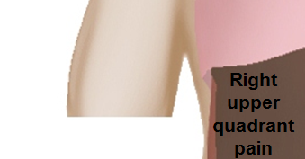
[[Image:Right_upper_quadrant.PNG|link=Right upper quadrant abdominal pain resident survival guide|339x339px]] | <figure-inline>[[Image:Right_upper_quadrant.PNG|link=Right upper quadrant abdominal pain resident survival guide|339x339px]]</figure-inline> | ||
| | | | ||
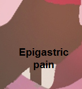
[[Image:Epigastric_quadrant_pain.PNG|link=Epigastric pain resident survival guide|179x179px]] | <figure-inline>[[Image:Epigastric_quadrant_pain.PNG|link=Epigastric pain resident survival guide|179x179px]]</figure-inline> | ||
| | | | ||
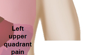
[[Image:Left_upper_quadrant.PNG|link=Left upper quadrant abdominal pain resident survival guide|329x329px]] | <figure-inline>[[Image:Left_upper_quadrant.PNG|link=Left upper quadrant abdominal pain resident survival guide|329x329px]]</figure-inline> | ||
|- | |- | ||
| | | | ||
[[Image:Right_flank_quadrant.PNG|link=Right flank pain resident survival guide|338x338px]] | <figure-inline>[[Image:Right_flank_quadrant.PNG|link=Right flank pain resident survival guide|338x338px]]</figure-inline> | ||
| | | | ||
[[Image:Umbilical_pain.PNG|link=Umbilical region pain resident survival guide|165x165px]] | <figure-inline>[[Image:Umbilical_pain.PNG|link=Umbilical region pain resident survival guide|165x165px]]</figure-inline> | ||
| | | | ||
[[Image:Left_flank_quadrant.PNG|link=Left flank quadrant abdominal pain resident survival guide|335x335px]] | <figure-inline>[[Image:Left_flank_quadrant.PNG|link=Left flank quadrant abdominal pain resident survival guide|335x335px]]</figure-inline> | ||
|- | |- | ||
| | | | ||
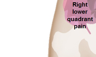
[[Image:Right_lower_quadrant.PNG|link=Right lower quadrant abdominal pain resident survival guide|338x338px]] | <figure-inline>[[Image:Right_lower_quadrant.PNG|link=Right lower quadrant abdominal pain resident survival guide|338x338px]]</figure-inline> | ||
| | | | ||
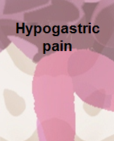
[[Image:Hypogastric.PNG|link=Hypogastric pain resident survival guide|199x199px]] | <figure-inline>[[Image:Hypogastric.PNG|link=Hypogastric pain resident survival guide|199x199px]]</figure-inline> | ||
| | | | ||
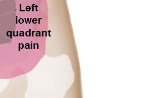
[[Image:Left_lower_quadrant.PNG|link=Left lower quadrant abdominal pain resident survival guide|335x335px]] | <figure-inline>[[Image:Left_lower_quadrant.PNG|link=Left lower quadrant abdominal pain resident survival guide|335x335px]]</figure-inline> | ||
|} | |} | ||
Revision as of 14:08, 29 November 2017
Abbreviations: RUQ= Right upper quadrant of the abdomen, LUQ= Left upper quadrant, LLQ= Left lower quadrant, RLQ= Right lower quadrant, LFT= Liver function test, SIRS= Systemic inflammatory response syndrome, ERCP= Endoscopic retrograde cholangiopancreatography, IV= Intravenous, N= Normal, AMA= Anti mitochondrial antibodies, LDH= Lactate dehydrogenase, GI= Gastrointestinal, CXR= Chest X ray, IgA= Immunoglobulin A, IgG= Immunoglobulin G, IgM= Immunoglobulin M, CT= Computed tomography, PMN= Polymorphonuclear cells, ESR= Erythrocyte sedimentation rate, CRP= C-reactive protein, TS= Transferrin saturation, SF= Serum Ferritin, SMA= Superior mesenteric artery, SMV= Superior mesenteric vein, ECG= Electrocardiogram
| ||||||||||||||||||||||||||||||||||||||||||||||||||||||||||||||||||||||||||||||||||||||||||||||||||||||||||||||||||||||||||||||||||||||||||||||||||||||||||||||||||||||||||||||||||||||||||||||||||||||||||||||||||||||||||||||||||||||||||||||||||||||||||||||||||||||||||||||||||||||||||||||||||||||||||||||||||||||||||||||||||||||||||||||||||||||||||||||||||||||||||||||||||||||||||||||||||||||||||||||||||||||||||||||||||||||||||||||||||||||||||||||||||||||||||||||||||||||||||||||||||||||||||||||||||||||||||||||||||||||||||||||||||||||||||||||||||||||||||||||||||||||||||||||||||||||||||||||||||||||||||||||||||||||||||||||||||||||||||||||||||||||||||||||||||||||||||||||||||||||||||||||||||||||||||||||||||||||||||||||||||||||||||||||||||||||||||||||||||||||||||||
|
<figure-inline> |
<figure-inline> |
<figure-inline> |
|
<figure-inline> |
<figure-inline> |
<figure-inline> |
|
<figure-inline> |
<figure-inline> |
<figure-inline> |