Rheumatoid arthritis physical examination: Difference between revisions
No edit summary |
(→Skin) |
||
| Line 15: | Line 15: | ||
*[[Sweet's syndrome]], a neutrophilic dermatosis usually associated with myeloproliferative disorders | *[[Sweet's syndrome]], a neutrophilic dermatosis usually associated with myeloproliferative disorders | ||
*viral infections | *viral infections | ||
*drug reactions | *drug reactions | ||
*[[erythema nodosum]] | *[[erythema nodosum]] | ||
*lobular [[panniculitis]] | *lobular [[panniculitis]] | ||
| Line 22: | Line 22: | ||
*diffuse thinning (rice paper skin), and skin fragility. | *diffuse thinning (rice paper skin), and skin fragility. | ||
*beading of the nails | *beading of the nails | ||
==== Eyes ==== | ==== Eyes ==== | ||
*Dry eyes ([[Sjogren's syndrome]]) | *Dry eyes ([[Sjogren's syndrome]]) | ||
Revision as of 17:15, 5 November 2012
|
Rheumatoid arthritis Microchapters | |
|
Diagnosis | |
|---|---|
|
Treatment | |
|
Case Studies | |
|
Rheumatoid arthritis physical examination On the Web | |
|
American Roentgen Ray Society Images of Rheumatoid arthritis physical examination | |
|
Risk calculators and risk factors for Rheumatoid arthritis physical examination | |
Editor-In-Chief: C. Michael Gibson, M.S., M.D. [1]; Associate Editor(s)-in-Chief: Aarti Narayan, M.B.B.S [2]
Overview
Rheumatoid arthritis (RA) is a chronic, inflammatory, autoimmune disorder affecting the joints and sometimes other organs as well. It is by definition polyarticular; that is, it affects many joints. Most commonly, the small joints in the hands and feet are affected, but larger joints (shoulders, knees etc) can also be affected; the pattern of joint involvement can differ from patient to patient.[1]
Physical examination
Appearance of the Patient
- General fatigue and lassitude
Skin
- The rheumatoid nodule is the cutaneous (strictly speaking subcutaneous) feature most characteristic of rheumatoid arthritis. The initial pathologic process in nodule formation is unknown but is thought to be related to small-vessel inflammation. The mature lesion is defined by an area of central necrosis surrounded by palisading macrophages and fibroblasts and a cuff of cellular connective tissue and chronic inflammatory cells. The typical rheumatoid nodule may be a few millimetres to a few centimetres in diameter and is usually found over bony prominences, such as the olecranon, the calcaneal tuberosity, the metacarpophalangeal joints, or other areas that sustain repeated mechanical stress. Nodules are associated with a positive RF titer and severe erosive arthritis. They can rarely occur throughout the body in internal organs.
- A variety of forms of vasculitis is also a cutaneous manifestation associated with rheumatoid arthritis. A benign form occurs as microinfarcts around the nailfolds. More severe forms include livedo reticularis, which is a network (reticulum) of erythematous to purplish discoloration of the skin due to the presence of an obliterative cutaneous capillaropathy. (This rash is also otherwise associated with the antiphospholipid-antibody syndrome, a hypercoagulable state linked to antiphospholipid antibodies and characterized by recurrent vascular thrombosis and second trimester miscarriages.
Other, rather rare, cutaneous features include:
- pyoderma gangrenosum, a necrotizing, ulcerative, noninfectious neutrophilic dermatosis.
- Sweet's syndrome, a neutrophilic dermatosis usually associated with myeloproliferative disorders
- viral infections
- drug reactions
- erythema nodosum
- lobular panniculitis
- atrophy of digital skin
- palmar erythema
- diffuse thinning (rice paper skin), and skin fragility.
- beading of the nails
Eyes
- Dry eyes (Sjogren's syndrome)
- Scleritis
- Keratoconjunctivitis sicca (dry eyes)
- Episcleritis and scleromalacia
Lungs
- Palpation:
- Breathing movements may be decreased on both sides
- Auscultation:
- Decreased breath sounds on both sides
- Crackles may be present
Abdomen
Extremities
- The small joints of the cervical spine can also be involved.
- Inflammation in the joints manifests itself as a soft, "doughy" swelling, pain, tenderness to palpation and movement, local warmth, and functional impairment.
- In RA, the joints are usually affected in a fairly symmetrical fashion although the initial presentation may be asymmetrical.
Deformity
- The fingers are typically deviated towards the little finger (ulnar deviation) and can assume unnatural shapes. Classical deformities in rheumatoid arthritis are the Boutonniere deformity (Hyperflexion at the proximal interphalangeal joint with hyperextension at the distal interphalangeal joint), swan neck deformity (Hyperextension at the proximal interphalangeal joint, hyperflexion at the distal interphalangeal joint).
- The thumb may develop a "Z-Thumb" deformity with fixed flexion and subluxation at the metacarpophalangeal joint, and hyperextension at the IP joint.
-

-

-

-

-
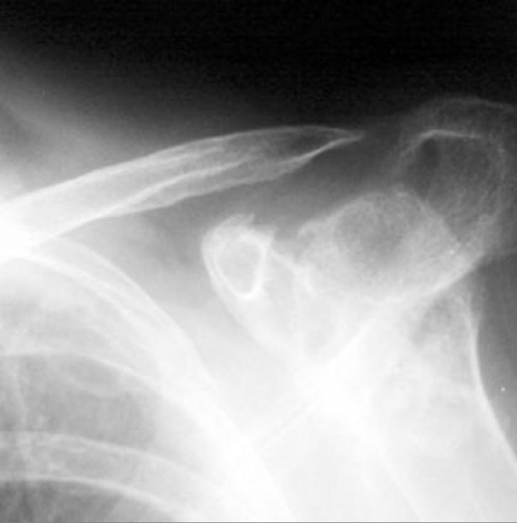
Distal clavicle erosion


Neurologic
Extra-articular (elsewhere)
Patients with RA usually exhibit signs of systemic inflammation, that is, the inflammatory process in the joint leaves its marks on other organs as well (and this may also help distinguish it from osteoarthritis). Examples are a general tiredness and lassitude, sometimes low-grade fever, and some abnormalities on blood tests such as an elevated erythrocyte sedimentation rate (ESR), and anemia, which is often seen as a consequence of the disease itself (anaemia of chronic disease) although it may also be caused by gastrointestinal bleeding as a side effect of drugs used in treatment, especially NSAIDs used for analgesia. Extra-articular manifestations (manifestations outside the musculoskeletal system) occur in about 15% of patients with rheumatoid arthritis.[2] Examples are Hepatosplenomegaly which may occur with concurrent leukopenia and is then referred to as
Sources
Copyleft images obtained courtesy of RadsWiki [3]
References
- ↑ Majithia V, Geraci SA (2007). "Rheumatoid arthritis: diagnosis and management". Am. J. Med. 120 (11): 936–9. doi:10.1016/j.amjmed.2007.04.005. PMID 17976416.
- ↑ Turesson C, O'Fallon WM, Crowson CS, Gabriel SE, Matteson EL (2003). "Extra-articular disease manifestations in rheumatoid arthritis: incidence trends and risk factors over 46 years". Ann. Rheum. Dis. 62 (8): 722–7. PMID 12860726.