Pericarditis pathophysiology: Difference between revisions
| Line 25: | Line 25: | ||
==Acute Versus Chronic Pericarditis== | ==Acute Versus Chronic Pericarditis== | ||
Depending on the timing of presentation and duration, pericarditis is divided into "acute" and "chronic" forms. Clinically, acute pericarditis presents within 6 weeks of the disease onset; subacute pericarditis presents within 6 weeks to 6 months of the disease onset; and chronic pericarditis manifests after 6 months of the disease onset.[[Acute pericarditis]] is more common than chronic pericarditis, and often occurs as a complication of viral infections, immunologic conditions, or as a result of a [[heart attack]] ([[myocardial infarction]]). Chronic pericarditis is less common, which may manifest as scarring of the pericardium a condition known as [[constrictive pericarditis]]. | Depending on the timing of presentation and duration, pericarditis is divided into "acute" and "chronic" forms. Clinically, acute pericarditis presents within 6 weeks of the disease onset; subacute pericarditis presents within 6 weeks to 6 months of the disease onset; and chronic pericarditis manifests after 6 months of the disease onset.[[Acute pericarditis]] is more common than chronic pericarditis, and often occurs as a complication of viral infections, immunologic conditions, or as a result of a [[heart attack]] ([[myocardial infarction]]). Chronic pericarditis is less common, which may manifest as scarring of the pericardium a condition known as [[constrictive pericarditis]]. | ||
==Gross Pathology Images== | |||
[http://www.peir.net Images courtesy of Professor Peter Anderson DVM PhD and published with permission © PEIR, University of Alabama at Birmingham, Department of Pathology] | |||
<div align="left"> | |||
<gallery heights="175" widths="175"> | |||
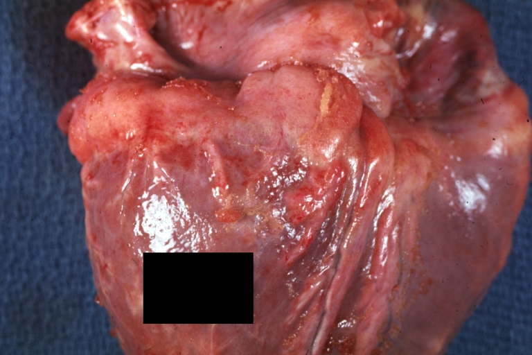
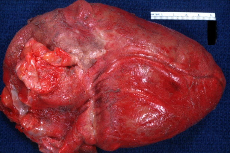
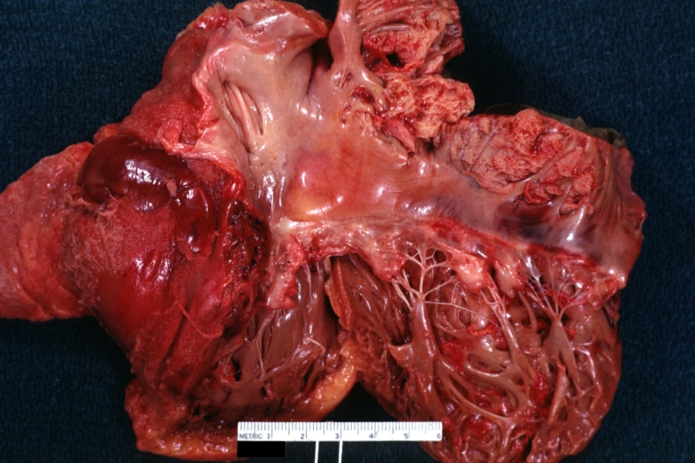
Image:Pericarditis 0001.jpg|Fibrinous pericarditis: Gross, natural color, an excellent example of bread and butter appearance. Uremia, chronic glomerulonephritis and sepsis. | |||
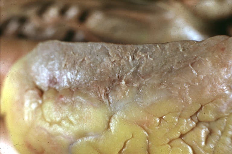
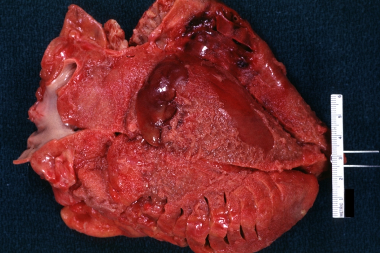
Image:Pericarditis 0002.jpg|Fibrinous pericarditis: Gross, a good example (bread and butter appearance). | |||
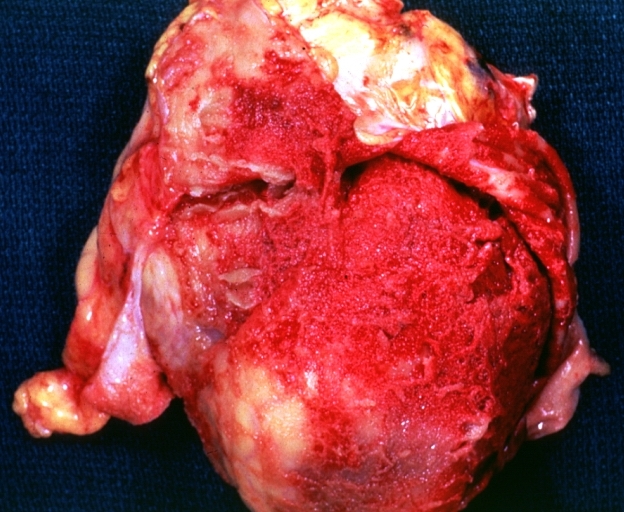
Image:Pericarditis 0003.jpg|Fibrinous pericarditis: Gross, an excellent example. | |||
</gallery> | |||
</div> | |||
<div align="left"> | |||
<gallery heights="175" widths="175"> | |||
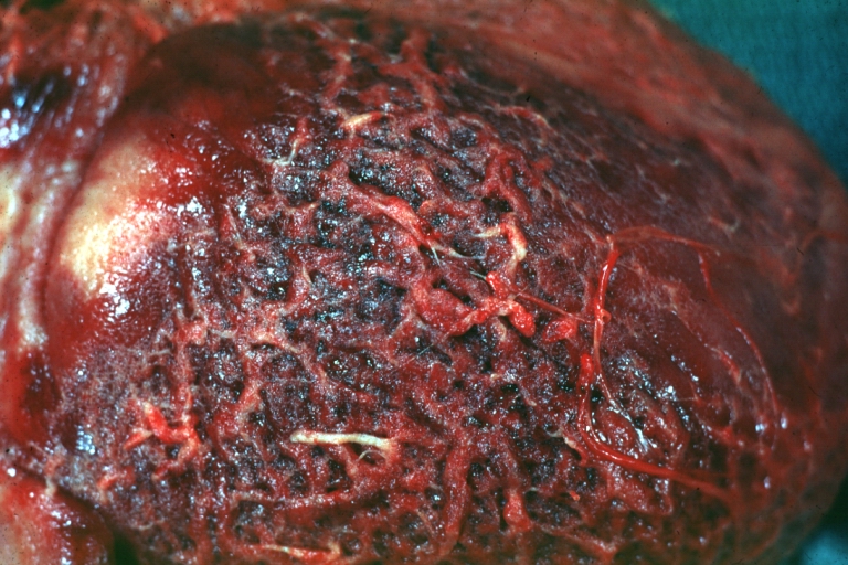
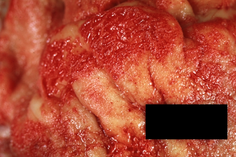
Image:Pericarditis 0004.jpg|Fibrinous pericarditis: Gross, an excellent example, close-up view of fibrin. | |||
Image:Pericarditis 0005.jpg|Fibrinous pericarditis: Gross, an excellent example, close-up view. | |||
Image:Pericarditis 0006.jpg|Fibrinous pericarditis: Gross, an excellent example. | |||
</gallery> | |||
</div> | |||
<div align="left"> | |||
<gallery heights="175" widths="175"> | |||
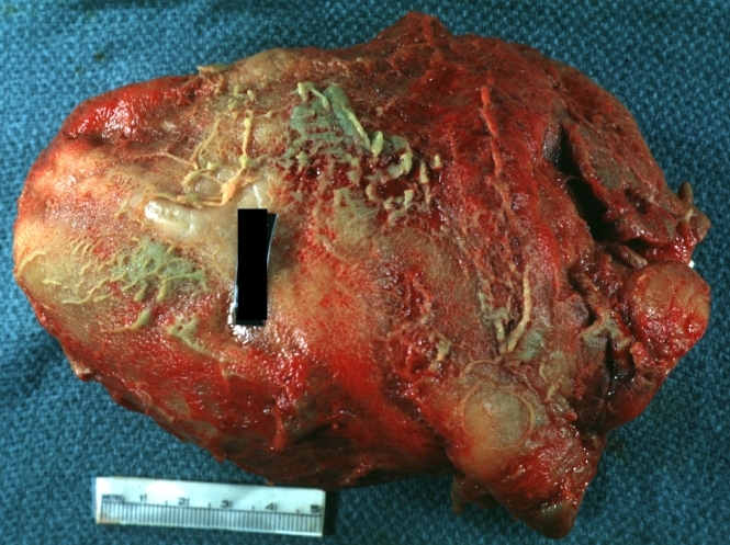
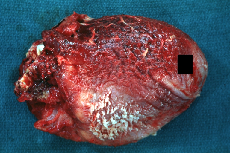
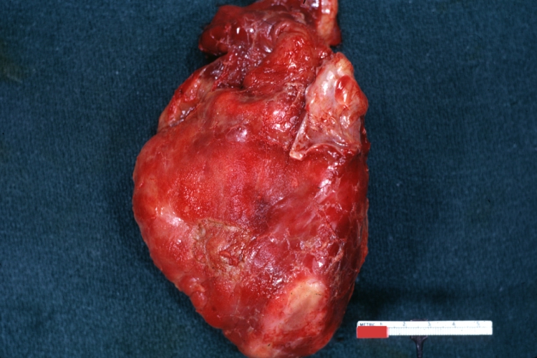
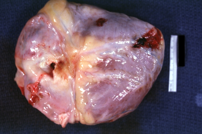
Image:Pericarditis 0007.jpg|Fibrinous pericarditis: Gross, external view of localized pericarditis over an acute infarction | |||
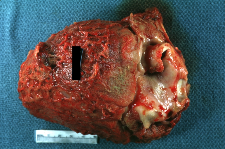
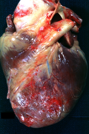
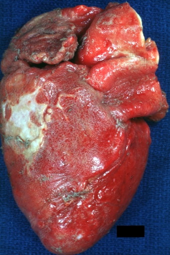
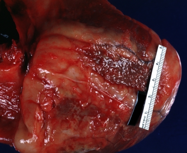
Image:Pericarditis 0008.jpg|Fibrinous pericarditis: Gross, intact heart, good example | |||
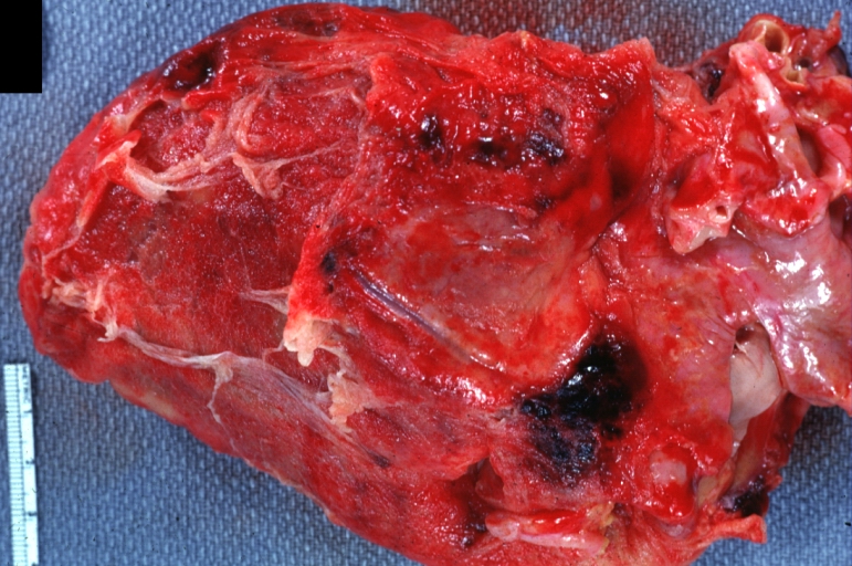
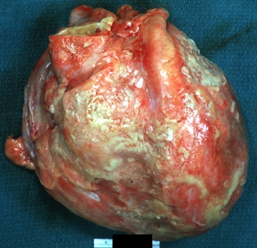
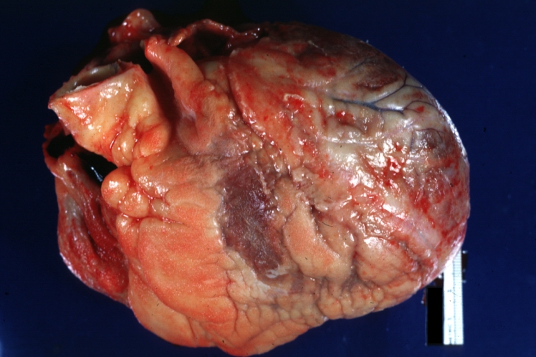
Image:Pericarditis 0009.jpg|Fibrinous pericarditis: Gross, good example, mild, with small amount of fibrin | |||
</gallery> | |||
</div> | |||
<div align="left"> | |||
<gallery heights="175" widths="175"> | |||
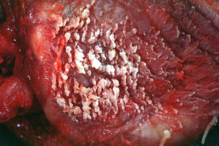
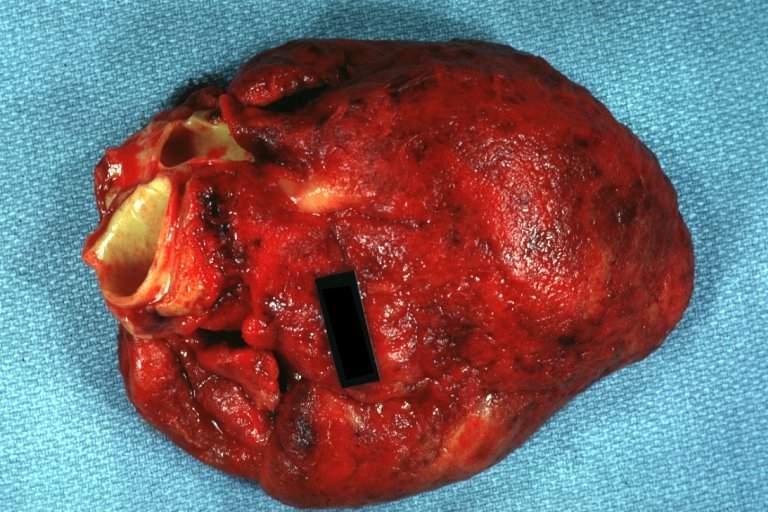
Image:Pericarditis 0010.jpg|Fibrinous pericarditis: Gross, close-up, an excellent example of color and detail | |||
Image:Pericarditis 0011.jpg|Fibrinous pericarditis: Gross, a good example | |||
Image:Pericarditis 0012.jpg|Fibrinous pericarditis: Gross, a good example, very mild case | |||
</gallery> | |||
</div> | |||
<div align="left"> | |||
<gallery heights="175" widths="175"> | |||
Image:Pericarditis 0013.jpg|Fibrinous pericarditis: Gross, an excellent example. | |||
Image:Pericarditis 0014.jpg|Fibrinous pericarditis: Gross, a close-up view, an excellent illustration of fibrinous exudate. | |||
Image:Pericarditis 0015.jpg|Pericarditis in [[uremia]] | |||
</gallery> | |||
</div> | |||
<div align="left"> | |||
<gallery heights="175" widths="175"> | |||
Image:Pericarditis 0016.jpg|Fibrinous pericarditis: Gross, fixed tissue (note to color changes), a close-up view of fibrin on epicardial surface of heart. A typical example. | |||
Image:Pericarditis 0017.jpg|Fibrinous pericarditis: Gross, natural color, large right atrial thrombus and fibrinous pericarditis. Normal [[tricuspid valve]] with some aging changes (good example) | |||
Image:Pericarditis 0018.jpg|Fibrinous pericarditis: Gross, natural color | |||
</gallery> | |||
</div> | |||
<div align="left"> | |||
<gallery heights="175" widths="175"> | |||
Image:Pericarditis 0019.jpg|Fibrinous pericarditis: Gross, natural color, an excellent example | |||
Image:Pericarditis 0020.jpg|Fibrinous pericarditis: Gross, natural color, very close-up photo showing fibrinous exudate simulating frost (an excellent example) | |||
Image:Pericarditis 0021.jpg|Rheumatoid fibrinous pericarditis: Gross, natural color, a typical lesion in 22 years old white female due to juvenile rheumatoid arthritis. | |||
</gallery> | |||
</div> | |||
<div align="left"> | |||
<gallery heights="175" widths="175"> | |||
Image:Pericarditis 0022.jpg|Fibrinous pericarditis: Gross, natural color, close-up view of minimal fibrinous exudate on epicardial surface due to terminal renal failure | |||
Image:Pericarditis 0023.jpg|Fibrinous pericarditis: Gross, natural color, anterior view of heart with mild fibrinous exudate over epicardium due to terminal renal failure | |||
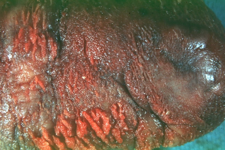
Image:Pericarditis 0024.jpg|Tuberculous pericarditis: Gross, natural color, shaggy hemorrhagic exudate. This case is much more hemorrhagic than the typical tuberculous pericarditis. | |||
</gallery> | |||
</div> | |||
<div align="left"> | |||
<gallery heights="175" widths="175"> | |||
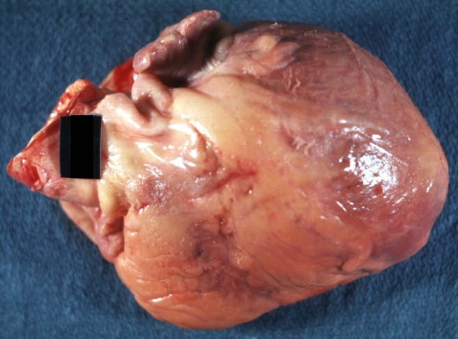
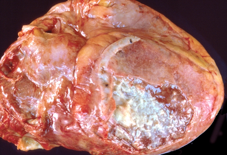
Image:Pericarditis 0025.jpg|Heart transplant: Gross, natural color, external view of heart. Two months after transplantation with fibrinous pericarditis | |||
Image:Pericarditis 0026.jpg|Neoplastic pericarditis: Gross, natural color, shaggy pericarditis. Primer is adenocarcinoma of the lung. | |||
Image:Pericarditis 0027.jpg|Heart: Septic pericarditis | |||
</gallery> | |||
</div> | |||
<div align="left"> | |||
<gallery heights="175" widths="175"> | |||
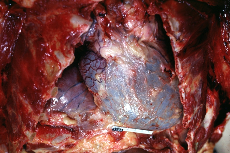
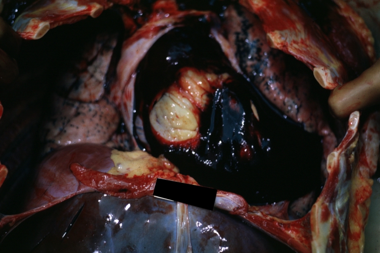
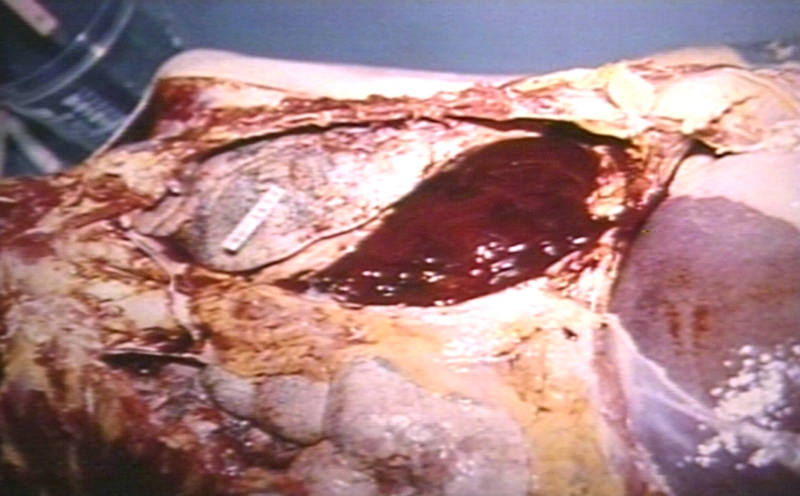
Image:Hemopericardium 001.jpg|Hemopericardium: Gross, an excellent in situ view | |||
Image:Hemopericardium 002.jpg|Hemopericardium: Gross, in situ, unopened pericardium (a very good example) | |||
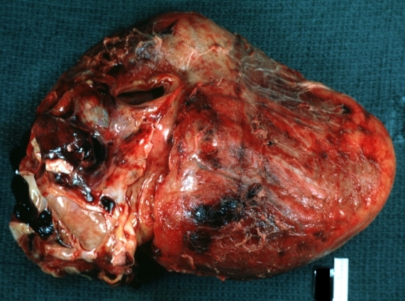
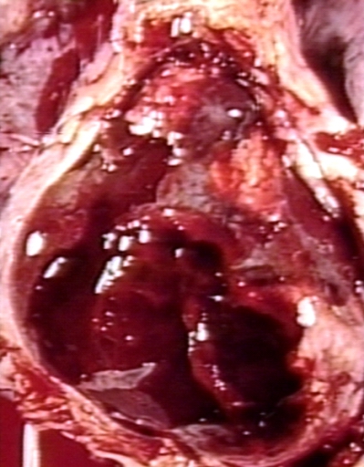
Image:Hemopericardium 003.jpg|Hemopericardium: Gross, natural color, heart in situ with opened pericardium and filled with red blood clot (quite good example) dissecting aneurysm | |||
</gallery> | |||
</div> | |||
<div align="left"> | |||
<gallery heights="175" widths="175"> | |||
Image:Hemopericardium 004.jpg|Hemopericardium due to Needle Puncture: Gross, natural color, external view of heart covered by blood | |||
Image:Hemopericardium 005.jpg|Needle Puncture Mark in Epicardium: Gross, natural color, close-up of needle puncture marks tap resulted in hemopericardium | |||
Image:Hemopericardium 006.jpg|Hemopericardium: Hemopericardium caused by pericardiocentesis: Gross, natural color, close-up view of apex of the heart. Needle apparently entered the distal posterior descending artery. | |||
</gallery> | |||
</div> | |||
<div align="left"> | |||
<gallery heights="175" widths="175"> | |||
Image:Hemopericardium 007.jpg|Hemopericardium: Hemopericardium caused by pericardiocentesis: Gross, natural color, view of apex of the heart. Needle apparently entered the distal posterior descending artery | |||
Image:Hemopericardium 008.jpg|Hemopericardium: Hemopericardium due to pericardiocentesis: Gross, fixed tissue, close-up view of slice through distal posterior descending artery showing periarterial hemorrhage | |||
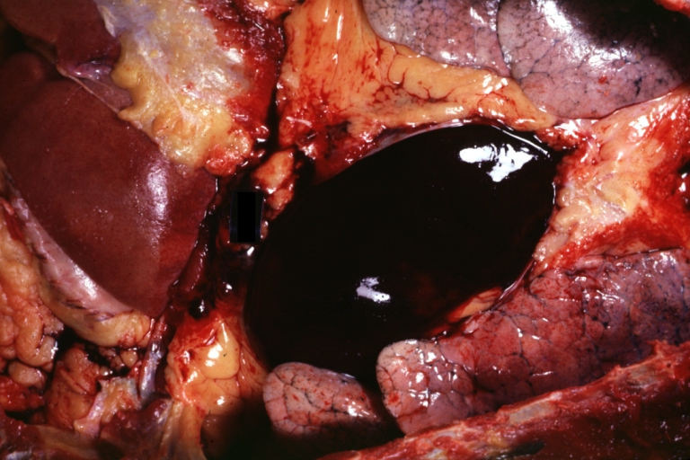
Image:Hemopericardium 009.jpg|Hemopericardium: Liver: Gross, natural color, typical shock liver case of death due to hemopericardium secondary to pericardiocentesis | |||
</gallery> | |||
</div> | |||
<div align="left"> | |||
<gallery heights="175" widths="175"> | |||
Image:Hemopericardium 010.jpg|Hemopericardium in newborn: Gross, natural color, opened body with large collection blood in pericardial sac. Cause uncertain. A 26 week premature with hyaline membrane disease and DIC | |||
Image:Hemopericardium 011.jpg|Hemopericardium: Myocardial Infarction and Ventricular Rupture | |||
</gallery> | |||
</div> | |||
<div align="left"> | |||
<gallery heights="175" widths="175"> | |||
Image:Hemopericardium 012.jpg|Hemopericardium: Infarct rupture after 7 days of chest pain onset. | |||
Image:Hemopericardium 013.jpg|Hemopericardium in dissecting aneurysm: Gross, heart with root of aorta to show hemorrhage into pericardium (very good example) | |||
</gallery> | |||
</div> | |||
==Microscopic Pathology Images== | |||
[http://www.peir.net Images courtesy of Professor Peter Anderson DVM PhD and published with permission © PEIR, University of Alabama at Birmingham, Department of Pathology] | |||
<div align="left"> | |||
<gallery heights="175" widths="175"> | |||
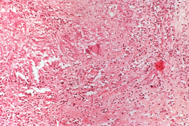
Image:Pericarditis 0028.jpg|Tuberculous pericarditis. | |||
Image:Pericarditis 0029.jpg|Tuberculous pericarditis. | |||
Image:Pericarditis 0030.jpg|Tuberculous pericarditis: Micro oil acid fast stain. The organism easily seen. | |||
</gallery> | |||
</div> | |||
<div align="left"> | |||
<gallery heights="175" widths="175"> | |||
Image:Pericarditis 0031.jpg|Tuberculous pericarditis: Micro oil acid fast stain. The organism easily seen. | |||
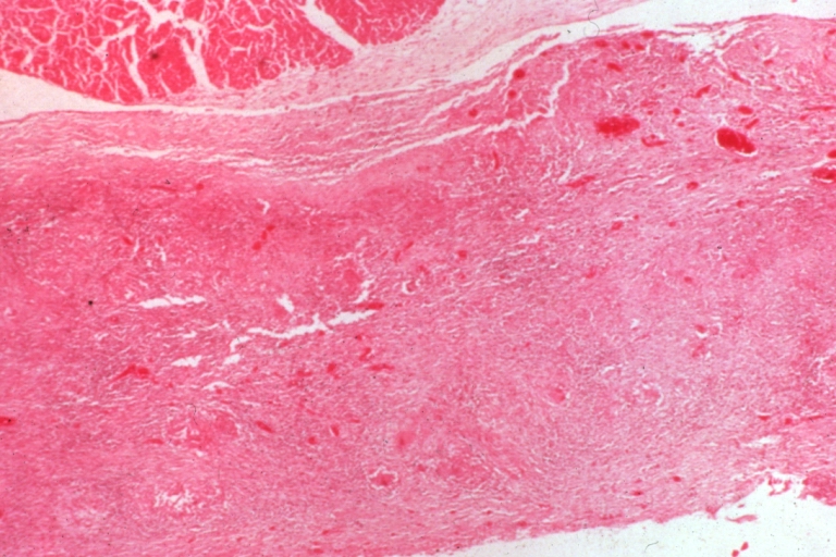
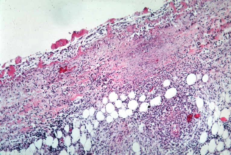
Image:Pericarditis 0032.jpg|Uremic pericarditis: Micro med mag, H&E. A good example | |||
Image:Pericarditis 0033.jpg|Tuberculous pericarditis: Micro med mag, H&E, a typical lesion | |||
</gallery> | |||
</div> | |||
<div align="left"> | |||
<gallery heights="175" widths="175"> | |||
Image:Pericarditis 0034.jpg|Fibrinous pericarditis. | |||
Image:Pericarditis fibrinosa.jpg|Pericarditis fibrinosa (Fibrinous pericarditis). | |||
Image:Heart in mesothelial tumors 16.jpg|Malignant Mesothelioma, Biphasic Type: Pericardium: This tumor has epithelioid cells (lower half) surrounded by spindled cells. The patient was a 46-year-old woman with constrictive pericarditis; the pericardium was studded with coalescing tumor nodules. | |||
</gallery> | |||
</div> | |||
* <Youtube v=AKS7kSl4x5k/> | |||
* Acute fibrinous pericarditis | |||
<Youtube v=5fz_W1YxbC8/> | |||
{{Reflist|2}} | {{Reflist|2}} | ||
Revision as of 13:55, 26 June 2011
|
Pericarditis Microchapters |
|
Diagnosis |
|---|
|
Treatment |
|
Surgery |
|
Case Studies |
|
Pericarditis pathophysiology On the Web |
|
American Roentgen Ray Society Images of Pericarditis pathophysiology |
|
Risk calculators and risk factors for Pericarditis pathophysiology |
Editor-In-Chief: C. Michael Gibson, M.S., M.D. [1]
Overview
Pericarditis may:
- Present as part of a generalized disease
- Present as an isolated disease
- Present as part of a disease that affects nearby organs
- Present as part of multiple diseases
Classification
Pericarditis can be classified according to the composition of the inflammatory exudate or the composition of the fluid that accumulates around the heart.
See also: Pericarditis pathology
Types include:
Acute Versus Chronic Pericarditis
Depending on the timing of presentation and duration, pericarditis is divided into "acute" and "chronic" forms. Clinically, acute pericarditis presents within 6 weeks of the disease onset; subacute pericarditis presents within 6 weeks to 6 months of the disease onset; and chronic pericarditis manifests after 6 months of the disease onset.Acute pericarditis is more common than chronic pericarditis, and often occurs as a complication of viral infections, immunologic conditions, or as a result of a heart attack (myocardial infarction). Chronic pericarditis is less common, which may manifest as scarring of the pericardium a condition known as constrictive pericarditis.
Gross Pathology Images
-
Fibrinous pericarditis: Gross, natural color, an excellent example of bread and butter appearance. Uremia, chronic glomerulonephritis and sepsis.
-
Fibrinous pericarditis: Gross, a good example (bread and butter appearance).
-
Fibrinous pericarditis: Gross, an excellent example.


-
Fibrinous pericarditis: Gross, an excellent example, close-up view of fibrin.
-
Fibrinous pericarditis: Gross, an excellent example, close-up view.
-
Fibrinous pericarditis: Gross, an excellent example.


-
Fibrinous pericarditis: Gross, external view of localized pericarditis over an acute infarction
-
Fibrinous pericarditis: Gross, intact heart, good example
-
Fibrinous pericarditis: Gross, good example, mild, with small amount of fibrin


-
Fibrinous pericarditis: Gross, close-up, an excellent example of color and detail
-
Fibrinous pericarditis: Gross, a good example
-
Fibrinous pericarditis: Gross, a good example, very mild case


-
Fibrinous pericarditis: Gross, an excellent example.
-
Fibrinous pericarditis: Gross, a close-up view, an excellent illustration of fibrinous exudate.
-
Pericarditis in uremia


-
Fibrinous pericarditis: Gross, fixed tissue (note to color changes), a close-up view of fibrin on epicardial surface of heart. A typical example.
-
Fibrinous pericarditis: Gross, natural color, large right atrial thrombus and fibrinous pericarditis. Normal tricuspid valve with some aging changes (good example)
-
Fibrinous pericarditis: Gross, natural color


-
Fibrinous pericarditis: Gross, natural color, an excellent example
-
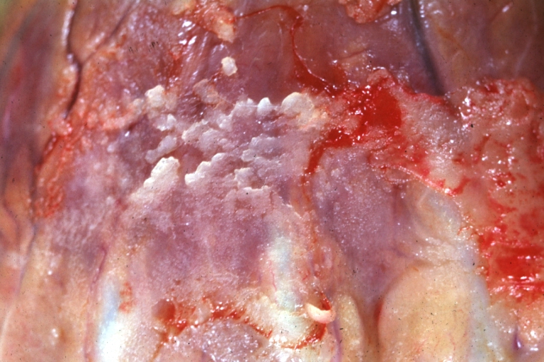
Fibrinous pericarditis: Gross, natural color, very close-up photo showing fibrinous exudate simulating frost (an excellent example)
-
Rheumatoid fibrinous pericarditis: Gross, natural color, a typical lesion in 22 years old white female due to juvenile rheumatoid arthritis.


-
Fibrinous pericarditis: Gross, natural color, close-up view of minimal fibrinous exudate on epicardial surface due to terminal renal failure
-
Fibrinous pericarditis: Gross, natural color, anterior view of heart with mild fibrinous exudate over epicardium due to terminal renal failure
-
Tuberculous pericarditis: Gross, natural color, shaggy hemorrhagic exudate. This case is much more hemorrhagic than the typical tuberculous pericarditis.


-

Heart transplant: Gross, natural color, external view of heart. Two months after transplantation with fibrinous pericarditis
-
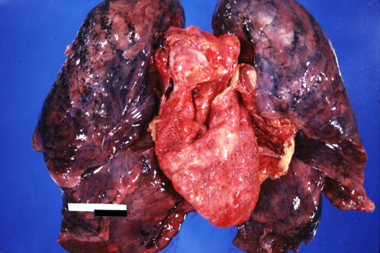
Neoplastic pericarditis: Gross, natural color, shaggy pericarditis. Primer is adenocarcinoma of the lung.
-
Heart: Septic pericarditis


-
Hemopericardium: Gross, an excellent in situ view
-
Hemopericardium: Gross, in situ, unopened pericardium (a very good example)
-
Hemopericardium: Gross, natural color, heart in situ with opened pericardium and filled with red blood clot (quite good example) dissecting aneurysm


-
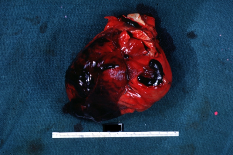
Hemopericardium due to Needle Puncture: Gross, natural color, external view of heart covered by blood
-
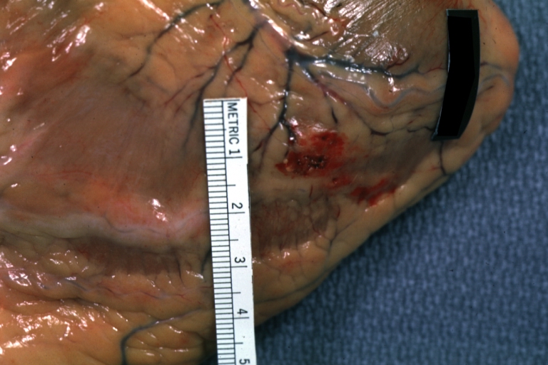
Needle Puncture Mark in Epicardium: Gross, natural color, close-up of needle puncture marks tap resulted in hemopericardium
-
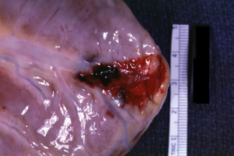
Hemopericardium: Hemopericardium caused by pericardiocentesis: Gross, natural color, close-up view of apex of the heart. Needle apparently entered the distal posterior descending artery.


-
Hemopericardium: Hemopericardium caused by pericardiocentesis: Gross, natural color, view of apex of the heart. Needle apparently entered the distal posterior descending artery
-
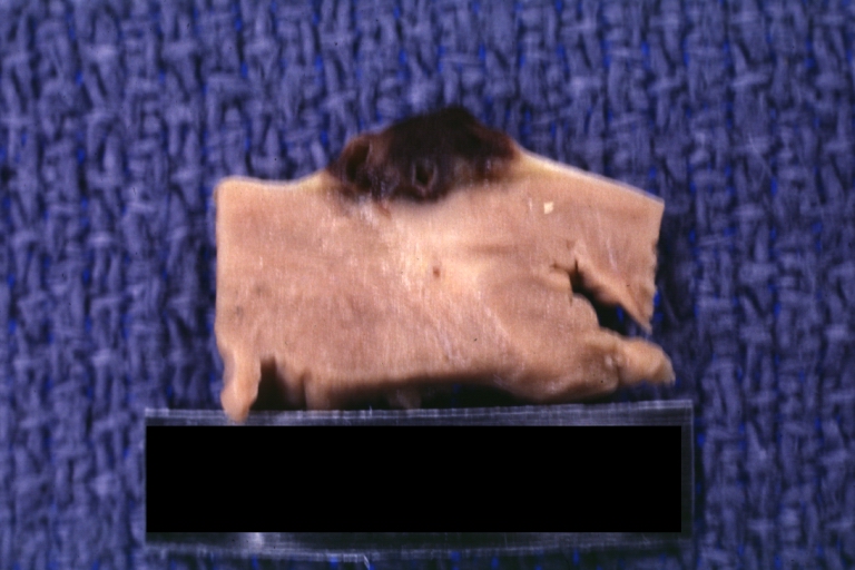
Hemopericardium: Hemopericardium due to pericardiocentesis: Gross, fixed tissue, close-up view of slice through distal posterior descending artery showing periarterial hemorrhage
-
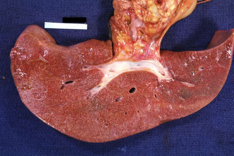
Hemopericardium: Liver: Gross, natural color, typical shock liver case of death due to hemopericardium secondary to pericardiocentesis


-
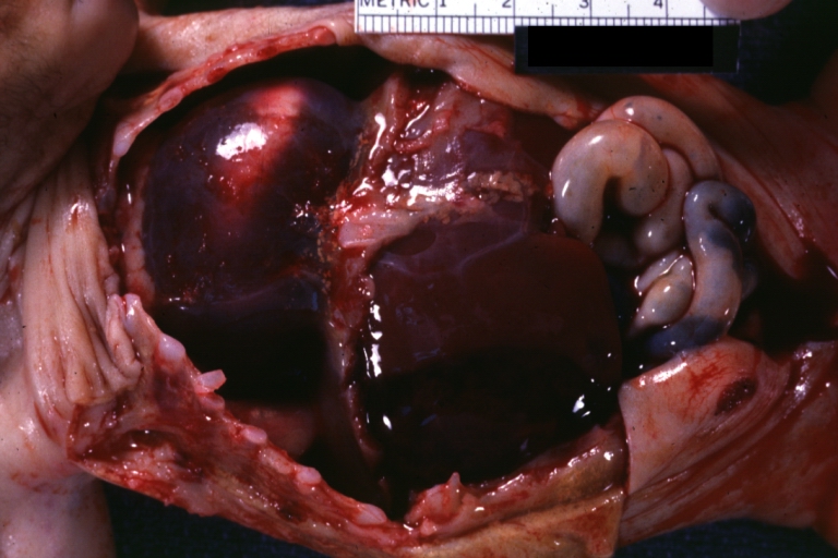
Hemopericardium in newborn: Gross, natural color, opened body with large collection blood in pericardial sac. Cause uncertain. A 26 week premature with hyaline membrane disease and DIC
-
Hemopericardium: Myocardial Infarction and Ventricular Rupture


-
Hemopericardium: Infarct rupture after 7 days of chest pain onset.
-
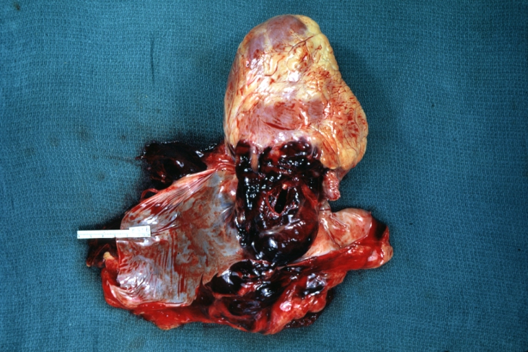
Hemopericardium in dissecting aneurysm: Gross, heart with root of aorta to show hemorrhage into pericardium (very good example)


Microscopic Pathology Images
-
Tuberculous pericarditis.
-
Tuberculous pericarditis.
-
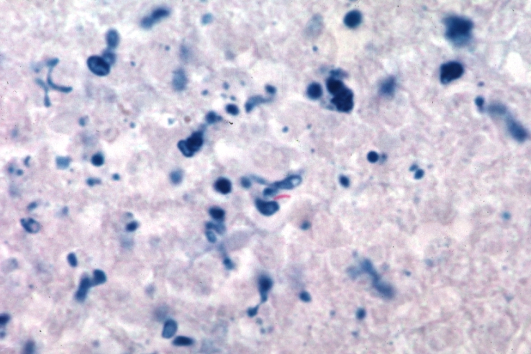
Tuberculous pericarditis: Micro oil acid fast stain. The organism easily seen.


-
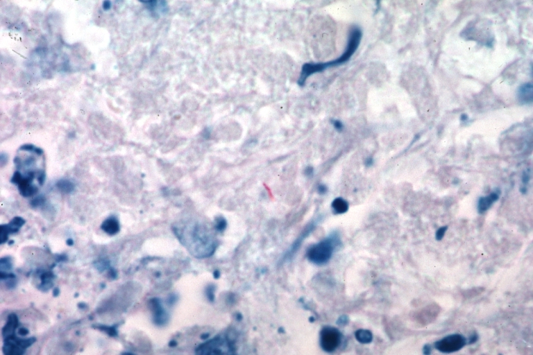
Tuberculous pericarditis: Micro oil acid fast stain. The organism easily seen.
-
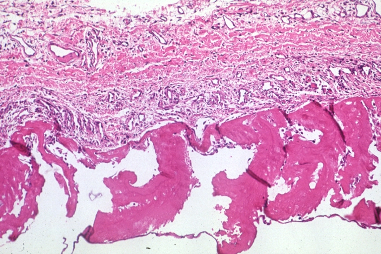
Uremic pericarditis: Micro med mag, H&E. A good example
-
Tuberculous pericarditis: Micro med mag, H&E, a typical lesion


-
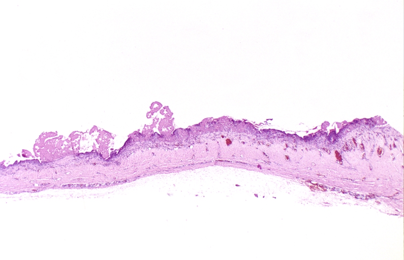
Fibrinous pericarditis.
-
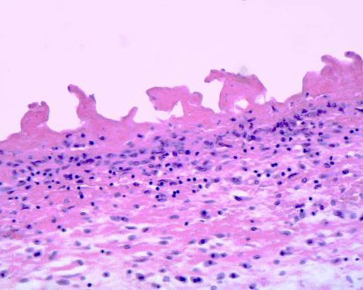
Pericarditis fibrinosa (Fibrinous pericarditis).
-
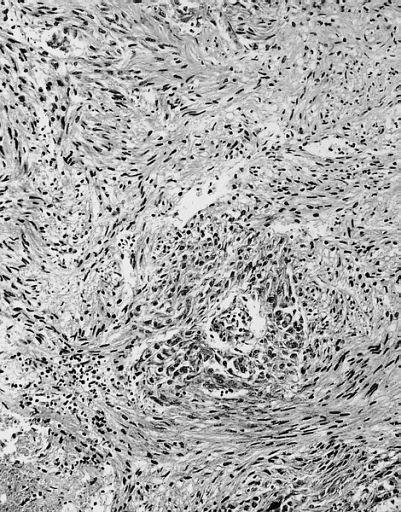
Malignant Mesothelioma, Biphasic Type: Pericardium: This tumor has epithelioid cells (lower half) surrounded by spindled cells. The patient was a 46-year-old woman with constrictive pericarditis; the pericardium was studded with coalescing tumor nodules.


- <Youtube v=AKS7kSl4x5k/>
- Acute fibrinous pericarditis
<Youtube v=5fz_W1YxbC8/>