Mast cell tumor pathophysiology: Difference between revisions
No edit summary |
|||
| (26 intermediate revisions by 2 users not shown) | |||
| Line 3: | Line 3: | ||
{{CMG}};{{AE}} {{PSK}} | {{CMG}};{{AE}} {{PSK}} | ||
==Overview== | ==Overview== | ||
Mast cell [[tumor]] arises from the mast cell, which is a type of [[White blood cells|white blood cell]] involved in the [[inflammatory]] process. The progression to mast cell tumor usually involves the uncontrolled stimulation of the [[receptor]] for [[stem cell factor]] following mutation of [[C-kit]] cell surface receptor. On microscopic histopathological analysis, mast cells in the superficial and mid dermis that are lymphocyte like with dense granular cytoplasm which tend to be more abundant around [[blood vessels]] is characteristic finding of mast cell tumor. | Mast cell [[tumor]] arises from the mast cell, which is a type of [[White blood cells|white blood cell]] involved in the [[inflammatory]] process. The progression to mast cell [[tumor]] usually involves the uncontrolled stimulation of the [[receptor]] for [[stem cell factor]] following [[mutation]] of [[C-kit]] [[cell surface receptor]]. On [[microscopic]] histopathological analysis, mast cells in the [[Dermis|superficial and mid dermis]] that are [[lymphocyte]] like with dense granular [[cytoplasm]] which tend to be more abundant around [[blood vessels]] is characteristic finding of mast cell [[tumor]]. | ||
==Pathophysiology== | ==Pathophysiology== | ||
===Mast Cell=== | ===Mast Cell=== | ||
*Mast cells are bone marrow derived multi-functional immune cells and are normally found throughout the [[connective tissue]] of the body. | *Mast cells are [[bone marrow]] derived multi-functional [[immune]] cells and are normally found throughout the [[connective tissue]] of the body. | ||
*It is a normal component of the [[immune system]] and as it releases [[histamine]] it is associated with [[allergy|allergic reactions]]. | *It is a normal component of the [[immune system]] and as it releases [[histamine]] it is associated with [[allergy|allergic reactions]]. | ||
*Mast cell [[Granule|granules]] contain [[histamine]], [[heparin]], [[platelet-activating factor]], [[leukotrienes]], [[prostaglandins]], cytokines and [[proteases]].<ref name="pmid25452755">{{cite journal |vauthors=Moon TC, Befus AD, Kulka M |title=Mast cell mediators: their differential release and the secretory pathways involved |journal=Front Immunol |volume=5 |issue= |pages=569 |date=2014 |pmid=25452755 |doi=10.3389/fimmu.2014.00569 |url=}}</ref><ref name="pmid26779180">{{cite journal |vauthors=Krystel-Whittemore M, Dileepan KN, Wood JG |title=Mast Cell: A Multi-Functional Master Cell |journal=Front Immunol |volume=6 |issue= |pages=620 |date=2015 |pmid=26779180 |doi=10.3389/fimmu.2015.00620 |url=}}</ref> | *Mast cell [[Granule|granules]] contain [[histamine]], [[heparin]], [[platelet-activating factor]], [[leukotrienes]], [[prostaglandins]], [[cytokines]] and [[proteases]].<ref name="pmid25452755">{{cite journal |vauthors=Moon TC, Befus AD, Kulka M |title=Mast cell mediators: their differential release and the secretory pathways involved |journal=Front Immunol |volume=5 |issue= |pages=569 |date=2014 |pmid=25452755 |doi=10.3389/fimmu.2014.00569 |url=}}</ref><ref name="pmid26779180">{{cite journal |vauthors=Krystel-Whittemore M, Dileepan KN, Wood JG |title=Mast Cell: A Multi-Functional Master Cell |journal=Front Immunol |volume=6 |issue= |pages=620 |date=2015 |pmid=26779180 |doi=10.3389/fimmu.2015.00620 |url=}}</ref> | ||
*It is thought that the effects of mast cell tumor relate at least in part to mediator release. | *It is thought that the effects of mast cell [[tumor]] relate at least in part to [[mediator]] release. | ||
*The clinical features of mast cell tumor arise from release of mast cell mediators, inflitration of tissues by mast cells, local build-up of mast cells and associated neoplasms.<ref name="pmid">{{cite journal |vauthors=Metcalfe DD |title=Regulation of normal and neoplastic human mast cell development in mastocytosis |journal=Trans. Am. Clin. Climatol. Assoc. |volume=116 |issue= |pages=185–203; discussion 203–4 |date=2005 |pmid= |doi= |url=}}</ref> | *The clinical features of mast cell tumor arise from release of mast cell [[Mediator|mediators]], inflitration of tissues by mast cells, local build-up of mast cells and associated [[neoplasms]].<ref name="pmid">{{cite journal |vauthors=Metcalfe DD |title=Regulation of normal and neoplastic human mast cell development in mastocytosis |journal=Trans. Am. Clin. Climatol. Assoc. |volume=116 |issue= |pages=185–203; discussion 203–4 |date=2005 |pmid= |doi= |url=}}</ref> | ||
* | * | ||
*In systemic mastocytosis, abnormal proliferation and microscopic infiltration of mast cells involves skin, [[bone marrow]], [[gastrointestinal tract]], [[liver]], and [[spleen]].<ref name="pmid21301631">{{cite journal |vauthors=Ramsay DB, Stephen S, Borum M, Voltaggio L, Doman DB |title=Mast cells in gastrointestinal disease |journal=Gastroenterol Hepatol (N Y) |volume=6 |issue=12 |pages=772–7 |date=December 2010 |pmid=21301631 |pmc=3033552 |doi= |url=}}</ref><ref name="pmid27433408">{{cite journal |vauthors=Ahmed M, Kesavan M, Jilani BN, Ahmed S, Deeb L |title=Systemic Mastocytosis as an Unconventional Cause of Variceal Bleeding: Think Outside the Box |journal=Cureus |volume=8 |issue=6 |pages=e629 |date=June 2016 |pmid=27433408 |pmc=4935436 |doi=10.7759/cureus.629 |url=}}</ref> | *In [[systemic]] mastocytosis, abnormal [[proliferation]] and [[microscopic]] infiltration of mast cells involves skin, [[bone marrow]], [[gastrointestinal tract]], [[liver]], and [[spleen]].<ref name="pmid21301631">{{cite journal |vauthors=Ramsay DB, Stephen S, Borum M, Voltaggio L, Doman DB |title=Mast cells in gastrointestinal disease |journal=Gastroenterol Hepatol (N Y) |volume=6 |issue=12 |pages=772–7 |date=December 2010 |pmid=21301631 |pmc=3033552 |doi= |url=}}</ref><ref name="pmid27433408">{{cite journal |vauthors=Ahmed M, Kesavan M, Jilani BN, Ahmed S, Deeb L |title=Systemic Mastocytosis as an Unconventional Cause of Variceal Bleeding: Think Outside the Box |journal=Cureus |volume=8 |issue=6 |pages=e629 |date=June 2016 |pmid=27433408 |pmc=4935436 |doi=10.7759/cureus.629 |url=}}</ref> | ||
* | * | ||
===Genetics=== | ===Genetics=== | ||
*Mutations in kinases (especially in the tyrosine kinase Kit) and in enzymes and receptors (histamine H4 receptor, PDGFRα, JAK2, RASGRP4, Src-kinases, c-Cbl-encoded E3 ligase) which are essentially involved in the regulation of | *[[Mutations]] in kinases (especially in the [[Tyrosine kinase|tyrosine kinase Kit]]) and in [[enzymes]] and [[receptors]] ([[histamine H4 receptor]], [[Platelet-derived growth factor receptor|PDGFRα]], [[JAK2]], [[RASGRP4]], Src-kinases, c-Cbl-encoded E3 ligase) which are essentially involved in the regulation of [[proliferation]] and [[differentiation]] of mast cell, and are required to establish a clonal mast cell population.<ref name="MolderingsBrettner2011">{{cite journal|last1=Molderings|first1=Gerhard J|last2=Brettner|first2=Stefan|last3=Homann|first3=Jürgen|last4=Afrin|first4=Lawrence B|title=Mast cell activation disease: a concise practical guide for diagnostic workup and therapeutic options|journal=Journal of Hematology & Oncology|volume=4|issue=1|year=2011|pages=10|issn=1756-8722|doi=10.1186/1756-8722-4-10}}</ref> | ||
*Mast cells express a cell surface receptor, [[C-kit]] ([[CD117]]), which is the [[receptor]] for [[stem cell factor]]. In laboratory studies, stem cell factor appears to be important for the proliferation of [[mast cells]].<ref name="pmid7682288">{{cite journal |vauthors=Longley BJ, Morganroth GS, Tyrrell L, Ding TG, Anderson DM, Williams DE, Halaban R |title=Altered metabolism of mast-cell growth factor (c-kit ligand) in cutaneous mastocytosis |journal=N. Engl. J. Med. |volume=328 |issue=18 |pages=1302–7 |date=May 1993 |pmid=7682288 |doi=10.1056/NEJM199305063281803 |url=}}</ref><ref name="pmid7682764">{{cite journal |vauthors=Galli SJ, Tsai M, Wershil BK |title=The c-kit receptor, stem cell factor, and mast cells. What each is teaching us about the others |journal=Am. J. Pathol. |volume=142 |issue=4 |pages=965–74 |date=April 1993 |pmid=7682764 |pmc=1886888 |doi= |url=}}</ref> | *Mast cells express a [[Cell surface receptor|cell surface receptor,]] [[C-kit]] ([[CD117]]), which is the [[receptor]] for [[stem cell factor]]. In laboratory studies, [[stem cell factor]] appears to be important for the [[proliferation]] of [[mast cells]].<ref name="pmid7682288">{{cite journal |vauthors=Longley BJ, Morganroth GS, Tyrrell L, Ding TG, Anderson DM, Williams DE, Halaban R |title=Altered metabolism of mast-cell growth factor (c-kit ligand) in cutaneous mastocytosis |journal=N. Engl. J. Med. |volume=328 |issue=18 |pages=1302–7 |date=May 1993 |pmid=7682288 |doi=10.1056/NEJM199305063281803 |url=}}</ref><ref name="pmid7682764">{{cite journal |vauthors=Galli SJ, Tsai M, Wershil BK |title=The c-kit receptor, stem cell factor, and mast cells. What each is teaching us about the others |journal=Am. J. Pathol. |volume=142 |issue=4 |pages=965–74 |date=April 1993 |pmid=7682764 |pmc=1886888 |doi= |url=}}</ref> | ||
*[[Mutations]] of the [[C-kit|C-kit receptor]], leading to uncontrolled stimulation of the receptor, is a cause for the disease.<ref name="pmid26158763">{{cite journal |vauthors=Chatterjee A, Ghosh J, Kapur R |title=Mastocytosis: a mutated KIT receptor induced myeloproliferative disorder |journal=Oncotarget |volume=6 |issue=21 |pages=18250–64 |date=July 2015 |pmid=26158763 |doi=10.18632/oncotarget.4213 |url=}}</ref> | *[[Mutations]] of the [[C-kit|C-kit receptor]], leading to uncontrolled stimulation of the [[receptor]], is a [[Causes|cause]] for the [[disease]].<ref name="pmid26158763">{{cite journal |vauthors=Chatterjee A, Ghosh J, Kapur R |title=Mastocytosis: a mutated KIT receptor induced myeloproliferative disorder |journal=Oncotarget |volume=6 |issue=21 |pages=18250–64 |date=July 2015 |pmid=26158763 |doi=10.18632/oncotarget.4213 |url=}}</ref> | ||
*The D816V [[point mutation]] within the tyrosine kinase Kit (C-kit) that is detected in 80% of cases is considered a driver mutation causing the permanent receptor activation and consequent proliferation, and thus neoplastic expansion of the mutated mast cell clone.<ref name="pmid21354053">{{cite journal |vauthors=Kristensen T, Vestergaard H, Møller MB |title=Improved detection of the KIT D816V mutation in patients with systemic mastocytosis using a quantitative and highly sensitive real-time qPCR assay |journal=J Mol Diagn |volume=13 |issue=2 |pages=180–8 |date=March 2011 |pmid=21354053 |pmc=3279709 |doi=10.1016/j.jmoldx.2010.10.004 |url=}}</ref> | *The D816V [[point mutation]] within the [[tyrosine kinase]] Kit (C-kit) that is detected in 80% of cases is considered a driver mutation causing the permanent receptor activation and consequent [[proliferation]], and thus [[neoplastic]] expansion of the [[mutated]] mast cell clone.<ref name="pmid21354053">{{cite journal |vauthors=Kristensen T, Vestergaard H, Møller MB |title=Improved detection of the KIT D816V mutation in patients with systemic mastocytosis using a quantitative and highly sensitive real-time qPCR assay |journal=J Mol Diagn |volume=13 |issue=2 |pages=180–8 |date=March 2011 |pmid=21354053 |pmc=3279709 |doi=10.1016/j.jmoldx.2010.10.004 |url=}}</ref> | ||
*The following genes are involved in the pathogenesis of mast cell tumor:<ref name="pmid23958953">{{cite journal |vauthors=Schwaab J, Schnittger S, Sotlar K, Walz C, Fabarius A, Pfirrmann M, Kohlmann A, Grossmann V, Meggendorfer M, Horny HP, Valent P, Jawhar M, Teichmann M, Metzgeroth G, Erben P, Ernst T, Hochhaus A, Haferlach T, Hofmann WK, Cross NC, Reiter A |title=Comprehensive mutational profiling in advanced systemic mastocytosis |journal=Blood |volume=122 |issue=14 |pages=2460–6 |date=October 2013 |pmid=23958953 |doi=10.1182/blood-2013-04-496448 |url=}}</ref><ref name="pmid22905207">{{cite journal |vauthors=Traina F, Visconte V, Jankowska AM, Makishima H, O'Keefe CL, Elson P, Han Y, Hsieh FH, Sekeres MA, Mali RS, Kalaycio M, Lichtin AE, Advani AS, Duong HK, Copelan E, Kapur R, Olalla Saad ST, Maciejewski JP, Tiu RV |title=Single nucleotide polymorphism array lesions, TET2, DNMT3A, ASXL1 and CBL mutations are present in systemic mastocytosis |journal=PLoS ONE |volume=7 |issue=8 |pages=e43090 |date=2012 |pmid=22905207 |pmc=3419680 |doi=10.1371/journal.pone.0043090 |url=}}</ref><ref name="pmid23743299">{{cite journal |vauthors=Chan EC, Bai Y, Bandara G, Simakova O, Brittain E, Scott L, Dyer KD, Klion AD, Maric I, Gilfillan AM, Metcalfe DD, Wilson TM |title=KIT GNNK splice variants: expression in systemic mastocytosis and influence on the activating potential of the D816V mutation in mast cells |journal=Exp. Hematol. |volume=41 |issue=10 |pages=870–881.e2 |date=October 2013 |pmid=23743299 |doi=10.1016/j.exphem.2013.05.005 |url=}}</ref> | *The following genes are involved in the [[pathogenesis]] of mast cell tumor:<ref name="pmid23958953">{{cite journal |vauthors=Schwaab J, Schnittger S, Sotlar K, Walz C, Fabarius A, Pfirrmann M, Kohlmann A, Grossmann V, Meggendorfer M, Horny HP, Valent P, Jawhar M, Teichmann M, Metzgeroth G, Erben P, Ernst T, Hochhaus A, Haferlach T, Hofmann WK, Cross NC, Reiter A |title=Comprehensive mutational profiling in advanced systemic mastocytosis |journal=Blood |volume=122 |issue=14 |pages=2460–6 |date=October 2013 |pmid=23958953 |doi=10.1182/blood-2013-04-496448 |url=}}</ref><ref name="pmid22905207">{{cite journal |vauthors=Traina F, Visconte V, Jankowska AM, Makishima H, O'Keefe CL, Elson P, Han Y, Hsieh FH, Sekeres MA, Mali RS, Kalaycio M, Lichtin AE, Advani AS, Duong HK, Copelan E, Kapur R, Olalla Saad ST, Maciejewski JP, Tiu RV |title=Single nucleotide polymorphism array lesions, TET2, DNMT3A, ASXL1 and CBL mutations are present in systemic mastocytosis |journal=PLoS ONE |volume=7 |issue=8 |pages=e43090 |date=2012 |pmid=22905207 |pmc=3419680 |doi=10.1371/journal.pone.0043090 |url=}}</ref><ref name="pmid23743299">{{cite journal |vauthors=Chan EC, Bai Y, Bandara G, Simakova O, Brittain E, Scott L, Dyer KD, Klion AD, Maric I, Gilfillan AM, Metcalfe DD, Wilson TM |title=KIT GNNK splice variants: expression in systemic mastocytosis and influence on the activating potential of the D816V mutation in mast cells |journal=Exp. Hematol. |volume=41 |issue=10 |pages=870–881.e2 |date=October 2013 |pmid=23743299 |doi=10.1016/j.exphem.2013.05.005 |url=}}</ref> | ||
:*''KIT''<ref name="pmid23807778">{{cite journal |vauthors=Berezowska S, Flaig MJ, Ruëff F, Walz C, Haferlach T, Krokowski M, Kerler R, Petat-Dutter K, Horny HP, Sotlar K |title=Adult-onset mastocytosis in the skin is highly suggestive of systemic mastocytosis |journal=Mod. Pathol. |volume=27 |issue=1 |pages=19–29 |date=January 2014 |pmid=23807778 |doi=10.1038/modpathol.2013.117 |url=}}</ref> | :*''[[KIT]]''<ref name="pmid23807778">{{cite journal |vauthors=Berezowska S, Flaig MJ, Ruëff F, Walz C, Haferlach T, Krokowski M, Kerler R, Petat-Dutter K, Horny HP, Sotlar K |title=Adult-onset mastocytosis in the skin is highly suggestive of systemic mastocytosis |journal=Mod. Pathol. |volume=27 |issue=1 |pages=19–29 |date=January 2014 |pmid=23807778 |doi=10.1038/modpathol.2013.117 |url=}}</ref> | ||
:**Exon 17 ''KIT'' mutations | :**Exon 17 ''KIT'' mutations | ||
:** | :** | ||
:*''RAS'' | :*''[[RAS]]'' | ||
:*''JAK2'' | :*''[[JAK2]]'' | ||
:*''TET2''<ref name="pmid19295549">{{cite journal |vauthors=Tefferi A, Lim KH, Abdel-Wahab O, Lasho TL, Patel J, Patnaik MM, Hanson CA, Pardanani A, Gilliland DG, Levine RL |title=Detection of mutant TET2 in myeloid malignancies other than myeloproliferative neoplasms: CMML, MDS, MDS/MPN and AML |journal=Leukemia |volume=23 |issue=7 |pages=1343–5 |date=July 2009 |pmid=19295549 |pmc=4654626 |doi=10.1038/leu.2009.59 |url=}}</ref> | :*''TET2''<ref name="pmid19295549">{{cite journal |vauthors=Tefferi A, Lim KH, Abdel-Wahab O, Lasho TL, Patel J, Patnaik MM, Hanson CA, Pardanani A, Gilliland DG, Levine RL |title=Detection of mutant TET2 in myeloid malignancies other than myeloproliferative neoplasms: CMML, MDS, MDS/MPN and AML |journal=Leukemia |volume=23 |issue=7 |pages=1343–5 |date=July 2009 |pmid=19295549 |pmc=4654626 |doi=10.1038/leu.2009.59 |url=}}</ref> | ||
:*''DNMT3A'' | :*''DNMT3A'' | ||
:*''ASXL1'' | :*''[[ASXL1]]'' | ||
:*''CBL'' | :*''[[CBL (gene)|CBL]]'' | ||
:*''SRSF2'' | :*''SRSF2'' | ||
:*''RUNX1'' | :*''[[RUNX1]]'' | ||
==Microscopic Pathology== | ==Microscopic Pathology== | ||
*[[Cells]] in the superficial/mid [[dermis]] that are: | *[[Cells]] in the superficial/mid [[dermis]] that are: | ||
:*[[Lymphocyte]]-like with more [[cytoplasm]] that is granular | :*[[Lymphocyte]]-like with more [[cytoplasm]] that is granular | ||
:*Cells may have spindled or stellate morphology | :*Cells may have spindled or [[Stellate cell|stellate]] [[morphology]] | ||
*Tend to be more abundant around vessels | *Tend to be more abundant around vessels | ||
*[[Eosinophils]] may present | *[[Eosinophils]] may present | ||
<gallery> | <gallery> | ||
Mastocytosis Histology.jpg|Micrograph showing a mast cell tumor. | Mastocytosis Histology.jpg|Micrograph showing a mast cell tumor. | ||
</gallery> | </gallery> | ||
Latest revision as of 14:35, 13 May 2019
|
Mast cell tumor Microchapters |
|
Diagnosis |
|---|
|
Treatment |
|
Case Studies |
|
Mast cell tumor pathophysiology On the Web |
|
American Roentgen Ray Society Images of Mast cell tumor pathophysiology |
|
Risk calculators and risk factors for Mast cell tumor pathophysiology |
Editor-In-Chief: C. Michael Gibson, M.S., M.D. [1];Associate Editor(s)-in-Chief: Suveenkrishna Pothuru, M.B,B.S. [2]
Overview
Mast cell tumor arises from the mast cell, which is a type of white blood cell involved in the inflammatory process. The progression to mast cell tumor usually involves the uncontrolled stimulation of the receptor for stem cell factor following mutation of C-kit cell surface receptor. On microscopic histopathological analysis, mast cells in the superficial and mid dermis that are lymphocyte like with dense granular cytoplasm which tend to be more abundant around blood vessels is characteristic finding of mast cell tumor.
Pathophysiology
Mast Cell
- Mast cells are bone marrow derived multi-functional immune cells and are normally found throughout the connective tissue of the body.
- It is a normal component of the immune system and as it releases histamine it is associated with allergic reactions.
- Mast cell granules contain histamine, heparin, platelet-activating factor, leukotrienes, prostaglandins, cytokines and proteases.[1][2]
- It is thought that the effects of mast cell tumor relate at least in part to mediator release.
- The clinical features of mast cell tumor arise from release of mast cell mediators, inflitration of tissues by mast cells, local build-up of mast cells and associated neoplasms.[3]
- In systemic mastocytosis, abnormal proliferation and microscopic infiltration of mast cells involves skin, bone marrow, gastrointestinal tract, liver, and spleen.[4][5]
Genetics
- Mutations in kinases (especially in the tyrosine kinase Kit) and in enzymes and receptors (histamine H4 receptor, PDGFRα, JAK2, RASGRP4, Src-kinases, c-Cbl-encoded E3 ligase) which are essentially involved in the regulation of proliferation and differentiation of mast cell, and are required to establish a clonal mast cell population.[6]
- Mast cells express a cell surface receptor, C-kit (CD117), which is the receptor for stem cell factor. In laboratory studies, stem cell factor appears to be important for the proliferation of mast cells.[7][8]
- Mutations of the C-kit receptor, leading to uncontrolled stimulation of the receptor, is a cause for the disease.[9]
- The D816V point mutation within the tyrosine kinase Kit (C-kit) that is detected in 80% of cases is considered a driver mutation causing the permanent receptor activation and consequent proliferation, and thus neoplastic expansion of the mutated mast cell clone.[10]
- The following genes are involved in the pathogenesis of mast cell tumor:[11][12][13]
Microscopic Pathology
- Lymphocyte-like with more cytoplasm that is granular
- Cells may have spindled or stellate morphology
- Tend to be more abundant around vessels
- Eosinophils may present
-
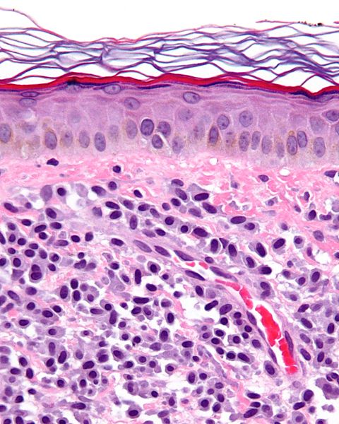
Micrograph showing a mast cell tumor.

References
- ↑ Moon TC, Befus AD, Kulka M (2014). "Mast cell mediators: their differential release and the secretory pathways involved". Front Immunol. 5: 569. doi:10.3389/fimmu.2014.00569. PMID 25452755.
- ↑ Krystel-Whittemore M, Dileepan KN, Wood JG (2015). "Mast Cell: A Multi-Functional Master Cell". Front Immunol. 6: 620. doi:10.3389/fimmu.2015.00620. PMID 26779180.
- ↑ Metcalfe DD (2005). "Regulation of normal and neoplastic human mast cell development in mastocytosis". Trans. Am. Clin. Climatol. Assoc. 116: 185–203, discussion 203–4.
- ↑ Ramsay DB, Stephen S, Borum M, Voltaggio L, Doman DB (December 2010). "Mast cells in gastrointestinal disease". Gastroenterol Hepatol (N Y). 6 (12): 772–7. PMC 3033552. PMID 21301631.
- ↑ Ahmed M, Kesavan M, Jilani BN, Ahmed S, Deeb L (June 2016). "Systemic Mastocytosis as an Unconventional Cause of Variceal Bleeding: Think Outside the Box". Cureus. 8 (6): e629. doi:10.7759/cureus.629. PMC 4935436. PMID 27433408.
- ↑ Molderings, Gerhard J; Brettner, Stefan; Homann, Jürgen; Afrin, Lawrence B (2011). "Mast cell activation disease: a concise practical guide for diagnostic workup and therapeutic options". Journal of Hematology & Oncology. 4 (1): 10. doi:10.1186/1756-8722-4-10. ISSN 1756-8722.
- ↑ Longley BJ, Morganroth GS, Tyrrell L, Ding TG, Anderson DM, Williams DE, Halaban R (May 1993). "Altered metabolism of mast-cell growth factor (c-kit ligand) in cutaneous mastocytosis". N. Engl. J. Med. 328 (18): 1302–7. doi:10.1056/NEJM199305063281803. PMID 7682288.
- ↑ Galli SJ, Tsai M, Wershil BK (April 1993). "The c-kit receptor, stem cell factor, and mast cells. What each is teaching us about the others". Am. J. Pathol. 142 (4): 965–74. PMC 1886888. PMID 7682764.
- ↑ Chatterjee A, Ghosh J, Kapur R (July 2015). "Mastocytosis: a mutated KIT receptor induced myeloproliferative disorder". Oncotarget. 6 (21): 18250–64. doi:10.18632/oncotarget.4213. PMID 26158763.
- ↑ Kristensen T, Vestergaard H, Møller MB (March 2011). "Improved detection of the KIT D816V mutation in patients with systemic mastocytosis using a quantitative and highly sensitive real-time qPCR assay". J Mol Diagn. 13 (2): 180–8. doi:10.1016/j.jmoldx.2010.10.004. PMC 3279709. PMID 21354053.
- ↑ Schwaab J, Schnittger S, Sotlar K, Walz C, Fabarius A, Pfirrmann M, Kohlmann A, Grossmann V, Meggendorfer M, Horny HP, Valent P, Jawhar M, Teichmann M, Metzgeroth G, Erben P, Ernst T, Hochhaus A, Haferlach T, Hofmann WK, Cross NC, Reiter A (October 2013). "Comprehensive mutational profiling in advanced systemic mastocytosis". Blood. 122 (14): 2460–6. doi:10.1182/blood-2013-04-496448. PMID 23958953.
- ↑ Traina F, Visconte V, Jankowska AM, Makishima H, O'Keefe CL, Elson P, Han Y, Hsieh FH, Sekeres MA, Mali RS, Kalaycio M, Lichtin AE, Advani AS, Duong HK, Copelan E, Kapur R, Olalla Saad ST, Maciejewski JP, Tiu RV (2012). "Single nucleotide polymorphism array lesions, TET2, DNMT3A, ASXL1 and CBL mutations are present in systemic mastocytosis". PLoS ONE. 7 (8): e43090. doi:10.1371/journal.pone.0043090. PMC 3419680. PMID 22905207.
- ↑ Chan EC, Bai Y, Bandara G, Simakova O, Brittain E, Scott L, Dyer KD, Klion AD, Maric I, Gilfillan AM, Metcalfe DD, Wilson TM (October 2013). "KIT GNNK splice variants: expression in systemic mastocytosis and influence on the activating potential of the D816V mutation in mast cells". Exp. Hematol. 41 (10): 870–881.e2. doi:10.1016/j.exphem.2013.05.005. PMID 23743299.
- ↑ Berezowska S, Flaig MJ, Ruëff F, Walz C, Haferlach T, Krokowski M, Kerler R, Petat-Dutter K, Horny HP, Sotlar K (January 2014). "Adult-onset mastocytosis in the skin is highly suggestive of systemic mastocytosis". Mod. Pathol. 27 (1): 19–29. doi:10.1038/modpathol.2013.117. PMID 23807778.
- ↑ Tefferi A, Lim KH, Abdel-Wahab O, Lasho TL, Patel J, Patnaik MM, Hanson CA, Pardanani A, Gilliland DG, Levine RL (July 2009). "Detection of mutant TET2 in myeloid malignancies other than myeloproliferative neoplasms: CMML, MDS, MDS/MPN and AML". Leukemia. 23 (7): 1343–5. doi:10.1038/leu.2009.59. PMC 4654626. PMID 19295549.