Hemophilia x ray
|
Hemophilia Microchapters |
|
Diagnosis |
|---|
|
Treatment |
|
Case Studies |
|
Hemophilia x ray On the Web |
|
American Roentgen Ray Society Images of Hemophilia x ray |
Editor-In-Chief: C. Michael Gibson, M.S., M.D. [1];Associate Editor(s)-in-Chief: Simrat Sarai, M.D. [2]
Overview
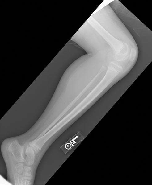
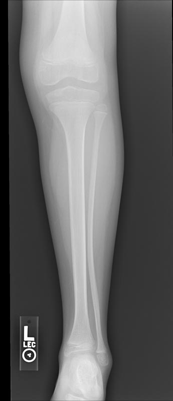
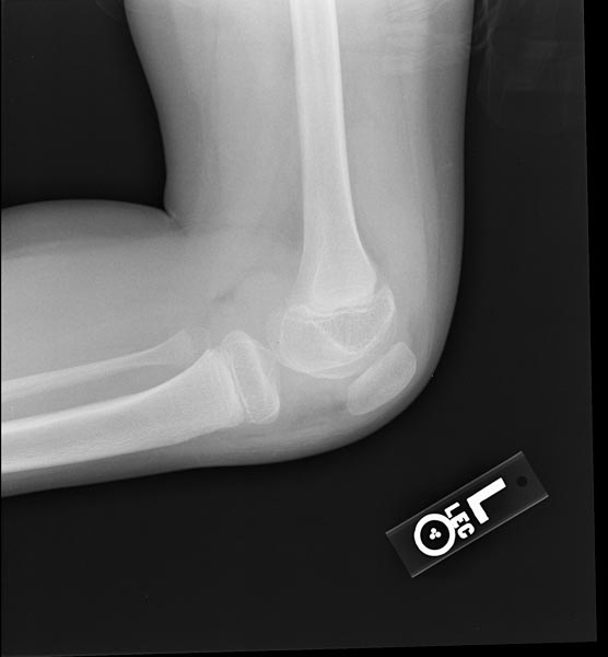
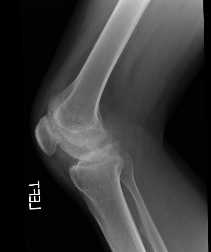
An x-ray of the joints in the case of hemophilic arthropathy may be helpful in the diagnosis of hemophilia. Pettersson scoring system, designed in 1980, is widely applied for the classification of osteochondral changes of hemophilic arthropathy in elbows, knees, and ankles. This scoring system is based on typical findings of hemophilic arthropathy on posterior-anterior and lateral x-rays.
X Ray
- An x-ray of the joints in the case of hemophilic arthropathy may be helpful in the diagnosis of hemophilia.
- Pettersson scoring system, designed in 1980 and based on typical findings of hemophilic arthropathy on posterior-anterior and lateral x-rays, is widely applied for the classification of osteochondral changes of hemophilic arthropathy.[1]
Classification of hemophilic arthropathy according to the Pettersson score[1]
| Finding | Interpretation |
|---|---|
| Osteoporosis |
|
| Enlargement of epiphysis |
|
| Irregularity of subchondral surface |
|
| Narrowing of joint space |
|
| Subchondral cysts formation |
|
| Erosion of joint margins |
|
| Gross incongruence of articulating bone ends |
|
| Joint deformity (angulation and/or displacement) |
|
Images
-
Hemophilic arthropathy involving the bilateral knees - Source: radswiki.net
-
Hemophilic arthropathy involving the bilateral knees
-
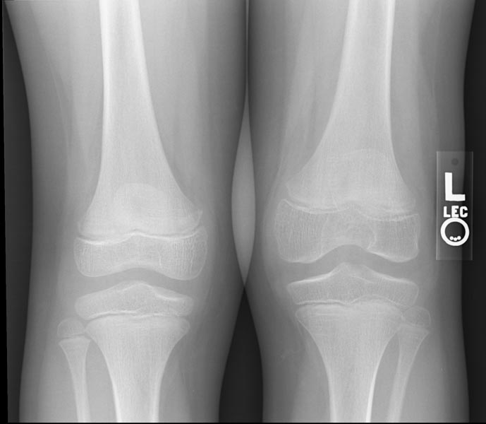
Hemophilic arthropathy involving the bilateral knees
-
Hemophilic arthropathy involving the bilateral knees


-
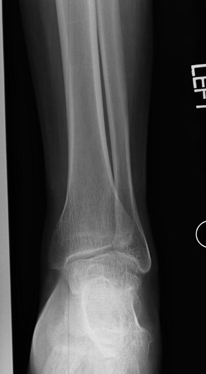
X-ray of the ankle in a patient with Hemophilia
-
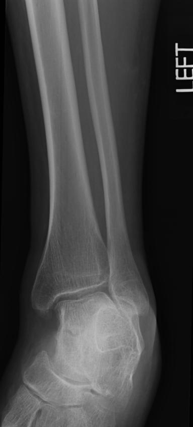
X-ray of the ankle in a patient with Hemophilia
-
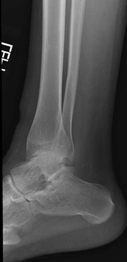
X-ray of the ankle in a patient with Hemophilia


-
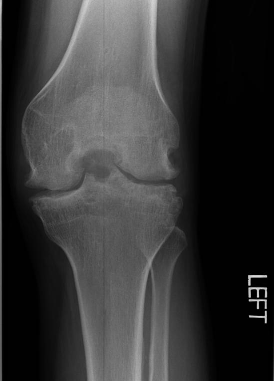
X-ray of the knee in a patient with Hemophilia
-
X-ray of the knee in a patient with Hemophilia

