Hairy cell leukemia laboratory tests
|
Hairy cell leukemia Microchapters |
|
Diagnosis |
|---|
|
Treatment |
|
Case Studies |
|
Hairy cell leukemia laboratory tests On the Web |
|
American Roentgen Ray Society Images of Hairy cell leukemia laboratory tests |
|
Risk calculators and risk factors for Hairy cell leukemia laboratory tests |
Editor-In-Chief: C. Michael Gibson, M.S., M.D. [1]
Overview
Hairy cell leukemia is commonly diagnosed after a routine blood count shows unexpectedly low numbers for one or more kinds of blood cells, or after unexplained bruises or unexplained infections, such as repeated bouts of pneumonia in an otherwise apparently healthy patient.
Laboratory Findings
Platelet function may be somewhat impaired in HCL patients, although this does not appear to have any significant practical effect.[1] It may result in somewhat more mild bruises than would otherwise be expected for a given platelet count or a mildly increased bleeding time for a minor cut. It is likely the result of producing slightly abnormal platelets in the overstressed bone marrow tissue.
Patients with a high tumor burden may also have somewhat reduced levels of cholesterol,[2] especially in patients with an enlarged spleen.[3] Cholesterol levels return to more normal values with successful treatment of HCL.
-
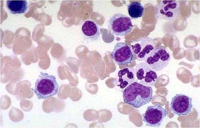
Hairy cell leukemia
-
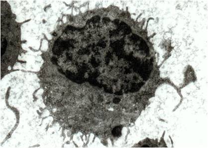
Hairy cell leukemia TEM


References
- ↑ Zuzel M, Cawley JC, Paton RC, Burns GF, McNicol GP (1979). "Platelet function in hairy-cell leukaemia". J. Clin. Pathol. 32 (8): 814–21. PMID 512041.
- ↑ "wiley.com". Retrieved 2007-09-07.
- ↑ "Mechanisms behind hypocholesterolaemia in hairy cell leukaemia -- Juliusson et al. 311 (6996): 27 -- BMJ". Retrieved 2007-09-07.