Entamoeba histolytica
|
Amoebiasis Microchapters |
|
Diagnosis |
|---|
|
Treatment |
|
Case Studies |
|
Entamoeba histolytica On the Web |
|
American Roentgen Ray Society Images of Entamoeba histolytica |
| style="background:#Template:Taxobox colour;"|Entamoeba histolytica | ||||||||||
|---|---|---|---|---|---|---|---|---|---|---|
 Entamoeba histolytica cyst
| ||||||||||
| style="background:#Template:Taxobox colour;" | Scientific classification | ||||||||||
|
Editor-In-Chief: C. Michael Gibson, M.S., M.D. [1]; Associate Editor(s)-in-Chief: Jesus Rosario Hernandez, M.D. [2]
Overview
Entamoeba histolytica is an anaerobic parasitic protozoan, part of the genus Entamoeba. It infects predominantly humans and other primates. It is estimated that about 50 million people are infected with the parasite worldwide. Diverse mammals such as dogs and cats can become infected but are not thought to contribute significantly to transmission. The active (trophozoite) stage exists only in the host and in fresh loose feces; cysts survive outside the host in water, soils and on foods, especially under moist conditions on the latter. When cysts are swallowed they cause infections by excysting (releasing the trophozoite stage) in the digestive tract.
E. histolytica, as its name suggests (histo–lytic = tissue destroying), causes disease; infection can lead to amoebic dysentery or amoebic liver abscess. Symptoms can include fulminating dysentery, diarrhea, weight loss, fatigue, abdominal pain, and amebomas. The amoeba can actually 'bore' into the intestinal wall, causing lesions and intestinal symptoms, and it may reach the blood stream. From there, it can reach different vital organs of the human body, usually the liver, but sometimes the lungs, brain, spleen, etc. A common outcome of this invasion of tissues is a liver abscess, which can be fatal if untreated. Ingested red blood cells are sometimes seen in the amoeba cell cytoplasm.
It can be diagnosed by stool samples but it is important to note that certain other species are impossible to distinguish by microscopy alone. Trophozoites may be seen in a fresh fecal smear and cysts in an ordinary stool sample. ELISA or RIA can also be used.
|
Amoebiasis Microchapters |
|
Diagnosis |
|---|
|
Treatment |
|
Case Studies |
|
Entamoeba histolytica On the Web |
|
American Roentgen Ray Society Images of Entamoeba histolytica |
Editor-In-Chief: C. Michael Gibson, M.S., M.D. [3]
Overview
Entamoeba histolytica can live in the large intestine (colon) without causing disease. However, sometimes, it invades the colon wall, causing colitis, acute dysentery, or long-term (chronic) diarrhea. The infection can also spread through the blood to the liver and, rarely, to the lungs, brain or other organs.
Amoebiasis can be seen anywhere in the world, but it is most common in tropical areas with crowded living conditions and poor sanitation. Africa, Mexico, parts of South America, and India have significant health problems associated with this disease. Entamoeba histolytica is spread through food or water contaminated with stools. This is common when human waste is used as fertilizer. It can also be spread from person to person -- particularly by contact with the mouth or rectal area of an infected person.
Life Cycle
Cysts and trophozoites are passed in feces (1). Cysts are typically found in formed stool, whereas trophozoites are typically found in diarrheal stool. Infection by Entamoeba histolytica occurs by ingestion of mature cysts (2) in fecally contaminated food, water, or hands. Excystation (3) occurs in the small intestine and trophozoites (4) are released, which migrate to the large intestine. The trophozoites multiply by binary fission and produce cysts (5), and both stages are passed in the feces (1). Because of the protection conferred by their walls, the cysts can survive days to weeks in the external environment and are responsible for transmission. Trophozoites passed in the stool are rapidly destroyed once outside the body, and if ingested would not survive exposure to the gastric environment. In many cases, the trophozoites remain confined to the intestinal lumen (A: noninvasive infection) of individuals who are asymptomatic carriers, passing cysts in their stool. In some patients the trophozoites invade the intestinal mucosa (B: intestinal disease), or, through the bloodstream, extraintestinal sites such as the liver, brain, and lungs (C: extraintestinal disease), with resultant pathologic manifestations. It has been established that the invasive and noninvasive forms represent two separate species, respectively E. histolytica and E. dispar. These two species are morphologically indistinguishable unless E. histolytica is observed with ingested red blood cells (erythrophagocystosis). Transmission can also occur through exposure to fecal matter during sexual contact (in which case not only cysts, but also trophozoites could prove infective).
-
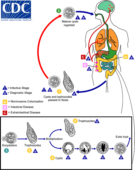
Life cycle of Amebiasis
Adapted from CDC

Life Cycle
| Genus and Species | Entamoeba histolytica |
| Etiologic Agent of: | Amoebiasis; Amoebic dysentery; Extraintestinal Amoebiasis, usually Amoebic Liver Abscess = “anchovy sauce”); Amoeba Cutis; Amoebic Lung Abscess (“liver-colored sputum”) |
| Infective stage | Cyst |
| Definitive Host | Human |
| Portal of Entry | Mouth |
| Mode of Transmission | Ingestion of mature cyst through contaminated food or water |
| Habitat | Colon and Cecum |
| Pathogenic Stage | Trophozoite |
| Locomotive apparatus | Pseudopodia (“False Foot”) |
| Motility | Active, Progressive and Directional |
| Nucleus | 'Ring and dot' appearance: peripheral chromatin and central karyosome |
| Mode of Reproduction | Binary Fission |
| Pathogenesis | Lytic necrosis (it looks like “flask-shaped” holes in Gastrointestinal tract sections (GIT) |
| Type of Encystment | Protective and Reproductive |
| Lab Diagnosis | Most common is Direct Fecal Smear (DFS) and staining (but does not allow identification to species level); Enzyme immunoassay (EIA); Indirect Hemagglutination (IHA); Antigen detection – monoclonal antibody; PCR for species identification. Culture: From faecal samples - Robinson's medium, Jones' medium |
| Treatment | Metronidazole for the invasive trophozoites PLUS a lumenal amoebicide for those still in the intestine (Paromomycin is the most widely used) |
| Trophozoite Stage | |
| Pathognomonic/Diagnostic Feature | Ingested RBC; distinctive nucleus |
| Cyst Stage | |
| Chromatoidal Body | 'Cigar' shaped bodies (made up of crystalline ribosomes) |
| Number of Nuclei | 1 in early stages, 4 when mature |
| Pathognomonic/Diagnostic Feature | 'Ring and dot' nucleus and chromatoid bodies |
Gallery
-
![Entamoeba histolytica. From Public Health Image Library (PHIL). [1]](/images/5/5d/Entamoeba_histolytica01.jpeg)
Entamoeba histolytica. From Public Health Image Library (PHIL). [1]
-
![Entamoeba histolytica. From Public Health Image Library (PHIL). [1]](/images/1/11/Entamoeba_histolytica02.jpeg)
Entamoeba histolytica. From Public Health Image Library (PHIL). [1]
-
![Entamoeba histolytica. From Public Health Image Library (PHIL). [1]](/images/8/8d/Entamoeba_histolytica03.jpeg)
Entamoeba histolytica. From Public Health Image Library (PHIL). [1]
-
![Entamoeba histolytica. From Public Health Image Library (PHIL). [1]](/images/e/ec/Entamoeba_histolytica04.jpeg)
Entamoeba histolytica. From Public Health Image Library (PHIL). [1]
-
![Entamoeba histolytica. From Public Health Image Library (PHIL). [1]](/images/7/74/Entamoeba_histolytica05.jpeg)
Entamoeba histolytica. From Public Health Image Library (PHIL). [1]
-
![Entamoeba histolytica. From Public Health Image Library (PHIL). [1]](/images/d/d9/Entamoeba_histolytica06.jpeg)
Entamoeba histolytica. From Public Health Image Library (PHIL). [1]
-
![Entamoeba histolytica. From Public Health Image Library (PHIL). [1]](/images/b/bc/Entamoeba_histolytica07.jpeg)
Entamoeba histolytica. From Public Health Image Library (PHIL). [1]
-
![Entamoeba histolytica. From Public Health Image Library (PHIL). [1]](/images/2/24/Entamoeba_histolytica08.jpeg)
Entamoeba histolytica. From Public Health Image Library (PHIL). [1]
-
![Entamoeba histolytica. From Public Health Image Library (PHIL). [1]](/images/e/e0/Entamoeba_histolytica09.jpeg)
Entamoeba histolytica. From Public Health Image Library (PHIL). [1]
-
![Entamoeba histolytica. From Public Health Image Library (PHIL). [1]](/images/0/0b/Entamoeba_histolytica11.jpeg)
Entamoeba histolytica. From Public Health Image Library (PHIL). [1]
-
![Entamoeba histolytica. From Public Health Image Library (PHIL). [1]](/images/e/e9/Entamoeba_histolytica12.jpeg)
Entamoeba histolytica. From Public Health Image Library (PHIL). [1]
-
![Entamoeba histolytica. From Public Health Image Library (PHIL). [1]](/images/2/2b/Entamoeba_histolytica13.jpeg)
Entamoeba histolytica. From Public Health Image Library (PHIL). [1]
-
![Entamoeba histolytica. From Public Health Image Library (PHIL). [1]](/images/1/10/Entamoeba_histolytica14.jpeg)
Entamoeba histolytica. From Public Health Image Library (PHIL). [1]
![Entamoeba histolytica. From Public Health Image Library (PHIL). [1]](/index.php/File:Entamoeba_histolytica01.jpeg)
![Entamoeba histolytica. From Public Health Image Library (PHIL). [1]](/index.php/File:Entamoeba_histolytica02.jpeg)
![Entamoeba histolytica. From Public Health Image Library (PHIL). [1]](/index.php/File:Entamoeba_histolytica03.jpeg)
![Entamoeba histolytica. From Public Health Image Library (PHIL). [1]](/index.php/File:Entamoeba_histolytica04.jpeg)
![Entamoeba histolytica. From Public Health Image Library (PHIL). [1]](/index.php/File:Entamoeba_histolytica05.jpeg)
![Entamoeba histolytica. From Public Health Image Library (PHIL). [1]](/index.php/File:Entamoeba_histolytica06.jpeg)
![Entamoeba histolytica. From Public Health Image Library (PHIL). [1]](/index.php/File:Entamoeba_histolytica07.jpeg)
![Entamoeba histolytica. From Public Health Image Library (PHIL). [1]](/index.php/File:Entamoeba_histolytica08.jpeg)
![Entamoeba histolytica. From Public Health Image Library (PHIL). [1]](/index.php/File:Entamoeba_histolytica09.jpeg)
![Entamoeba histolytica. From Public Health Image Library (PHIL). [1]](/index.php/File:Entamoeba_histolytica11.jpeg)
![Entamoeba histolytica. From Public Health Image Library (PHIL). [1]](/index.php/File:Entamoeba_histolytica12.jpeg)
![Entamoeba histolytica. From Public Health Image Library (PHIL). [1]](/index.php/File:Entamoeba_histolytica13.jpeg)
![Entamoeba histolytica. From Public Health Image Library (PHIL). [1]](/index.php/File:Entamoeba_histolytica14.jpeg)