Choroid plexus papilloma: Difference between revisions
No edit summary |
|||
| Line 109: | Line 109: | ||
*A [positive/negative] [test name] is diagnostic of [disease name]. | *A [positive/negative] [test name] is diagnostic of [disease name]. | ||
*An [elevated/reduced] concentration of [serum/blood/urinary/CSF/other] [lab test] is diagnostic of [disease name]. | *An [elevated/reduced] concentration of [serum/blood/urinary/CSF/other] [lab test] is diagnostic of [disease name]. | ||
*Other laboratory findings consistent with the diagnosis of [disease name] include | *Other laboratory findings consistent with the diagnosis of [disease name] include immunohistochemistry, [abnormal test 2], and [abnormal test 3]. | ||
====Immunohistochemistry==== | |||
*Choroid plexus papilloma is demonstrated by positivity to tumor marker such as [[cytokeratin]] (CK7).<ref name=clinicalpresentationcpp1>Clinical presentation of choroid plexus papilloma. Dr Tim Luijkx and Dr Paresh K Desai et al. Radiopaedia 2016. http://radiopaedia.org/articles/choroid-plexus-papilloma-1. Accessed on January 13, 2016</ref> | |||
*Some reactivity for [[GFAP]] and epithelial membrane antigen may be present.<ref name="pmid26500797">{{cite journal| author=Prasad GL, Mahapatra AK| title=Case series of choroid plexus papilloma in children at uncommon locations and review of the literature. | journal=Surg Neurol Int | year= 2015 | volume= 6 | issue= | pages= 151 | pmid=26500797 | doi=10.4103/2152-7806.166167 | pmc=PMC4596056 | url=http://www.ncbi.nlm.nih.gov/entrez/eutils/elink.fcgi?dbfrom=pubmed&tool=sumsearch.org/cite&retmode=ref&cmd=prlinks&id=26500797 }} </ref> | |||
===Imaging Findings=== | ===Imaging Findings=== | ||
*There are no [imaging study] findings associated with [disease name]. | *There are no [imaging study] findings associated with [disease name]. | ||
Revision as of 15:25, 11 April 2016
For patient information, click here
Template:Choroid plexus papilloma Editor-In-Chief: C. Michael Gibson, M.S., M.D. [1]Associate Editor(s)-in-Chief: Sujit Routray, M.D. [2]
Synonyms and keywords: Choroid plexus papillomas; Papilloma of choroid plexus; Papilloma of the choroid plexus; CPP
Overview
Choroid plexus papilloma was first described by Guerard, in 1832.[1]Choroid plexus papilloma may be classified into two groups: typical and atypical.[2]
Historical Perspective
Choroid plexus papilloma was first described by Guerard, in 1832.[1]
Classification
Choroid plexus papilloma may be classified into two groups:[2]
Choroid plexus papilloma | |||||||||||||||||||||||||||||||||||||||||||||||||||||||||||||||||||||||
Typical | Atypical | ||||||||||||||||||||||||||||||||||||||||||||||||||||||||||||||||||||||
WHO grade I | WHO grade II | ||||||||||||||||||||||||||||||||||||||||||||||||||||||||||||||||||||||
Pathophysiology
- Choroid plexus papilloma is neuroectodermal in origin and similar in structure to a normal choroid plexus. They may be created by epithelial cells of the choroid plexus.[3]
- It may be a true congenital intracranial neoplasm.
Choroid plexus papilloma may be associated with:[4][5]
- Aicardi syndrome
- Von Hippel-Lindau disease
- Li-Fraumeni syndrome
- On gross pathology, choroid plexus papilloma is characterized by a soft, solid, pink to red, capsulated, vascular, and friable cauliflower-like mass.[4]
- Unlike most other brain tumors which are more common in the posterior fossa in children and supratentorial compartment in adults, the relationship is reversed for choroid plexus papillomas:
- Adults: 70% occur in the fourth ventricle
- Children: most often occur in the lateral ventricles, with a predilection for the trigone
- Third ventricular, cerebellopontine angle, parenchymal and even pineal region tumors have also been described.
Gallery
-
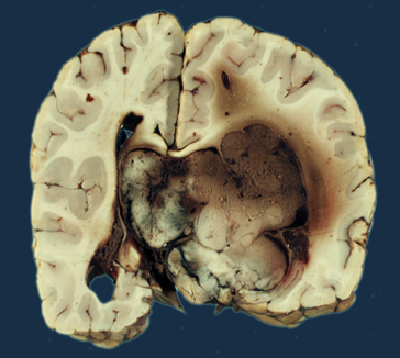
Gross pathological specimen of choroid plexus papilloma.

Microscopic Pathology
- On microscopic histopathological analysis, choroid plexus papilloma is characterized by papillary structures with a delicate fibrovascular core lined by columnar or cuboidal epithelial cells with vesicular nuclei. Its appearance is very similar to normal choroid plexus.[4]
- If two or more mitoses are present per 10 high power field (HPF), then the tumor is designated an atypical choroid plexus papilloma and is classified as a WHO grade II tumor.
Gallery
-
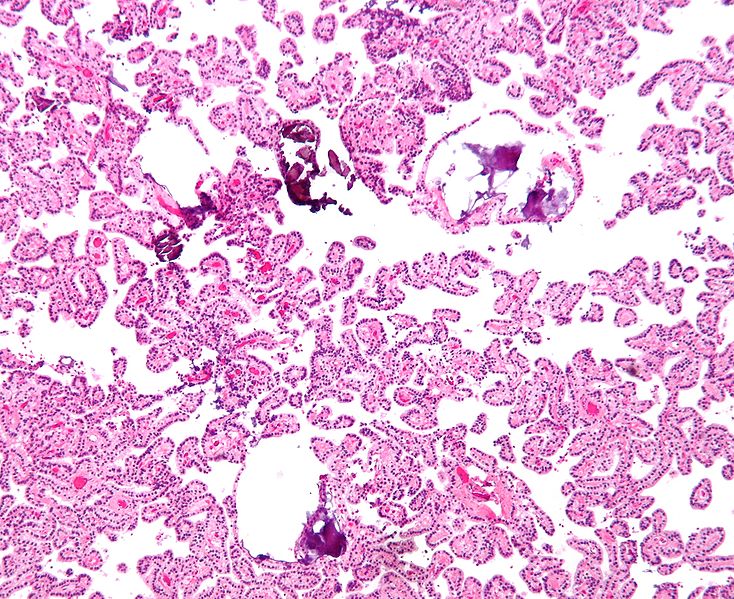
Shown above is a micrograph of choroid plexus papilloma (H&E stain).

Causes
- [Disease name] may be caused by either [cause1], [cause2], or [cause3].
- [Disease name] is caused by a mutation in the [gene1], [gene2], or [gene3] gene[s].
- There are no established causes for [disease name].
Differentiating [disease name] from other Diseases
- [Disease name] must be differentiated from other diseases that cause [clinical feature 1], [clinical feature 2], and [clinical feature 3], such as:
- [Differential dx1]
- [Differential dx2]
- [Differential dx3]
Epidemiology and Demographics
- The prevalence of [disease name] is approximately [number or range] per 100,000 individuals worldwide.
- In [year], the incidence of [disease name] was estimated to be [number or range] cases per 100,000 individuals in [location].
Age
- Patients of all age groups may develop [disease name].
- [Disease name] is more commonly observed among patients aged [age range] years old.
- [Disease name] is more commonly observed among [elderly patients/young patients/children].
Gender
- [Disease name] affects men and women equally.
- [Gender 1] are more commonly affected with [disease name] than [gender 2].
- The [gender 1] to [Gender 2] ratio is approximately [number > 1] to 1.
Race
- There is no racial predilection for [disease name].
- [Disease name] usually affects individuals of the [race 1] race.
- [Race 2] individuals are less likely to develop [disease name].
Risk Factors
- Common risk factors in the development of [disease name] are [risk factor 1], [risk factor 2], [risk factor 3], and [risk factor 4].
Natural History, Complications and Prognosis
- The majority of patients with [disease name] remain asymptomatic for [duration/years].
- Early clinical features include [manifestation 1], [manifestation 2], and [manifestation 3].
- If left untreated, [#%] of patients with [disease name] may progress to develop [manifestation 1], [manifestation 2], and [manifestation ].
- Common complications of [disease name] include [complication 1], [complication 2], and [complication 3].
- Prognosis is generally [excellent/good/poor], and the [1/5/10year mortality/survival rate] of patients with [disease name] is approximately [#%].
Diagnosis
Diagnostic Criteria
- The diagnosis of [disease name] is made when at least [number] of the following [number] diagnostic criteria are met:
- [criterion 1]
- [criterion 2]
- [criterion 3]
- [criterion 4]
Symptoms
- [Disease name] is usually asymptomatic.
- Symptoms of [disease name] may include the following:
- [symptom 1]
- [symptom 2]
- [symptom 3]
- [symptom 4]
- [symptom 5]
- [symptom 6]
Physical Examination
- Patients with [disease name] usually appear [general appearance].
- Physical examination may be remarkable for:
- [finding 1]
- [finding 2]
- [finding 3]
- [finding 4]
- [finding 5]
- [finding 6]
Laboratory Findings
- There are no specific laboratory findings associated with [disease name].
- A [positive/negative] [test name] is diagnostic of [disease name].
- An [elevated/reduced] concentration of [serum/blood/urinary/CSF/other] [lab test] is diagnostic of [disease name].
- Other laboratory findings consistent with the diagnosis of [disease name] include immunohistochemistry, [abnormal test 2], and [abnormal test 3].
Immunohistochemistry
- Choroid plexus papilloma is demonstrated by positivity to tumor marker such as cytokeratin (CK7).[4]
- Some reactivity for GFAP and epithelial membrane antigen may be present.[1]
Imaging Findings
- There are no [imaging study] findings associated with [disease name].
- [Imaging study 1] is the imaging modality of choice for [disease ame].
- On [imaging study 1], [disease name] is characterized by [finding 1], [finding 2], and [finding 3].
- [Imaging study 2] may demonstrate [finding 1], [finding 2], and [finding 3].
Other Diagnostic Studies
- [Disease name] may also be diagnosed using [diagnostic study name].
- Findings on [diagnostic study name] include [finding 1], [finding 2], and [finding 3].
Treatment
Medical Therapy
- There is no treatment for [disease name]; the mainstay of therapy is supportive care.
- The mainstay of therapy for [disease name] is [medical therapy 1] and [medical therapy 2].
- [Medical therapy 1] acts by [mechanism of action1].
- Response to [medical therapy 1] can be monitored with [test/physical finding/imaging] every [frequency/duration].
Surgery
- Surgery is the mainstay of therapy for [disease name].
- [Surgical procedure] in conjunction with [chemotherapy/radiation] is he most common approach to the treatment of [disease name].
- [Surgical procedure] can only be performed for patients with [disease tage] [disease name].
Prevention
- There are no primary preventive measures available for [disease name].
- Effective measures for the primary prevention of [disease name] include [measure1], [measure2], and [measure3].
- Once diagnosed and successfully treated, patients with [disease name] are followedup every [duration]. Followup testing includes [test 1], [test 2], and [test 3].
References
- ↑ 1.0 1.1 1.2 Prasad GL, Mahapatra AK (2015). "Case series of choroid plexus papilloma in children at uncommon locations and review of the literature". Surg Neurol Int. 6: 151. doi:10.4103/2152-7806.166167. PMC 4596056. PMID 26500797.
- ↑ 2.0 2.1 Choroid plexus papillomas. Dr Tim Luijkx and Dr Paresh K Desai et al. Radiopaedia 2016. http://radiopaedia.org/articles/choroid-plexus-papilloma-1. Accessed on January 13, 2016
- ↑ Choroid plexus papilloma. Wikipedia 2016. https://en.wikipedia.org/wiki/Choroid_plexus_papilloma. Accessed on January 13, 2016
- ↑ 4.0 4.1 4.2 4.3 Clinical presentation of choroid plexus papilloma. Dr Tim Luijkx and Dr Paresh K Desai et al. Radiopaedia 2016. http://radiopaedia.org/articles/choroid-plexus-papilloma-1. Accessed on January 13, 2016
- ↑ Gozali AE, Britt B, Shane L, Gonzalez I, Gilles F, McComb JG; et al. (2012). "Choroid plexus tumors; management, outcome, and association with the Li-Fraumeni syndrome: the Children's Hospital Los Angeles (CHLA) experience, 1991-2010". Pediatr Blood Cancer. 58 (6): 905–9. doi:10.1002/pbc.23349. PMID 21990040.