Choroid plexus papilloma: Difference between revisions
| (34 intermediate revisions by 2 users not shown) | |||
| Line 1: | Line 1: | ||
__NOTOC__ | __NOTOC__ | ||
{{SI}} | |||
{{CMG}}{{AE}}{{SR}}; {{Ammu}} | |||
{{ | |||
{{CMG}}{{AE}}{{SR}} | |||
{{SK}} Choroid plexus papillomas; Papilloma of choroid plexus; Papilloma of the choroid plexus; CPP | {{SK}} Choroid plexus papillomas; Papilloma of choroid plexus; Papilloma of the choroid plexus; CPP | ||
==Overview== | ==Overview== | ||
Choroid plexus papilloma | Choroid plexus papilloma is a a rare, slow-growing, histologically benign intracranial [[tumor]] that is commonly located in the [[ventricular system]] of the [[choroid plexus]]. It may obstruct the [[cerebrospinal fluid]] flow, causing elevated [[intracranial pressure]]. Choroid plexus papilloma is [[neuroectoderm|neuroectodermal]] in origin and similar in structure to a normal [[choroid plexus]]. They may be created by [[epithelial cell]]s of the [[choroid plexus]].<ref name=cpp1ovebkj>Choroid plexus papilloma. Wikipedia 2016. https://en.wikipedia.org/wiki/Choroid_plexus_papilloma. Accessed on January 13, 2016</ref> Choroid plexus papilloma may be associated with [[Aicardi syndrome]], [[Von Hippel-Lindau disease]], and [[Li-Fraumeni syndrome]].<ref name=clinicalpresentationcpp1>Clinical presentation of choroid plexus papilloma. Dr Tim Luijkx and Dr Paresh K Desai et al. Radiopaedia 2016. http://radiopaedia.org/articles/choroid-plexus-papilloma-1. Accessed on January 13, 2016</ref><ref name="pmid21990040">{{cite journal| author=Gozali AE, Britt B, Shane L, Gonzalez I, Gilles F, McComb JG et al.| title=Choroid plexus tumors; management, outcome, and association with the Li-Fraumeni syndrome: the Children's Hospital Los Angeles (CHLA) experience, 1991-2010. | journal=Pediatr Blood Cancer | year= 2012 | volume= 58 | issue= 6 | pages= 905-9 | pmid=21990040 | doi=10.1002/pbc.23349 | pmc= | url=http://www.ncbi.nlm.nih.gov/entrez/eutils/elink.fcgi?dbfrom=pubmed&tool=sumsearch.org/cite&retmode=ref&cmd=prlinks&id=21990040 }} </ref> On gross pathology, choroid plexus papilloma is characterized by a soft, solid, pink to red, capsulated, vascular, and friable cauliflower-like mass.<ref name=clinicalpresentationcpp1>Clinical presentation of choroid plexus papilloma. Dr Tim Luijkx and Dr Paresh K Desai et al. Radiopaedia 2016. http://radiopaedia.org/articles/choroid-plexus-papilloma-1. Accessed on January 13, 2016</ref> On microscopic histopathological analysis, choroid plexus papilloma is characterized by papillary structures with a delicate fibrovascular core lined by [[columnar epithelium|columnar]] or [[cuboidal epithelium|cuboidal epithelial cells]] with vesicular nuclei. Its appearance is very similar to normal [[choroid plexus]].<ref name=clinicalpresentationcpp1>Clinical presentation of choroid plexus papilloma. Dr Tim Luijkx and Dr Paresh K Desai et al. Radiopaedia 2016. http://radiopaedia.org/articles/choroid-plexus-papilloma-1. Accessed on January 13, 2016</ref> Choroid plexus papilloma is demonstrated by positivity to tumor marker such as [[cytokeratin]] (CK7).<ref name=clinicalpresentationcpp1>Clinical presentation of choroid plexus papilloma. Dr Tim Luijkx and Dr Paresh K Desai et al. Radiopaedia 2016. http://radiopaedia.org/articles/choroid-plexus-papilloma-1. Accessed on January 13, 2016</ref> Choroid plexus papilloma may be differentiated from choroid plexus carcinoma, [[intracerebral metastasis|choroid plexus metastasis]], [[medulloblastoma]], and [[meningioma|intraventricular meningioma]].<ref name=ddxcpp1>Differential diagnosis of choroid plexus. Dr Tim Luijkx and Dr Paresh K Desai et al. Radiopaedia 2016. http://radiopaedia.org/articles/choroid-plexus-papilloma-1. Accessed on January 13, 2016</ref> Choroid plexus papilloma accounts for approximately 1% of all brain tumors, 2-6% of all pediatric brain tumors, and 0.5% of the adult brain tumors.<ref name=cppepidemiology1>Epidemiology of choroid plexus papilloma. Dr Tim Luijkx and Dr Paresh K Desai et al. Radiopaedia 2016. http://radiopaedia.org/articles/choroid-plexus-papilloma-1. Accessed on January 13, 2016</ref> Choroid plexus papilloma is a rare disease that tends to affect children and adults.<ref name=cppepidemiology1>Epidemiology of choroid plexus papilloma. Dr Tim Luijkx and Dr Paresh K Desai et al. Radiopaedia 2016. http://radiopaedia.org/articles/choroid-plexus-papilloma-1. Accessed on January 13, 2016</ref> Males are more commonly affected with choroid plexus papilloma than females. The male to female ratio is approximately 2.8 to 1.<ref name=epicpp1>Epidemiology of choroid plexus papilloma. DR.SAGAR JUNG RANA 2016. http://drsagarjungrana.blogspot.com/2012/11/choroid-plexus-papilloma.html. Accessed on January 14, 2016</ref> If left untreated, patients with choroid plexus papilloma may progress to develop [[seizures]], [[hydrocephalus]], focal neurological deficits, [[nerve palsy|cranioneuropathies]], [[psychosis]], and [[coma]].<ref name="pmid26500797">{{cite journal| author=Prasad GL, Mahapatra AK| title=Case series of choroid plexus papilloma in children at uncommon locations and review of the literature. | journal=Surg Neurol Int | year= 2015 | volume= 6 | issue= | pages= 151 | pmid=26500797 | doi=10.4103/2152-7806.166167 | pmc=PMC4596056 | url=http://www.ncbi.nlm.nih.gov/entrez/eutils/elink.fcgi?dbfrom=pubmed&tool=sumsearch.org/cite&retmode=ref&cmd=prlinks&id=26500797 }} </ref><ref name="KoellerSandberg2002">{{cite journal|last1=Koeller|first1=Kelly K.|last2=Sandberg|first2=Glenn D.|title=From the Archives of the AFIP|journal=RadioGraphics|volume=22|issue=6|year=2002|pages=1473–1505|issn=0271-5333|doi=10.1148/rg.226025118}}</ref> Common complications of choroid plexus papilloma include CSF seeding, [[obstructive hydrocephalus]], [[blindness]], [[psychosis]], and [[coma]]. The prognosis is excellent, and the 5-year survival rate after complete excision of choroid plexus papilloma is approximately 100%.<ref name="KoellerSandberg2002">{{cite journal|last1=Koeller|first1=Kelly K.|last2=Sandberg|first2=Glenn D.|title=From the Archives of the AFIP|journal=RadioGraphics|volume=22|issue=6|year=2002|pages=1473–1505|issn=0271-5333|doi=10.1148/rg.226025118}}</ref><ref name="pmid9488307">{{cite journal| author=Pencalet P, Sainte-Rose C, Lellouch-Tubiana A, Kalifa C, Brunelle F, Sgouros S et al.| title=Papillomas and carcinomas of the choroid plexus in children. | journal=J Neurosurg | year= 1998 | volume= 88 | issue= 3 | pages= 521-8 | pmid=9488307 | doi=10.3171/jns.1998.88.3.0521 | pmc= | url=http://www.ncbi.nlm.nih.gov/entrez/eutils/elink.fcgi?dbfrom=pubmed&tool=sumsearch.org/cite&retmode=ref&cmd=prlinks&id=9488307 }} </ref> Symptoms of choroid plexus papilloma include [[headache]], [[vomiting]], [[seizures]], and [[vision loss]].<ref name=sympbjhbcpp1>Signs and symptoms of choroid plexus papilloma. Wikipedia 2016. https://en.wikipedia.org/wiki/Choroid_plexus_papilloma. Accessed on January 13, 2016</ref><ref name="pmid26500797">{{cite journal| author=Prasad GL, Mahapatra AK| title=Case series of choroid plexus papilloma in children at uncommon locations and review of the literature. | journal=Surg Neurol Int | year= 2015 | volume= 6 | issue= | pages= 151 | pmid=26500797 | doi=10.4103/2152-7806.166167 | pmc=PMC4596056 | url=http://www.ncbi.nlm.nih.gov/entrez/eutils/elink.fcgi?dbfrom=pubmed&tool=sumsearch.org/cite&retmode=ref&cmd=prlinks&id=26500797 }} </ref> Common physical examination findings of choroid plexus papilloma include bulging fontanelle, enlarged head size, [[papilledema]], [[altered mental status]], and [[Cranial Nerve Palsy|cranioneuropathies]] ([[abducens nerve palsy|6<sup>th</sup>]] and [[facial nerve palsy|7<sup>th</sup> nerve palsy]]).<ref name=sympbjhbcpp1>Signs and symptoms of choroid plexus papilloma. Wikipedia 2016. https://en.wikipedia.org/wiki/Choroid_plexus_papilloma. Accessed on January 13, 2016</ref><ref name="pmid26500797">{{cite journal| author=Prasad GL, Mahapatra AK| title=Case series of choroid plexus papilloma in children at uncommon locations and review of the literature. | journal=Surg Neurol Int | year= 2015 | volume= 6 | issue= | pages= 151 | pmid=26500797 | doi=10.4103/2152-7806.166167 | pmc=PMC4596056 | url=http://www.ncbi.nlm.nih.gov/entrez/eutils/elink.fcgi?dbfrom=pubmed&tool=sumsearch.org/cite&retmode=ref&cmd=prlinks&id=26500797 }} </ref><ref name="KoellerSandberg2002">{{cite journal|last1=Koeller|first1=Kelly K.|last2=Sandberg|first2=Glenn D.|title=From the Archives of the AFIP|journal=RadioGraphics|volume=22|issue=6|year=2002|pages=1473–1505|issn=0271-5333|doi=10.1148/rg.226025118}}</ref> Head CT scan and brain MRI are helpful in the diagnosis of choroid plexus papilloma. On CT scan, choroid plexus papilloma is characterized by iso- to hyperdense well-defined lobulated masses with intense homogenous enhancement and associated [[hydrocephalus]]. If there is markedly heterogeneous contrast enhancement, a choroid plexus carcinoma should be suspected.<ref name=radgraphiccpp1>Radiographic features of choroid plexus papilloma. Dr Tim Luijkx and Dr Paresh K Desai et al. Radiopaedia 2016. http://radiopaedia.org/articles/choroid-plexus-papilloma-1. Accessed on January 13, 2016</ref><ref name="pmid26500797">{{cite journal| author=Prasad GL, Mahapatra AK| title=Case series of choroid plexus papilloma in children at uncommon locations and review of the literature. | journal=Surg Neurol Int | year= 2015 | volume= 6 | issue= | pages= 151 | pmid=26500797 | doi=10.4103/2152-7806.166167 | pmc=PMC4596056 | url=http://www.ncbi.nlm.nih.gov/entrez/eutils/elink.fcgi?dbfrom=pubmed&tool=sumsearch.org/cite&retmode=ref&cmd=prlinks&id=26500797 }} </ref> On MRI, choroid plexus papilloma is characterized by a well demarcated, extra-axial mass that is iso- to hypointense on T1-weighted imaging and iso- to hyperintense on T2-weighted imaging. On contrast administration, there is generally marked homogenous enhancement.<ref name=radgraphiccpp1>Radiographic features of choroid plexus papilloma. Dr Tim Luijkx and Dr Paresh K Desai et al. Radiopaedia 2016. http://radiopaedia.org/articles/choroid-plexus-papilloma-1. Accessed on January 13, 2016</ref><ref name="pmid26500797">{{cite journal| author=Prasad GL, Mahapatra AK| title=Case series of choroid plexus papilloma in children at uncommon locations and review of the literature. | journal=Surg Neurol Int | year= 2015 | volume= 6 | issue= | pages= 151 | pmid=26500797 | doi=10.4103/2152-7806.166167 | pmc=PMC4596056 | url=http://www.ncbi.nlm.nih.gov/entrez/eutils/elink.fcgi?dbfrom=pubmed&tool=sumsearch.org/cite&retmode=ref&cmd=prlinks&id=26500797 }} </ref> The predominant therapy for choroid plexus papilloma is [[surgery|surgical resection]]. [[Radiotherapy]] is indicated for the inoperable residual tumor or resistant/aggressive subtype, whereas [[chemotherapy]] is reserved for the recurrent tumors.<ref name="pmid26500797">{{cite journal| author=Prasad GL, Mahapatra AK| title=Case series of choroid plexus papilloma in children at uncommon locations and review of the literature. | journal=Surg Neurol Int | year= 2015 | volume= 6 | issue= | pages= 151 | pmid=26500797 | doi=10.4103/2152-7806.166167 | pmc=PMC4596056 | url=http://www.ncbi.nlm.nih.gov/entrez/eutils/elink.fcgi?dbfrom=pubmed&tool=sumsearch.org/cite&retmode=ref&cmd=prlinks&id=26500797 }} </ref><ref name=rxcpp1>Treatment and prognosis of choroid plexus papilloma. Dr Tim Luijkx and Dr Paresh K Desai et al. Radiopaedia 2016. http://radiopaedia.org/articles/choroid-plexus-papilloma-1. Accessed on January 14, 2016</ref> | ||
==Historical Perspective== | ==Historical Perspective== | ||
Choroid plexus papilloma was first described by Guerard, in 1832.<ref name="pmid26500797">{{cite journal| author=Prasad GL, Mahapatra AK| title=Case series of choroid plexus papilloma in children at uncommon locations and review of the literature. | journal=Surg Neurol Int | year= 2015 | volume= 6 | issue= | pages= 151 | pmid=26500797 | doi=10.4103/2152-7806.166167 | pmc=PMC4596056 | url=http://www.ncbi.nlm.nih.gov/entrez/eutils/elink.fcgi?dbfrom=pubmed&tool=sumsearch.org/cite&retmode=ref&cmd=prlinks&id=26500797 | Choroid plexus papilloma was first described by Guerard, in 1832.<ref name="pmid26500797">{{cite journal| author=Prasad GL, Mahapatra AK| title=Case series of choroid plexus papilloma in children at uncommon locations and review of the literature. | journal=Surg Neurol Int | year= 2015 | volume= 6 | issue= | pages= 151 | pmid=26500797 | doi=10.4103/2152-7806.166167 | pmc=PMC4596056 | url=http://www.ncbi.nlm.nih.gov/entrez/eutils/elink.fcgi?dbfrom=pubmed&tool=sumsearch.org/cite&retmode=ref&cmd=prlinks&id=26500797}}</ref> | ||
==Classification== | ==Classification== | ||
Choroid plexus papilloma may be classified into two groups:<ref name=cpp1>Choroid plexus papillomas. Dr Tim Luijkx and Dr Paresh K Desai et al. Radiopaedia 2016. http://radiopaedia.org/articles/choroid-plexus-papilloma-1. Accessed on January 13, 2016</ref> | Choroid plexus papilloma may be classified into two groups:<ref name=cpp1>Choroid plexus papillomas. Dr Tim Luijkx and Dr Paresh K Desai et al. Radiopaedia 2016. http://radiopaedia.org/articles/choroid-plexus-papilloma-1. Accessed on January 13, 2016</ref> | ||
| Line 22: | Line 20: | ||
==Pathophysiology== | ==Pathophysiology== | ||
* | *Choroid plexus papilloma is [[neuroectoderm|neuroectodermal]] in origin and similar in structure to a normal [[choroid plexus]]. They may be created by [[epithelial cell]]s of the [[choroid plexus]].<ref name=cpp1ovebkj>Choroid plexus papilloma. Wikipedia 2016. https://en.wikipedia.org/wiki/Choroid_plexus_papilloma. Accessed on January 13, 2016</ref> | ||
* | *It may be a true congenital intracranial [[neoplasm]]. | ||
*On gross pathology, [ | *Choroid plexus papilloma may be associated with:<ref name=clinicalpresentationcpp1>Clinical presentation of choroid plexus papilloma. Dr Tim Luijkx and Dr Paresh K Desai et al. Radiopaedia 2016. http://radiopaedia.org/articles/choroid-plexus-papilloma-1. Accessed on January 13, 2016</ref><ref name="pmid21990040">{{cite journal| author=Gozali AE, Britt B, Shane L, Gonzalez I, Gilles F, McComb JG et al.| title=Choroid plexus tumors; management, outcome, and association with the Li-Fraumeni syndrome: the Children's Hospital Los Angeles (CHLA) experience, 1991-2010. | journal=Pediatr Blood Cancer | year= 2012 | volume= 58 | issue= 6 | pages= 905-9 | pmid=21990040 | doi=10.1002/pbc.23349 | pmc= | url=http://www.ncbi.nlm.nih.gov/entrez/eutils/elink.fcgi?dbfrom=pubmed&tool=sumsearch.org/cite&retmode=ref&cmd=prlinks&id=21990040 }} </ref> | ||
*On microscopic histopathological analysis, [ | :*[[Aicardi syndrome]] | ||
:*[[Von Hippel-Lindau disease]] | |||
:*[[Li-Fraumeni syndrome]] | |||
*On gross pathology, choroid plexus papilloma is characterized by a soft, solid, pink to red, capsulated, vascular, and friable cauliflower-like mass.<ref name=clinicalpresentationcpp1>Clinical presentation of choroid plexus papilloma. Dr Tim Luijkx and Dr Paresh K Desai et al. Radiopaedia 2016. http://radiopaedia.org/articles/choroid-plexus-papilloma-1. Accessed on January 13, 2016</ref> | |||
*Unlike most other brain tumors which are more common in the [[posterior fossa]] in children and [[supratentorial]] compartment in adults, the relationship is reversed for choroid plexus papillomas: | |||
:*'''Adults''': 70% occur in the [[fourth ventricle]] | |||
:*'''Children''': most often occur in the [[lateral ventricles]], with a predilection for the trigone | |||
*Third ventricular, cerebellopontine angle, parenchymal and even pineal region tumors have also been described. | |||
===Gallery=== | |||
<gallery> | |||
Image:Plexuspapillom_Makroskopie.png|<sub>Gross pathological specimen of choroid plexus papilloma.</sub> | |||
</gallery> | |||
==Microscopic Pathology== | |||
*On microscopic histopathological analysis, choroid plexus papilloma is characterized by [[papillary]] structures with a delicate [[fibrovascular]] core lined by [[columnar epithelium|columnar]] or [[cuboidal epithelium|cuboidal epithelial cells]] with vesicular nuclei. Its appearance is very similar to normal [[choroid plexus]].<ref name=clinicalpresentationcpp1>Clinical presentation of choroid plexus papilloma. Dr Tim Luijkx and Dr Paresh K Desai et al. Radiopaedia 2016. http://radiopaedia.org/articles/choroid-plexus-papilloma-1. Accessed on January 13, 2016</ref> | |||
*If two or more [[mitoses]] are present per 10 high power field (HPF), then the tumor is designated an atypical choroid plexus papilloma and is classified as a ''WHO grade II'' tumor. | |||
===Gallery=== | |||
<gallery> | |||
Image:734px-Choroid_plexus_papilloma_micrograph.jpg|<sub>Shown above is a micrograph of choroid plexus papilloma (H&E stain).</sub> | |||
</gallery> | |||
==Causes== | ==Causes== | ||
* | * There are no established causes for choroid plexus papilloma.<ref name="KoellerSandberg2002">{{cite journal|last1=Koeller|first1=Kelly K.|last2=Sandberg|first2=Glenn D.|title=From the Archives of the AFIP|journal=RadioGraphics|volume=22|issue=6|year=2002|pages=1473–1505|issn=0271-5333|doi=10.1148/rg.226025118}}</ref> | ||
* [ | |||
* | ==Differentiating Choroid Plexus Papilloma from other Diseases== | ||
Choroid plexus papilloma may be differentiated from:<ref name=ddxcpp1>Differential diagnosis of choroid plexus. Dr Tim Luijkx and Dr Paresh K Desai et al. Radiopaedia 2016. http://radiopaedia.org/articles/choroid-plexus-papilloma-1. Accessed on January 13, 2016</ref><ref name="pmid26500797">{{cite journal| author=Prasad GL, Mahapatra AK| title=Case series of choroid plexus papilloma in children at uncommon locations and review of the literature. | journal=Surg Neurol Int | year= 2015 | volume= 6 | issue= | pages= 151 | pmid=26500797 | doi=10.4103/2152-7806.166167 | pmc=PMC4596056 | url=http://www.ncbi.nlm.nih.gov/entrez/eutils/elink.fcgi?dbfrom=pubmed&tool=sumsearch.org/cite&retmode=ref&cmd=prlinks&id=26500797 }} </ref> | |||
*[ | *Choroid plexus carcinoma | ||
*[[intracerebral metastasis|Choroid plexus metastasis]] | |||
'''Among children''', choroid plexus papilloma must be differentiated from:<ref name=ddxcpp1>Differential diagnosis of choroid plexus. Dr Tim Luijkx and Dr Paresh K Desai et al. Radiopaedia 2016. http://radiopaedia.org/articles/choroid-plexus-papilloma-1. Accessed on January 13, 2016</ref> | |||
*[[Medulloblastoma]] | |||
*[[Atypical teratoid rhabdoid tumor]] (AT/RT) | |||
*[[ependymoma|Papillary anaplastic ependymoma]] | |||
*[[Pineocytoma]] | |||
*[[Pineoblastoma]] | |||
'''Among adults''', choroid plexus papilloma must be differentiated from:<ref name=ddxcpp1>Differential diagnosis of choroid plexus. Dr Tim Luijkx and Dr Paresh K Desai et al. Radiopaedia 2016. http://radiopaedia.org/articles/choroid-plexus-papilloma-1. Accessed on January 13, 2016</ref> | |||
*[[meningioma|Intraventricular meningioma]] | |||
*[[Subependymoma]] | |||
*[[Adult brain tumors classification|Central neurocytoma]] | |||
==Epidemiology and Demographics== | ==Epidemiology and Demographics== | ||
* | ===Prevalence=== | ||
* | *Choroid plexus papilloma accounts for approximately 1% of all brain tumors, 2-6% of all pediatric brain tumors, and 0.5% of the adult brain tumors.<ref name=cppepidemiology1>Epidemiology of choroid plexus papilloma. Dr Tim Luijkx and Dr Paresh K Desai et al. Radiopaedia 2016. http://radiopaedia.org/articles/choroid-plexus-papilloma-1. Accessed on January 13, 2016</ref> | ||
*They are far more common than the more aggressive choroid plexus carcinoma (''WHO grade III''), which they outnumber 5:1.<ref name=cppoverviewy1>Choroid plexus papilloma. Dr Tim Luijkx and Dr Paresh K Desai et al. Radiopaedia 2016. http://radiopaedia.org/articles/choroid-plexus-papilloma-1. Accessed on January 13, 2016</ref> | |||
===Age=== | ===Age=== | ||
* | *Choroid plexus papilloma is a rare disease that tends to affect children and adults.<ref name=cppepidemiology1>Epidemiology of choroid plexus papilloma. Dr Tim Luijkx and Dr Paresh K Desai et al. Radiopaedia 2016. http://radiopaedia.org/articles/choroid-plexus-papilloma-1. Accessed on January 13, 2016</ref> | ||
* | *Approximately 85% of all choroid plexus papilloma occurs in children under the age of 5 years. | ||
===Gender=== | ===Gender=== | ||
* | *Males are more commonly affected with choroid plexus papilloma than females. The male to female ratio is approximately 2.8 to 1.<ref name=epicpp1>Epidemiology of choroid plexus papilloma. DR.SAGAR JUNG RANA 2016. http://drsagarjungrana.blogspot.com/2012/11/choroid-plexus-papilloma.html. Accessed on January 14, 2016</ref> | ||
= | |||
==Risk Factors== | ==Risk Factors== | ||
* | * There are no established risk factors for choroid plexus papilloma. | ||
==Screening== | |||
* There is insufficient evidence to recommend routine screening for choroid plexus papilloma.<ref name=screen>Early detection, diagnosis, and staging of brain tumors. American cancer society. http://www.cancer.org/cancer/braincnstumorsinadults/detailedguide/brain-and-spinal-cord-tumors-in-adults-detection</ref> | |||
== Natural History, Complications and Prognosis== | == Natural History, Complications and Prognosis== | ||
* | *If left untreated, patients with choroid plexus papilloma may progress to develop [[seizures]], [[hydrocephalus]], focal neurological deficits, [[nerve palsy|cranioneuropathies]], [[psychosis]], and [[coma]].<ref name="pmid26500797">{{cite journal| author=Prasad GL, Mahapatra AK| title=Case series of choroid plexus papilloma in children at uncommon locations and review of the literature. | journal=Surg Neurol Int | year= 2015 | volume= 6 | issue= | pages= 151 | pmid=26500797 | doi=10.4103/2152-7806.166167 | pmc=PMC4596056 | url=http://www.ncbi.nlm.nih.gov/entrez/eutils/elink.fcgi?dbfrom=pubmed&tool=sumsearch.org/cite&retmode=ref&cmd=prlinks&id=26500797 }} </ref><ref name="KoellerSandberg2002">{{cite journal|last1=Koeller|first1=Kelly K.|last2=Sandberg|first2=Glenn D.|title=From the Archives of the AFIP|journal=RadioGraphics|volume=22|issue=6|year=2002|pages=1473–1505|issn=0271-5333|doi=10.1148/rg.226025118}}</ref> | ||
* Common complications of choroid plexus papilloma include CSF seeding, [[obstructive hydrocephalus]], [[blindness]], [[psychosis]], and [[coma]]. | |||
* The prognosis is excellent, and the 5-year survival rate after complete excision of choroid plexus papilloma is approximately 100%.<ref name="KoellerSandberg2002">{{cite journal|last1=Koeller|first1=Kelly K.|last2=Sandberg|first2=Glenn D.|title=From the Archives of the AFIP|journal=RadioGraphics|volume=22|issue=6|year=2002|pages=1473–1505|issn=0271-5333|doi=10.1148/rg.226025118}}</ref><ref name="pmid9488307">{{cite journal| author=Pencalet P, Sainte-Rose C, Lellouch-Tubiana A, Kalifa C, Brunelle F, Sgouros S et al.| title=Papillomas and carcinomas of the choroid plexus in children. | journal=J Neurosurg | year= 1998 | volume= 88 | issue= 3 | pages= 521-8 | pmid=9488307 | doi=10.3171/jns.1998.88.3.0521 | pmc= | url=http://www.ncbi.nlm.nih.gov/entrez/eutils/elink.fcgi?dbfrom=pubmed&tool=sumsearch.org/cite&retmode=ref&cmd=prlinks&id=9488307 }} </ref> | |||
*Common complications of [ | |||
* | |||
== Diagnosis == | == Diagnosis == | ||
=== Symptoms === | === Symptoms === | ||
* | *When evaluating a patient for choroid plexus papilloma, you should take a detailed history of the presenting symptom (onset, duration, and progression), other associated symptoms, and a thorough family and past medical history review. | ||
* | *Other specific areas of focus when obtaining the history include the review of common associated conditions such as [[Aicardi syndrome]], [[Von Hippel-Lindau disease]], and [[Li-Fraumeni syndrome]].<ref name=clinicalpresentationcpp1>Clinical presentation of choroid plexus papilloma. Dr Tim Luijkx and Dr Paresh K Desai et al. Radiopaedia 2016. http://radiopaedia.org/articles/choroid-plexus-papilloma-1. Accessed on January 13, 2016</ref><ref name="pmid21990040">{{cite journal| author=Gozali AE, Britt B, Shane L, Gonzalez I, Gilles F, McComb JG et al.| title=Choroid plexus tumors; management, outcome, and association with the Li-Fraumeni syndrome: the Children's Hospital Los Angeles (CHLA) experience, 1991-2010. | journal=Pediatr Blood Cancer | year= 2012 | volume= 58 | issue= 6 | pages= 905-9 | pmid=21990040 | doi=10.1002/pbc.23349 | pmc= | url=http://www.ncbi.nlm.nih.gov/entrez/eutils/elink.fcgi?dbfrom=pubmed&tool=sumsearch.org/cite&retmode=ref&cmd=prlinks&id=21990040 }} </ref> | ||
*Symptoms of choroid plexus papilloma include [[headache]], [[vomiting]], [[seizures]], and [[vision loss]].<ref name=sympbjhbcpp1>Signs and symptoms of choroid plexus papilloma. Wikipedia 2016. https://en.wikipedia.org/wiki/Choroid_plexus_papilloma. Accessed on January 13, 2016</ref><ref name="pmid26500797">{{cite journal| author=Prasad GL, Mahapatra AK| title=Case series of choroid plexus papilloma in children at uncommon locations and review of the literature. | journal=Surg Neurol Int | year= 2015 | volume= 6 | issue= | pages= 151 | pmid=26500797 | doi=10.4103/2152-7806.166167 | pmc=PMC4596056 | url=http://www.ncbi.nlm.nih.gov/entrez/eutils/elink.fcgi?dbfrom=pubmed&tool=sumsearch.org/cite&retmode=ref&cmd=prlinks&id=26500797 }} </ref> | |||
: | |||
=== Physical Examination === | === Physical Examination === | ||
* | Common physical examination findings of choroid plexus papilloma include:<ref name=sympbjhbcpp1>Signs and symptoms of choroid plexus papilloma. Wikipedia 2016. https://en.wikipedia.org/wiki/Choroid_plexus_papilloma. Accessed on January 13, 2016</ref><ref name="pmid26500797">{{cite journal| author=Prasad GL, Mahapatra AK| title=Case series of choroid plexus papilloma in children at uncommon locations and review of the literature. | journal=Surg Neurol Int | year= 2015 | volume= 6 | issue= | pages= 151 | pmid=26500797 | doi=10.4103/2152-7806.166167 | pmc=PMC4596056 | url=http://www.ncbi.nlm.nih.gov/entrez/eutils/elink.fcgi?dbfrom=pubmed&tool=sumsearch.org/cite&retmode=ref&cmd=prlinks&id=26500797 }} </ref><ref name="KoellerSandberg2002">{{cite journal|last1=Koeller|first1=Kelly K.|last2=Sandberg|first2=Glenn D.|title=From the Archives of the AFIP|journal=RadioGraphics|volume=22|issue=6|year=2002|pages=1473–1505|issn=0271-5333|doi=10.1148/rg.226025118}}</ref> | ||
* | |||
===HEENT=== | |||
*Bulging fontanelle | |||
*Enlarged head size | |||
*[[Papilledema]] | |||
*[[Homonymous hemianopia]] | |||
:*[ | *Mild upward gaze [[paresis]] | ||
*[[Facial asymmetry]] | |||
===Neurological=== | |||
*[[Altered mental status]] | |||
*[[Gait ataxia]] | |||
*Focal neurological deficits | |||
*Cerebellar signs | |||
*[[Cranial Nerve Palsy|Cranioneuropathies]] | |||
:*[[abducens nerve palsy|6<sup>th</sup>]] and [[facial nerve palsy|7<sup>th</sup> nerve palsy]] | |||
=== Laboratory Findings === | === Laboratory Findings === | ||
*There are no specific laboratory findings associated with | *There are no specific laboratory findings associated with choroid plexus papilloma. | ||
* | *Other laboratory findings consistent with the diagnosis of choroid plexus papilloma is [[immunohistochemistry]]. | ||
* | ====Immunohistochemistry==== | ||
* | *Choroid plexus papilloma is demonstrated by positivity to tumor marker such as [[cytokeratin]] (CK7).<ref name=clinicalpresentationcpp1>Clinical presentation of choroid plexus papilloma. Dr Tim Luijkx and Dr Paresh K Desai et al. Radiopaedia 2016. http://radiopaedia.org/articles/choroid-plexus-papilloma-1. Accessed on January 13, 2016</ref> | ||
*Some reactivity for [[GFAP]] and epithelial membrane antigen may be present.<ref name="pmid26500797">{{cite journal| author=Prasad GL, Mahapatra AK| title=Case series of choroid plexus papilloma in children at uncommon locations and review of the literature. | journal=Surg Neurol Int | year= 2015 | volume= 6 | issue= | pages= 151 | pmid=26500797 | doi=10.4103/2152-7806.166167 | pmc=PMC4596056 | url=http://www.ncbi.nlm.nih.gov/entrez/eutils/elink.fcgi?dbfrom=pubmed&tool=sumsearch.org/cite&retmode=ref&cmd=prlinks&id=26500797 }} </ref> | |||
===Imaging Findings=== | ===Imaging Findings=== | ||
====CT==== | |||
* | *Head CT scan is helpful in the diagnosis of choroid plexus papilloma. | ||
*On | *On CT scan, choroid plexus papilloma is characterized by:<ref name=radgraphiccpp1>Radiographic features of choroid plexus papilloma. Dr Tim Luijkx and Dr Paresh K Desai et al. Radiopaedia 2016. http://radiopaedia.org/articles/choroid-plexus-papilloma-1. Accessed on January 13, 2016</ref><ref name="pmid26500797">{{cite journal| author=Prasad GL, Mahapatra AK| title=Case series of choroid plexus papilloma in children at uncommon locations and review of the literature. | journal=Surg Neurol Int | year= 2015 | volume= 6 | issue= | pages= 151 | pmid=26500797 | doi=10.4103/2152-7806.166167 | pmc=PMC4596056 | url=http://www.ncbi.nlm.nih.gov/entrez/eutils/elink.fcgi?dbfrom=pubmed&tool=sumsearch.org/cite&retmode=ref&cmd=prlinks&id=26500797 }} </ref> | ||
*[ | :*Well-defined lobulated masses | ||
=== Other | :*Iso- or somewhat hyperdense compared to the adjacent brain | ||
*[ | :*Associated [[hydrocephalus]] | ||
*Findings on [diagnostic | :*Fine, speckled [[calcification]] (25%) | ||
:*[[hemorrhage|Microhemorrhage]] | |||
:*Intense homogenous enhancement, with an irregular frond-like pattern | |||
::*If there is markedly heterogeneous contrast enhancement, a choroid plexus carcinoma should be suspected | |||
====Gallery==== | |||
<gallery> | |||
Image:CT scan of choroid plexus papilloma 1.jpg|<sub>The mass is relatively well circumscribed, slightly hyperdense compared to grey matter on CT, and following administration of contrast demonstrates vivid enhancement.<ref name=ctimage1>Image courtesy of Dr. Michael Sargent. Radiopaedia (original file [http://radiopaedia.org/cases/choroid-plexus-papilloma-2 here]). Creative Commons BY-SA-NC</ref></sub> | |||
</gallery> | |||
===MRI=== | |||
*Brain MRI is helpful in the diagnosis of choroid plexus papilloma. | |||
*On MRI, choroid plexus papilloma is characterized by:<ref name=radgraphiccpp1>Radiographic features of choroid plexus papilloma. Dr Tim Luijkx and Dr Paresh K Desai et al. Radiopaedia 2016. http://radiopaedia.org/articles/choroid-plexus-papilloma-1. Accessed on January 13, 2016</ref> | |||
{| style="border: 0px; font-size: 90%; margin: 3px; width:1000px" | |||
|valign=top| | |||
|+ | |||
! style="background: #4479BA; width: 200px;" | {{fontcolor|#FFF|MRI component}} | |||
! style="background: #4479BA; width: 800px;" | {{fontcolor|#FFF|Findings}} | |||
|- | |||
| style="padding: 5px 5px; background: #DCDCDC; font-weight: bold" align=center | | |||
T1 | |||
| style="padding: 5px 5px; background: #F5F5F5;" | | |||
*Iso- to hypointense compared to adjacent brain | |||
|- | |||
| style="padding: 5px 5px; background: #DCDCDC;font-weight: bold" align=center| | |||
T2 | |||
| style="padding: 5px 5px; background: #F5F5F5;" | | |||
*Iso- to hyperintense | |||
*Small flow-voids may be observed within the tumor | |||
|- | |||
| style="padding: 5px 5px; background: #DCDCDC;font-weight: bold" align=center| | |||
T1 with contrast | |||
| style="padding: 5px 5px; background: #F5F5F5;" | | |||
*Marked homogenous enhancement | |||
|} | |||
===Gallery=== | |||
<gallery> | |||
Image:MRI scan picture of choroid plexus papilloma image 1.jpg|<sub>Brain MRI T1 with contrast image demonstrates a vividly but heterogeneously enhancing mass in the right side of the 4th ventricle is associated with significant surrounding edema and mass effect. There is ventricular enlargement consistent with non-communicating hydrocephalus.<ref name=mriimage1>Image courtesy of Dr. Frank Gaillard. Radiopaedia (original file [http://radiopaedia.org/cases/choroid-plexus-papilloma here]). Creative Commons BY-SA-NC</ref></sub> | |||
Image:MRI scan picture of choroid plexus papilloma image 2.jpg|<sub>Axial T2-weighted MRI image demonstrates a 17 x 16 x 17 mm hyperintense mass within the floor of the fourth ventricle. No extension into the foramen of Luschka is observed.<ref name=mriimage2>Image courtesy of Dr. Frank Gaillard. Radiopaedia (original file [http://radiopaedia.org/cases/choroid-plexus-papilloma-6 here]). Creative Commons BY-SA-NC</ref></sub> | |||
</gallery> | |||
==Other Imaging Findings== | |||
*Other imaging studies for choroid plexus papilloma include [[Nuclear magnetic resonance spectroscopy|magnetic resonance spectroscopy]]. | |||
*Findings on MR spectroscopy include:<ref name=radgraphiccpp1>Radiographic features of choroid plexus papilloma. Dr Tim Luijkx and Dr Paresh K Desai et al. Radiopaedia 2016. http://radiopaedia.org/articles/choroid-plexus-papilloma-1. Accessed on January 13, 2016</ref> | |||
:*Elevated [[choline]] | |||
:*Decreased [[N-Acetylaspartate]] (NAA) | |||
==Other Diagnostic Studies== | |||
* Other diagnostic studies for choroid plexus papilloma include [[angiography]], which demonstrate intense vascular blush owing to the vascularity of the tumor. Enlarged [[choroid artery|choroidal arteries]] may be observed feeding the tumor, with shunting.<ref name=radgraphiccpp1>Radiographic features of choroid plexus papilloma. Dr Tim Luijkx and Dr Paresh K Desai et al. Radiopaedia 2016. http://radiopaedia.org/articles/choroid-plexus-papilloma-1. Accessed on January 13, 2016</ref> | |||
== Treatment == | == Treatment == | ||
=== Medical Therapy === | === Medical Therapy === | ||
The treatment options for choroid plexus papilloma include:<ref name="pmid26500797">{{cite journal| author=Prasad GL, Mahapatra AK| title=Case series of choroid plexus papilloma in children at uncommon locations and review of the literature. | journal=Surg Neurol Int | year= 2015 | volume= 6 | issue= | pages= 151 | pmid=26500797 | doi=10.4103/2152-7806.166167 | pmc=PMC4596056 | url=http://www.ncbi.nlm.nih.gov/entrez/eutils/elink.fcgi?dbfrom=pubmed&tool=sumsearch.org/cite&retmode=ref&cmd=prlinks&id=26500797 }} </ref> | |||
* | |||
* | {{familytree/start |summary=Treatment of choroid plexus papilloma}} | ||
* | {{familytree |boxstyle=background: #DCDCDC;| | | | | | | | | | | | | A01| | | | | | | | | | | | | |A01=<div style="width: 12em; padding:0.2em;">'''Treatment of choroid plexus papilloma'''</div>}} | ||
=== Surgery === | {{familytree |boxstyle=background: #DCDCDC;| | | | | | | |,|-|-|-|v|-|^|-|v|-|-|-|.| | | | | | | | | | | | | | |}} | ||
*Surgery is the mainstay of | {{familytree |boxstyle=background: #DCDCDC;| | | | | | | B01 | | B02 | | B03 | | B04 | | | | | | | | | | | | | |B01=<div style="width: 9em; padding:0.2em;">'''Surgery<br>(mainstay)<br>''' </div>|B02=<div style="width: 9em; padding:0.2em;">'''Radiotherapy'''</div>|B03=<div style="width: 9em; padding:0.2em;">'''Radiosurgery'''</div>|B04=<div style="width: 9em; padding:0.2em;">'''Chemotherapy'''</div>}} | ||
*[ | {{familytree/end}} | ||
* | |||
====Radiotherapy==== | |||
*If complete resection of choroid plexus papilloma is done, then no adjuvant [[radiotherapy]] is necessary. | |||
*Indications for radiotherapy include:<ref name="pmid26500797">{{cite journal| author=Prasad GL, Mahapatra AK| title=Case series of choroid plexus papilloma in children at uncommon locations and review of the literature. | journal=Surg Neurol Int | year= 2015 | volume= 6 | issue= | pages= 151 | pmid=26500797 | doi=10.4103/2152-7806.166167 | pmc=PMC4596056 | url=http://www.ncbi.nlm.nih.gov/entrez/eutils/elink.fcgi?dbfrom=pubmed&tool=sumsearch.org/cite&retmode=ref&cmd=prlinks&id=26500797 }} </ref> | |||
:*Inoperable residual tumor | |||
:*Resistant / aggressive tumor | |||
====Chemotherapy==== | |||
*Adjuvant [[chemotherapy]] is not required if choroid plexus papilloma is completely resected.<ref name="pmid26500797">{{cite journal| author=Prasad GL, Mahapatra AK| title=Case series of choroid plexus papilloma in children at uncommon locations and review of the literature. | journal=Surg Neurol Int | year= 2015 | volume= 6 | issue= | pages= 151 | pmid=26500797 | doi=10.4103/2152-7806.166167 | pmc=PMC4596056 | url=http://www.ncbi.nlm.nih.gov/entrez/eutils/elink.fcgi?dbfrom=pubmed&tool=sumsearch.org/cite&retmode=ref&cmd=prlinks&id=26500797 }} </ref> | |||
*Combination chemotherapy is used in the case of recurrent tumors. | |||
=== Surgery === | |||
*[[Surgery]] is the mainstay of treatment for choroid plexus papilloma.<ref name="pmid26500797">{{cite journal| author=Prasad GL, Mahapatra AK| title=Case series of choroid plexus papilloma in children at uncommon locations and review of the literature. | journal=Surg Neurol Int | year= 2015 | volume= 6 | issue= | pages= 151 | pmid=26500797 | doi=10.4103/2152-7806.166167 | pmc=PMC4596056 | url=http://www.ncbi.nlm.nih.gov/entrez/eutils/elink.fcgi?dbfrom=pubmed&tool=sumsearch.org/cite&retmode=ref&cmd=prlinks&id=26500797 }} </ref> | |||
*Complete resection of the tumor must be attempted in all cases. | |||
*Owing to the highly vascular nature of the tumor, care should be taken prior to and during surgery to prevent any life-threatening bleeding. | |||
*Techniques used to minimize this complication of surgery include:<ref name="pmid26500797">{{cite journal| author=Prasad GL, Mahapatra AK| title=Case series of choroid plexus papilloma in children at uncommon locations and review of the literature. | journal=Surg Neurol Int | year= 2015 | volume= 6 | issue= | pages= 151 | pmid=26500797 | doi=10.4103/2152-7806.166167 | pmc=PMC4596056 | url=http://www.ncbi.nlm.nih.gov/entrez/eutils/elink.fcgi?dbfrom=pubmed&tool=sumsearch.org/cite&retmode=ref&cmd=prlinks&id=26500797 }} </ref> | |||
:*Preoperative embolization of small caliber feeding vessels | |||
:*Intraoperative usage of [[tranexamic acid]] | |||
*[[Hydrocephalus]] may be managed either by the placement of an external ventricular drain (EVD), VP shunt, or endoscopic third ventriculostomy (ETV). | |||
*The approach to the surgery depends on the size, location, and extent of the tumor. | |||
**'''Tumor is in the posterior third ventricle''': infratentorial supracerebellar, transcallosal, occipital transtentorial, or transcortical-transforaminal approaches may be used for resection. | |||
**'''Tumor is in the fourth ventricle''': midline suboccipital approach is preferred. | |||
**'''Tumor is in the cerebellopontine angle''': midline cerebellomedullary fissure or the conventional retrosigmoid approach may be utilized. | |||
====Radiosurgery==== | |||
*If complete resection of choroid plexus papilloma is done, then no adjuvant radiosurgery is necessary. | |||
*Indications for [[radiotherapy]] include:<ref name="pmid26500797">{{cite journal| author=Prasad GL, Mahapatra AK| title=Case series of choroid plexus papilloma in children at uncommon locations and review of the literature. | journal=Surg Neurol Int | year= 2015 | volume= 6 | issue= | pages= 151 | pmid=26500797 | doi=10.4103/2152-7806.166167 | pmc=PMC4596056 | url=http://www.ncbi.nlm.nih.gov/entrez/eutils/elink.fcgi?dbfrom=pubmed&tool=sumsearch.org/cite&retmode=ref&cmd=prlinks&id=26500797 }} </ref> | |||
:*Inoperable residual tumor | |||
:*Resistant / aggressive tumor | |||
=== Prevention === | === Prevention === | ||
*There are no primary preventive measures available for | * There are no primary or secondary preventive measures available for choroid plexus papilloma. | ||
==References== | ==References== | ||
{{Reflist|2}} | {{Reflist|2}} | ||
Latest revision as of 18:53, 25 April 2016
Editor-In-Chief: C. Michael Gibson, M.S., M.D. [1]Associate Editor(s)-in-Chief: Sujit Routray, M.D. [2]; Ammu Susheela, M.D. [3]
Synonyms and keywords: Choroid plexus papillomas; Papilloma of choroid plexus; Papilloma of the choroid plexus; CPP
Overview
Choroid plexus papilloma is a a rare, slow-growing, histologically benign intracranial tumor that is commonly located in the ventricular system of the choroid plexus. It may obstruct the cerebrospinal fluid flow, causing elevated intracranial pressure. Choroid plexus papilloma is neuroectodermal in origin and similar in structure to a normal choroid plexus. They may be created by epithelial cells of the choroid plexus.[1] Choroid plexus papilloma may be associated with Aicardi syndrome, Von Hippel-Lindau disease, and Li-Fraumeni syndrome.[2][3] On gross pathology, choroid plexus papilloma is characterized by a soft, solid, pink to red, capsulated, vascular, and friable cauliflower-like mass.[2] On microscopic histopathological analysis, choroid plexus papilloma is characterized by papillary structures with a delicate fibrovascular core lined by columnar or cuboidal epithelial cells with vesicular nuclei. Its appearance is very similar to normal choroid plexus.[2] Choroid plexus papilloma is demonstrated by positivity to tumor marker such as cytokeratin (CK7).[2] Choroid plexus papilloma may be differentiated from choroid plexus carcinoma, choroid plexus metastasis, medulloblastoma, and intraventricular meningioma.[4] Choroid plexus papilloma accounts for approximately 1% of all brain tumors, 2-6% of all pediatric brain tumors, and 0.5% of the adult brain tumors.[5] Choroid plexus papilloma is a rare disease that tends to affect children and adults.[5] Males are more commonly affected with choroid plexus papilloma than females. The male to female ratio is approximately 2.8 to 1.[6] If left untreated, patients with choroid plexus papilloma may progress to develop seizures, hydrocephalus, focal neurological deficits, cranioneuropathies, psychosis, and coma.[7][8] Common complications of choroid plexus papilloma include CSF seeding, obstructive hydrocephalus, blindness, psychosis, and coma. The prognosis is excellent, and the 5-year survival rate after complete excision of choroid plexus papilloma is approximately 100%.[8][9] Symptoms of choroid plexus papilloma include headache, vomiting, seizures, and vision loss.[10][7] Common physical examination findings of choroid plexus papilloma include bulging fontanelle, enlarged head size, papilledema, altered mental status, and cranioneuropathies (6th and 7th nerve palsy).[10][7][8] Head CT scan and brain MRI are helpful in the diagnosis of choroid plexus papilloma. On CT scan, choroid plexus papilloma is characterized by iso- to hyperdense well-defined lobulated masses with intense homogenous enhancement and associated hydrocephalus. If there is markedly heterogeneous contrast enhancement, a choroid plexus carcinoma should be suspected.[11][7] On MRI, choroid plexus papilloma is characterized by a well demarcated, extra-axial mass that is iso- to hypointense on T1-weighted imaging and iso- to hyperintense on T2-weighted imaging. On contrast administration, there is generally marked homogenous enhancement.[11][7] The predominant therapy for choroid plexus papilloma is surgical resection. Radiotherapy is indicated for the inoperable residual tumor or resistant/aggressive subtype, whereas chemotherapy is reserved for the recurrent tumors.[7][12]
Historical Perspective
Choroid plexus papilloma was first described by Guerard, in 1832.[7]
Classification
Choroid plexus papilloma may be classified into two groups:[13]
Choroid plexus papilloma | |||||||||||||||||||||||||||||||||||||||||||||||||||||||||||||||||||||||
Typical | Atypical | ||||||||||||||||||||||||||||||||||||||||||||||||||||||||||||||||||||||
WHO grade I | WHO grade II | ||||||||||||||||||||||||||||||||||||||||||||||||||||||||||||||||||||||
Pathophysiology
- Choroid plexus papilloma is neuroectodermal in origin and similar in structure to a normal choroid plexus. They may be created by epithelial cells of the choroid plexus.[1]
- It may be a true congenital intracranial neoplasm.
- Choroid plexus papilloma may be associated with:[2][3]
- On gross pathology, choroid plexus papilloma is characterized by a soft, solid, pink to red, capsulated, vascular, and friable cauliflower-like mass.[2]
- Unlike most other brain tumors which are more common in the posterior fossa in children and supratentorial compartment in adults, the relationship is reversed for choroid plexus papillomas:
- Adults: 70% occur in the fourth ventricle
- Children: most often occur in the lateral ventricles, with a predilection for the trigone
- Third ventricular, cerebellopontine angle, parenchymal and even pineal region tumors have also been described.
Gallery
-
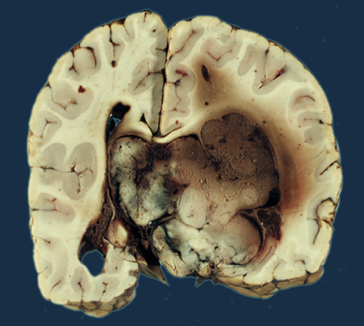
Gross pathological specimen of choroid plexus papilloma.

Microscopic Pathology
- On microscopic histopathological analysis, choroid plexus papilloma is characterized by papillary structures with a delicate fibrovascular core lined by columnar or cuboidal epithelial cells with vesicular nuclei. Its appearance is very similar to normal choroid plexus.[2]
- If two or more mitoses are present per 10 high power field (HPF), then the tumor is designated an atypical choroid plexus papilloma and is classified as a WHO grade II tumor.
Gallery
-
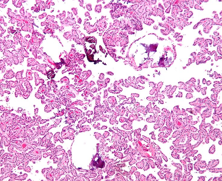
Shown above is a micrograph of choroid plexus papilloma (H&E stain).

Causes
- There are no established causes for choroid plexus papilloma.[8]
Differentiating Choroid Plexus Papilloma from other Diseases
Choroid plexus papilloma may be differentiated from:[4][7]
- Choroid plexus carcinoma
- Choroid plexus metastasis
Among children, choroid plexus papilloma must be differentiated from:[4]
- Medulloblastoma
- Atypical teratoid rhabdoid tumor (AT/RT)
- Papillary anaplastic ependymoma
- Pineocytoma
- Pineoblastoma
Among adults, choroid plexus papilloma must be differentiated from:[4]
Epidemiology and Demographics
Prevalence
- Choroid plexus papilloma accounts for approximately 1% of all brain tumors, 2-6% of all pediatric brain tumors, and 0.5% of the adult brain tumors.[5]
- They are far more common than the more aggressive choroid plexus carcinoma (WHO grade III), which they outnumber 5:1.[14]
Age
- Choroid plexus papilloma is a rare disease that tends to affect children and adults.[5]
- Approximately 85% of all choroid plexus papilloma occurs in children under the age of 5 years.
Gender
- Males are more commonly affected with choroid plexus papilloma than females. The male to female ratio is approximately 2.8 to 1.[6]
Risk Factors
- There are no established risk factors for choroid plexus papilloma.
Screening
- There is insufficient evidence to recommend routine screening for choroid plexus papilloma.[15]
Natural History, Complications and Prognosis
- If left untreated, patients with choroid plexus papilloma may progress to develop seizures, hydrocephalus, focal neurological deficits, cranioneuropathies, psychosis, and coma.[7][8]
- Common complications of choroid plexus papilloma include CSF seeding, obstructive hydrocephalus, blindness, psychosis, and coma.
- The prognosis is excellent, and the 5-year survival rate after complete excision of choroid plexus papilloma is approximately 100%.[8][9]
Diagnosis
Symptoms
- When evaluating a patient for choroid plexus papilloma, you should take a detailed history of the presenting symptom (onset, duration, and progression), other associated symptoms, and a thorough family and past medical history review.
- Other specific areas of focus when obtaining the history include the review of common associated conditions such as Aicardi syndrome, Von Hippel-Lindau disease, and Li-Fraumeni syndrome.[2][3]
- Symptoms of choroid plexus papilloma include headache, vomiting, seizures, and vision loss.[10][7]
Physical Examination
Common physical examination findings of choroid plexus papilloma include:[10][7][8]
HEENT
- Bulging fontanelle
- Enlarged head size
- Papilledema
- Homonymous hemianopia
- Mild upward gaze paresis
- Facial asymmetry
Neurological
- Altered mental status
- Gait ataxia
- Focal neurological deficits
- Cerebellar signs
- Cranioneuropathies
- 6th and 7th nerve palsy
Laboratory Findings
- There are no specific laboratory findings associated with choroid plexus papilloma.
- Other laboratory findings consistent with the diagnosis of choroid plexus papilloma is immunohistochemistry.
Immunohistochemistry
- Choroid plexus papilloma is demonstrated by positivity to tumor marker such as cytokeratin (CK7).[2]
- Some reactivity for GFAP and epithelial membrane antigen may be present.[7]
Imaging Findings
CT
- Head CT scan is helpful in the diagnosis of choroid plexus papilloma.
- On CT scan, choroid plexus papilloma is characterized by:[11][7]
- Well-defined lobulated masses
- Iso- or somewhat hyperdense compared to the adjacent brain
- Associated hydrocephalus
- Fine, speckled calcification (25%)
- Microhemorrhage
- Intense homogenous enhancement, with an irregular frond-like pattern
- If there is markedly heterogeneous contrast enhancement, a choroid plexus carcinoma should be suspected
Gallery
-
![The mass is relatively well circumscribed, slightly hyperdense compared to grey matter on CT, and following administration of contrast demonstrates vivid enhancement.[16]](/images/e/e1/CT_scan_of_choroid_plexus_papilloma_1.jpg)
The mass is relatively well circumscribed, slightly hyperdense compared to grey matter on CT, and following administration of contrast demonstrates vivid enhancement.[16]
![The mass is relatively well circumscribed, slightly hyperdense compared to grey matter on CT, and following administration of contrast demonstrates vivid enhancement.[16]](/index.php/File:CT_scan_of_choroid_plexus_papilloma_1.jpg)
MRI
- Brain MRI is helpful in the diagnosis of choroid plexus papilloma.
- On MRI, choroid plexus papilloma is characterized by:[11]
| MRI component | Findings |
|---|---|
|
T1 |
|
|
T2 |
|
|
T1 with contrast |
|
Gallery
-
![Brain MRI T1 with contrast image demonstrates a vividly but heterogeneously enhancing mass in the right side of the 4th ventricle is associated with significant surrounding edema and mass effect. There is ventricular enlargement consistent with non-communicating hydrocephalus.[17]](/images/d/db/MRI_scan_picture_of_choroid_plexus_papilloma_image_1.jpg)
Brain MRI T1 with contrast image demonstrates a vividly but heterogeneously enhancing mass in the right side of the 4th ventricle is associated with significant surrounding edema and mass effect. There is ventricular enlargement consistent with non-communicating hydrocephalus.[17]
-
![Axial T2-weighted MRI image demonstrates a 17 x 16 x 17 mm hyperintense mass within the floor of the fourth ventricle. No extension into the foramen of Luschka is observed.[18]](/images/5/50/MRI_scan_picture_of_choroid_plexus_papilloma_image_2.jpg)
Axial T2-weighted MRI image demonstrates a 17 x 16 x 17 mm hyperintense mass within the floor of the fourth ventricle. No extension into the foramen of Luschka is observed.[18]
![Brain MRI T1 with contrast image demonstrates a vividly but heterogeneously enhancing mass in the right side of the 4th ventricle is associated with significant surrounding edema and mass effect. There is ventricular enlargement consistent with non-communicating hydrocephalus.[17]](/index.php/File:MRI_scan_picture_of_choroid_plexus_papilloma_image_1.jpg)
![Axial T2-weighted MRI image demonstrates a 17 x 16 x 17 mm hyperintense mass within the floor of the fourth ventricle. No extension into the foramen of Luschka is observed.[18]](/index.php/File:MRI_scan_picture_of_choroid_plexus_papilloma_image_2.jpg)
Other Imaging Findings
- Other imaging studies for choroid plexus papilloma include magnetic resonance spectroscopy.
- Findings on MR spectroscopy include:[11]
- Elevated choline
- Decreased N-Acetylaspartate (NAA)
Other Diagnostic Studies
- Other diagnostic studies for choroid plexus papilloma include angiography, which demonstrate intense vascular blush owing to the vascularity of the tumor. Enlarged choroidal arteries may be observed feeding the tumor, with shunting.[11]
Treatment
Medical Therapy
The treatment options for choroid plexus papilloma include:[7]
Treatment of choroid plexus papilloma | |||||||||||||||||||||||||||||||||||||||||||||||||||||||||||||||||||||
Surgery (mainstay) | Radiotherapy | Radiosurgery | Chemotherapy | ||||||||||||||||||||||||||||||||||||||||||||||||||||||||||||||||||
Radiotherapy
- If complete resection of choroid plexus papilloma is done, then no adjuvant radiotherapy is necessary.
- Indications for radiotherapy include:[7]
- Inoperable residual tumor
- Resistant / aggressive tumor
Chemotherapy
- Adjuvant chemotherapy is not required if choroid plexus papilloma is completely resected.[7]
- Combination chemotherapy is used in the case of recurrent tumors.
Surgery
- Surgery is the mainstay of treatment for choroid plexus papilloma.[7]
- Complete resection of the tumor must be attempted in all cases.
- Owing to the highly vascular nature of the tumor, care should be taken prior to and during surgery to prevent any life-threatening bleeding.
- Techniques used to minimize this complication of surgery include:[7]
- Preoperative embolization of small caliber feeding vessels
- Intraoperative usage of tranexamic acid
- Hydrocephalus may be managed either by the placement of an external ventricular drain (EVD), VP shunt, or endoscopic third ventriculostomy (ETV).
- The approach to the surgery depends on the size, location, and extent of the tumor.
- Tumor is in the posterior third ventricle: infratentorial supracerebellar, transcallosal, occipital transtentorial, or transcortical-transforaminal approaches may be used for resection.
- Tumor is in the fourth ventricle: midline suboccipital approach is preferred.
- Tumor is in the cerebellopontine angle: midline cerebellomedullary fissure or the conventional retrosigmoid approach may be utilized.
Radiosurgery
- If complete resection of choroid plexus papilloma is done, then no adjuvant radiosurgery is necessary.
- Indications for radiotherapy include:[7]
- Inoperable residual tumor
- Resistant / aggressive tumor
Prevention
- There are no primary or secondary preventive measures available for choroid plexus papilloma.
References
- ↑ 1.0 1.1 Choroid plexus papilloma. Wikipedia 2016. https://en.wikipedia.org/wiki/Choroid_plexus_papilloma. Accessed on January 13, 2016
- ↑ 2.0 2.1 2.2 2.3 2.4 2.5 2.6 2.7 2.8 Clinical presentation of choroid plexus papilloma. Dr Tim Luijkx and Dr Paresh K Desai et al. Radiopaedia 2016. http://radiopaedia.org/articles/choroid-plexus-papilloma-1. Accessed on January 13, 2016
- ↑ 3.0 3.1 3.2 Gozali AE, Britt B, Shane L, Gonzalez I, Gilles F, McComb JG; et al. (2012). "Choroid plexus tumors; management, outcome, and association with the Li-Fraumeni syndrome: the Children's Hospital Los Angeles (CHLA) experience, 1991-2010". Pediatr Blood Cancer. 58 (6): 905–9. doi:10.1002/pbc.23349. PMID 21990040.
- ↑ 4.0 4.1 4.2 4.3 Differential diagnosis of choroid plexus. Dr Tim Luijkx and Dr Paresh K Desai et al. Radiopaedia 2016. http://radiopaedia.org/articles/choroid-plexus-papilloma-1. Accessed on January 13, 2016
- ↑ 5.0 5.1 5.2 5.3 Epidemiology of choroid plexus papilloma. Dr Tim Luijkx and Dr Paresh K Desai et al. Radiopaedia 2016. http://radiopaedia.org/articles/choroid-plexus-papilloma-1. Accessed on January 13, 2016
- ↑ 6.0 6.1 Epidemiology of choroid plexus papilloma. DR.SAGAR JUNG RANA 2016. http://drsagarjungrana.blogspot.com/2012/11/choroid-plexus-papilloma.html. Accessed on January 14, 2016
- ↑ 7.00 7.01 7.02 7.03 7.04 7.05 7.06 7.07 7.08 7.09 7.10 7.11 7.12 7.13 7.14 7.15 7.16 7.17 7.18 Prasad GL, Mahapatra AK (2015). "Case series of choroid plexus papilloma in children at uncommon locations and review of the literature". Surg Neurol Int. 6: 151. doi:10.4103/2152-7806.166167. PMC 4596056. PMID 26500797.
- ↑ 8.0 8.1 8.2 8.3 8.4 8.5 8.6 Koeller, Kelly K.; Sandberg, Glenn D. (2002). "From the Archives of the AFIP". RadioGraphics. 22 (6): 1473–1505. doi:10.1148/rg.226025118. ISSN 0271-5333.
- ↑ 9.0 9.1 Pencalet P, Sainte-Rose C, Lellouch-Tubiana A, Kalifa C, Brunelle F, Sgouros S; et al. (1998). "Papillomas and carcinomas of the choroid plexus in children". J Neurosurg. 88 (3): 521–8. doi:10.3171/jns.1998.88.3.0521. PMID 9488307.
- ↑ 10.0 10.1 10.2 10.3 Signs and symptoms of choroid plexus papilloma. Wikipedia 2016. https://en.wikipedia.org/wiki/Choroid_plexus_papilloma. Accessed on January 13, 2016
- ↑ 11.0 11.1 11.2 11.3 11.4 11.5 Radiographic features of choroid plexus papilloma. Dr Tim Luijkx and Dr Paresh K Desai et al. Radiopaedia 2016. http://radiopaedia.org/articles/choroid-plexus-papilloma-1. Accessed on January 13, 2016
- ↑ Treatment and prognosis of choroid plexus papilloma. Dr Tim Luijkx and Dr Paresh K Desai et al. Radiopaedia 2016. http://radiopaedia.org/articles/choroid-plexus-papilloma-1. Accessed on January 14, 2016
- ↑ Choroid plexus papillomas. Dr Tim Luijkx and Dr Paresh K Desai et al. Radiopaedia 2016. http://radiopaedia.org/articles/choroid-plexus-papilloma-1. Accessed on January 13, 2016
- ↑ Choroid plexus papilloma. Dr Tim Luijkx and Dr Paresh K Desai et al. Radiopaedia 2016. http://radiopaedia.org/articles/choroid-plexus-papilloma-1. Accessed on January 13, 2016
- ↑ Early detection, diagnosis, and staging of brain tumors. American cancer society. http://www.cancer.org/cancer/braincnstumorsinadults/detailedguide/brain-and-spinal-cord-tumors-in-adults-detection
- ↑ Image courtesy of Dr. Michael Sargent. Radiopaedia (original file here). Creative Commons BY-SA-NC
- ↑ Image courtesy of Dr. Frank Gaillard. Radiopaedia (original file here). Creative Commons BY-SA-NC
- ↑ Image courtesy of Dr. Frank Gaillard. Radiopaedia (original file here). Creative Commons BY-SA-NC