Oligodendroglioma pathophysiology
|
Oligodendroglioma Microchapters |
|
Diagnosis |
|---|
|
Treatment |
|
Case Studies |
|
Oligodendroglioma pathophysiology On the Web |
|
American Roentgen Ray Society Images of Oligodendroglioma pathophysiology |
|
Risk calculators and risk factors for Oligodendroglioma pathophysiology |
Editor-In-Chief: C. Michael Gibson, M.S., M.D. [1]Associate Editor(s)-in-Chief: Sara Mohsin, M.D.[2]Sujit Routray, M.D. [3]
Overview
Oligodendroglioma arises from the tripotential glial precursor cells and not from the bipotential oligodendrocytes. Genes associated with the pathogenesis of oligodendroglioma include t[1;19][q10;p10], NJDS, IDH1, IDH2, CIC, FUBP1, p53, Leu-7, TCF-12, MGMT, TP73, EGFR, and PTEN. On gross pathology, oligodendroglioma is characterized by a well-circumscribed, gelatinous, calcified, gray mass which may expand a gyrus and remodel the skull. On microscopic histopathological analysis, oligodendroglioma is characterized by diffuse growth pattern of highly cellular lesion with rounded nucleus with atypia and perinuclear halo resembling fried eggs, distinct cell borders, clear cytoplasm, and abundant calcification. Oligodendroglioma is demonstrated by positivity to tumor markers such as MAP2, GFAP, S-100, EMA, IDH1-R132H, ATRX, Ki-67, NSE, Synaptophysin, OLIG1, and OLIG2.
Pathophysiology
Pathogenesis
- Oligodendroglioma does not arise from the bipotential oligodendrocytes, although the tumor cells look very similiar[1]
- Oligodendroglioma arises from the tripotential glial precursor cells
Genetics
- Development of oligodendroglioma is the result of multiple genetic mutations
- Genes associated with the pathogenesis of oligodendroglioma include:[2][3][4][5][6][7][8][9][10][11]
- t(1;19)(q10;p10) (co-deletion of chromosomal arms 1p36 and 19q13; most common)[12][13]
- ATRX
- IDH1[14][15][16]
- IDH2[17]
- TERT promoter[11]
- H3 K27M mutations in either H3F3A (one of two genes encoding the histone H3.3 variant) or HIST1H3B/C (encoding the histone H3.1 variant)
- NJDS (A 2009 Oxford Neurosymposium study illustrated that there's a 69% correlation between NJDS gene mutation and tumor initiation)
- CIC
- FUBP1
- p53
- BRAF alterations
- KIAA1549-BRAF fusion
- BRAF V600E mutation
- Leu-7
- TCF-12
- MGMT
- TP73
- EGFR
- PTEN
- There is a strong association of oligodendroglioma with expression of receptor tyrosine kinases that activate PI3K/AKT, RAS/MAP, and PLC/PKC pathways[9]
Gross Pathology
- On gross pathology, oligodendroglioma is characterized by a well-circumscribed, gelatinous, gray mass which may expand a gyrus and remodel the skull[18]
- Other characteristic gross pathological features associated with oligodendroglioma include:[18][9]
- Calcification (70-90%; one of the most frequently calcifying tumors)
- Focal hemorrhage
- Cystic (20%)
- Common intracranial sites associated with oligodendroglioma include:[19]
- Cerebral hemispheres (cortex and white matter) - distribution between frontal (most common, > 50% of cases), parietal, temporal, and occipital lobe approximates 3:2:2:1
- Posterior fossa (rare)
- Intramedullary spinal cord (very rare, only 1.5% of oligodendrogliomas)
Gallery
-
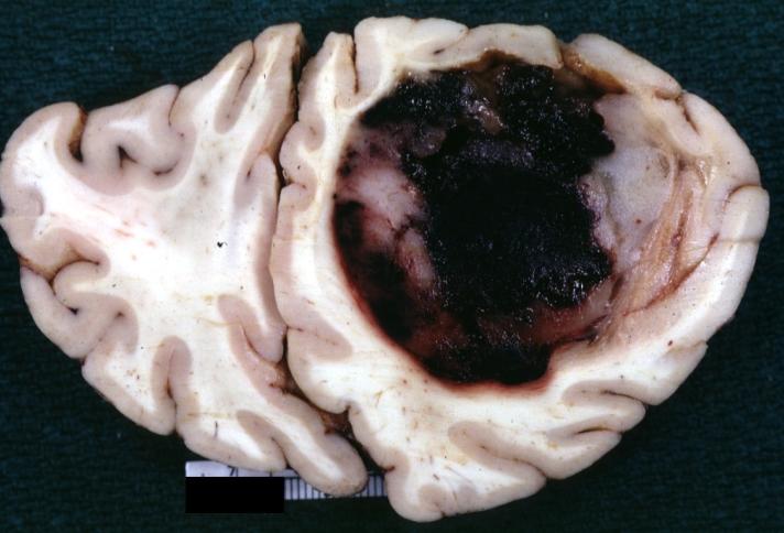
Gross specimen of oligodendroglioma demonstrate large, well circumscribed lesion in left frontal lobe.

Microscopic Pathology
On microscopic histopathological analysis, oligodendroglioma is characterized by:[9][20][21][22]
- Diffusely growing tumor
- Highly cellular lesion composed of cells resembling fried eggs with:
- Round nucleus - key feature
- Distinct cell borders
- Moderate-to-marked nuclear atypia with speckled "salt-and-pepper" chromatin pattern
- Clear cytoplasm
- Some oligodendrogliomas have eosinophilic cytoplasm with focal perinuclear clearing
- Acutely branched capillary sized vessels - "chicken-wire" like appearance[23]
- Abundant and delicate appearing; may vaguely resemble a paraganglioma at low power
- Calcifications - striking feature
- Perifocal edema - rare
- Few tumors may exhibit eosinophilic granular bodies
- Some tumors may show a spongioblastoma-like growth pattern
On microscopic histopathological analysis, anaplastic oligodendroglioma is characterized by:[20]
- Focal or diffusely increased cell density
- Atypical to frankly pleomorphic cells or multinucleated giant cells
- Tumor cells may be plasmacytoid (i.e. have a plasma cell-like appearance)
- Also called as minigemistocytes
- Significant or brisk mitotic activity (≥ 6 mitoses per 10 HPF)[24]
- Necrosis
- Apoptotic cells
- Microvacular proliferation
- Either in the form of 'glomeruloid' vessels or endothelial hyperplasia
Immunohistochemistry
Oligodendroglioma is demonstrated by positivity to tumor markers such as:[25][26][9]
- MAP2
- GFAP
- S-100
- EMA
- IDH1-R132H
- ATRX
- Ki-67
- NSE
- Synaptophysin
- OLIG1
- OLIG2
References
- ↑ General features of oligodendroglioma. Libre Pathology. http://librepathology.org/wiki/index.php/Oligodendroglioma#cite_note-1
- ↑ Molecular genetics of oligodendroglioma. https://en.wikipedia.org/wiki/Oligodendroglioma
- ↑ Bettegowda C, Agrawal N, Jiao Y, Sausen M, Wood LD, Hruban RH; et al. (2011). "Mutations in CIC and FUBP1 contribute to human oligodendroglioma". Science. 333 (6048): 1453–5. doi:10.1126/science.1210557. PMC 3170506. PMID 21817013.
- ↑ Prognosis and treatment of oligodendroglioma. Wikipedia 2015. https://en.wikipedia.org/wiki/Oligodendroglioma
- ↑ Yip S, Butterfield YS, Morozova O, Chittaranjan S, Blough MD, An J; et al. (2012). "Concurrent CIC mutations, IDH mutations, and 1p/19q loss distinguish oligodendrogliomas from other cancers". J Pathol. 226 (1): 7–16. doi:10.1002/path.2995. PMC 3246739. PMID 22072542.
- ↑ Labreche K, Simeonova I, Kamoun A, Gleize V, Chubb D, Letouzé E; et al. (2015). "TCF12 is mutated in anaplastic oligodendroglioma". Nat Commun. 6: 7207. doi:10.1038/ncomms8207. PMC 4490400. PMID 26068201.
- ↑ Suri V, Jha P, Agarwal S, Pathak P, Sharma MC, Sharma V; et al. (2011). "Molecular profile of oligodendrogliomas in young patients". Neuro Oncol. 13 (10): 1099–106. doi:10.1093/neuonc/nor146. PMC 3177666. PMID 21937591.
- ↑ Hagel C, Laking G, Laas R, Scheil S, Jung R, Milde-Langosch K; et al. (1996). "Demonstration of p53 protein and TP53 gene mutations in oligodendrogliomas". Eur J Cancer. 32A (13): 2242–8. PMID 9038605.
- ↑ 9.0 9.1 9.2 9.3 9.4 von Deimling, A; Hartmann, C (2005). "Oligodendrogliomas: Impact of molecular genetics on treatment". Neurology India. 53 (2): 140. doi:10.4103/0028-3886.16394. ISSN 0028-3886.
- ↑ Cancer Genome Atlas Research Network. Brat DJ, Verhaak RG, Aldape KD, Yung WK, Salama SR; et al. (2015). "Comprehensive, Integrative Genomic Analysis of Diffuse Lower-Grade Gliomas". N Engl J Med. 372 (26): 2481–98. doi:10.1056/NEJMoa1402121. PMC 4530011. PMID 26061751.
- ↑ 11.0 11.1 Eckel-Passow JE, Lachance DH, Molinaro AM, Walsh KM, Decker PA, Sicotte H; et al. (2015). "Glioma Groups Based on 1p/19q, IDH, and TERT Promoter Mutations in Tumors". N Engl J Med. 372 (26): 2499–508. doi:10.1056/NEJMoa1407279. PMC 4489704. PMID 26061753.
- ↑ McDonald JM, See SJ, Tremont IW, Colman H, Gilbert MR, Groves M; et al. (2005). "The prognostic impact of histology and 1p/19q status in anaplastic oligodendroglial tumors". Cancer. 104 (7): 1468–77. doi:10.1002/cncr.21338. PMID 16088966.
- ↑ Barbashina V, Salazar P, Holland EC, Rosenblum MK, Ladanyi M (2005). "Allelic losses at 1p36 and 19q13 in gliomas: correlation with histologic classification, definition of a 150-kb minimal deleted region on 1p36, and evaluation of CAMTA1 as a candidate tumor suppressor gene". Clin Cancer Res. 11 (3): 1119–28. PMID 15709179.
- ↑ Chen N, Yu T, Gong J, Nie L, Chen X, Zhang M; et al. (2016). "IDH1/2 gene hotspot mutations in central nervous system tumours: analysis of 922 Chinese patients". Pathology. 48 (7): 675–683. doi:10.1016/j.pathol.2016.07.010. PMID 27780605.
- ↑ Zhou YX, Wang JX, Feng M, Sun CM, Sun T, Chen GL; et al. (2012). "Analysis of isocitrate dehydrogenase 1 mutation in 97 patients with glioma". J Mol Neurosci. 47 (3): 442–7. doi:10.1007/s12031-011-9681-5. PMID 22113362.
- ↑ Capper D, Weissert S, Balss J, Habel A, Meyer J, Jäger D; et al. (2010). "Characterization of R132H mutation-specific IDH1 antibody binding in brain tumors". Brain Pathol. 20 (1): 245–54. doi:10.1111/j.1750-3639.2009.00352.x. PMID 19903171.
- ↑ Pan Y, Qi XL, Wang LM, Dong RF, Zhang M, Zheng DF; et al. (2013). "[Mutation of isocitrate dehydrogenase gene in Chinese patients with glioma]". Zhonghua Bing Li Xue Za Zhi. 42 (5): 292–8. doi:10.3760/cma.j.issn.0529-5807.2013.05.002. PMID 24004584.
- ↑ 18.0 18.1 Gross appearance of oligodendroglioma. Dr Henry Knipe and Dr Frank Gaillard et al. http://radiopaedia.org/articles/oligodendroglioma
- ↑ Gross/radiologic findings of oligodendroglioma. Libre Pathology. http://librepathology.org/wiki/index.php/Oligodendroglioma
- ↑ 20.0 20.1 Microscopic features of oligodendroglioma. Libre Pathology. http://librepathology.org/wiki/index.php/Oligodendroglioma
- ↑ Ersen, Ayca (2008), Pathology of malignant gliomas: Challenges of everyday practice and the WHO 2007, Turkish Journal of Pathology, retrieved 9 October, 2015 Check date values in:
|accessdate=(help) - ↑ Eskandar EN, Loeffler JS, O'Neill AM, Hunter GJ, Louis DN (2004). "Case records of the Massachusetts General Hospital. Weekly clinicopathological exercises. Case 33-2004. A 34-year-old man with a seizure and a frontal-lobe brain lesion". N Engl J Med. 351 (18): 1875–82. doi:10.1056/NEJMcpc049025. PMID 15509821.
- ↑ Images of microscopic appearance of oligodendroglioma. Wikipedia 2015. https://en.wikipedia.org/wiki/Oligodendroglioma
- ↑ Images of oligodendroglioma. Libre Pathology 2015. http://librepathology.org/wiki/index.php/Oligodendroglioma
- ↑ IHC of oligodendroglioma. Libre Pathology. http://librepathology.org/wiki/index.php/Oligodendroglioma
- ↑ Hilbig A, Barbosa-Coutinho LM, Netto GC, Bleil CB, Toscani NV (2006). "[Immunohistochemistry in oligodendrogliomas]". Arq Neuropsiquiatr. 64 (1): 67–71. doi:/S0004-282X2006000100014 Check
|doi=value (help). PMID 16622556.