Bursitis overview
|
Bursitis Microchapters |
|
Diagnosis |
|---|
|
Treatment |
|
Case Studies |
|
Bursitis overview On the Web |
|
American Roentgen Ray Society Images of Bursitis overview |
Editor-In-Chief: C. Michael Gibson, M.S., M.D. [1]; Associate Editor(s)-in-Chief: Sara Mehrsefat, M.D. [2]
Overview
Bursitis Classification
Based on the nature of inflammation bursitis may classified into 2 subtypes: septic and aseptic. Common anatomic location include shoulder, elbow, hip, knee, and ankle. Most common bursitis subtypes include subacromial, olecranon, trochanteric, prepatellar, and retrocalcaneal. Moreover, based on the location of the bursa from the skin, bursitis may be classified into 2 subtyopes: superficial and deep. Superficial bursa are more prone to get infected with bacteria and develop septic bursitis. common location of septic bursitis include knee (prepatellar bursitis), and elbow (olecranon bursitis).[1][2][3][4]
Bursitis Pathophysiology
Bursitis is characterized by acute or chronic inflammation of a bursa and buildup of fluid in the bursa sac. A bursa is a small, fluid-filled sac that acts as a cushion between a bone and other moving parts: muscles, tendons, or skin. Over 160 bursa are found throughout the body and only few of them can cause bursitis. Aseptic bursitis can be caused by overused and repetitive injuries to the joint, abdominal bony structure and crystal deposit in the bursa. It commonly affects knee or elbow, from kneeling or leaning on the elbows longer than usual. Moreover, septic bursitis can be caused by bacterial infection of the bursa through the skin injury following repetitive trauma.[2][5][6]
Images
The following are gross and microscopic images associated with bursitis:
-
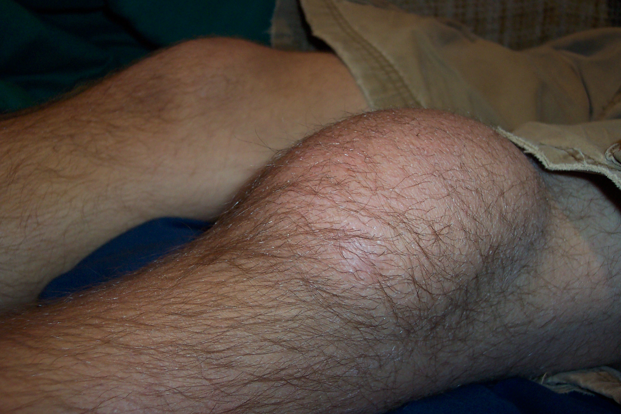
Prepatellar bursitis<ref> Wikipedia.Prepatellar bursitis. https://en.wikipedia.org/wiki/Prepatellar_bursitis#/media/File:Prepatellar_bursitis.JPG Accessed on August 25, 2016</red>
-
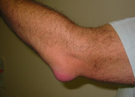
Olecranon bursitis<ref> Wikipedia.Prepatellar bursitis. https://en.wikipedia.org/wiki/Bursitis#/media/File:Bursitis_Elbow_WC.JPG Accessed on August 25, 2016</red>


===Bursitis
References
- ↑ Chatra PS (2012). "Bursae around the knee joints". Indian J Radiol Imaging. 22 (1): 27–30. doi:10.4103/0971-3026.95400. PMC 3354353. PMID 22623812.
- ↑ 2.0 2.1 Fauci, Anthony S., and Carol Langford. Harrison's rheumatology. McGraw Hill Professional, 2010.
- ↑ Walker‐Bone, Karen, et al. "Prevalence and impact of musculoskeletal disorders of the upper limb in the general population.
- ↑ Aaron, Daniel L., et al. "Four common types of bursitis: diagnosis and management." Journal of the American Academy of Orthopaedic Surgeons 19.6 (2011): 359-367.
- ↑ Hellmann DB, Imboden JB., Jr. Musculoskeletal and immunologic disorders. In: McPhee SJ, Papadakis MA, editors. Current Medical Diagnosis & Treatment. McGraw-Hill Lange; 2010. pp. 2056–2061.
- ↑ García-Porrúa C, González-Gay MA, Ibañez D, García-País MJ (1999). "The clinical spectrum of severe septic bursitis in northwestern Spain: a 10 year study". J Rheumatol. 26 (3): 663–7. PMID 10090179.