Cardiac tamponade anatomy: Difference between revisions
No edit summary |
No edit summary |
||
| Line 1: | Line 1: | ||
__NOTOC__ | |||
{{Infobox Anatomy | | {{Infobox Anatomy | | ||
Name = {{PAGENAME}} | | Name = {{PAGENAME}} | | ||
| Line 8: | Line 9: | ||
Image2 = Gray968.png | | Image2 = Gray968.png | | ||
Caption2 = A transverse section of the [[thorax]], showing the contents of the middle and the posterior [[mediastinum]]. The [[pleural cavity|pleural]] and [[pericardial cavity|pericardial cavities]] are exaggerated since normally there is no space between parietal and visceral pleura and between pericardium and [[heart]] Paricardium is also known as cariac epidemis. | | Caption2 = A transverse section of the [[thorax]], showing the contents of the middle and the posterior [[mediastinum]]. The [[pleural cavity|pleural]] and [[pericardial cavity|pericardial cavities]] are exaggerated since normally there is no space between parietal and visceral pleura and between pericardium and [[heart]] Paricardium is also known as cariac epidemis. | | ||
}} | }} | ||
{{Cardiac tamponade}} | {{Cardiac tamponade}} | ||
| Line 23: | Line 14: | ||
==Overview== | ==Overview== | ||
The pericardium is a double-walled sac that contains the [[heart]] and the roots of the [[great vessels]]. | |||
==Overview== | |||
===Layers=== | |||
==Layers== | |||
There are two layers to the [[pericardial sac]]: the [[fibrous pericardium]] and the [[serous pericardium]]. The serous pericardium, in turn, is divided into two layers, the ''parietal pericardium'', which is fused to and inseparable from the fibrous pericardium, and the ''visceral pericardium'', which is in fact the [[epicardium]], or the outer surface of the heart. | There are two layers to the [[pericardial sac]]: the [[fibrous pericardium]] and the [[serous pericardium]]. The serous pericardium, in turn, is divided into two layers, the ''parietal pericardium'', which is fused to and inseparable from the fibrous pericardium, and the ''visceral pericardium'', which is in fact the [[epicardium]], or the outer surface of the heart. | ||
In between the parietal and visceral pericardial layers there is a [[potential space]] called the [[pericardial cavity]]. It is normally lubricated by a film of pericardial fluid. Too much fluid in the cavity (such as in a [[pericardial effusion]]) can result in [[pericardial tamponade]], which refers to compression of the heart within the pericardial sac. | In between the parietal and visceral pericardial layers there is a [[potential space]] called the [[pericardial cavity]]. It is normally lubricated by a film of pericardial fluid. Too much fluid in the cavity (such as in a [[pericardial effusion]]) can result in [[pericardial tamponade]], which refers to compression of the heart within the pericardial sac. | ||
==Pericardial Sinuses== | ===Pericardial Sinuses=== | ||
There are two small chambers or sinuses are located where the visceral and parietal pericardia are continuous with one another within the pericardial cavity. | There are two small chambers or sinuses are located where the visceral and parietal pericardia are continuous with one another within the pericardial cavity. | ||
| Line 48: | Line 37: | ||
</div> | </div> | ||
==Additional Images== | ===Additional Images=== | ||
<gallery> | <gallery> | ||
Image:Gray806.png|The phrenic nerve and its relations with the vagus nerve. | Image:Gray806.png|The phrenic nerve and its relations with the vagus nerve. | ||
| Line 56: | Line 45: | ||
</gallery> | </gallery> | ||
==Diseases of the Pericardium== | ===Diseases of the Pericardium=== | ||
* [[Pericarditis]] is an inflammatory condition of the pericardium. | * [[Pericarditis]] is an inflammatory condition of the pericardium. | ||
| Line 62: | Line 51: | ||
* [[Constrictive pericarditis]] occurs when there is a scar encasing the heart that chronically constricts the filling of the heart. | * [[Constrictive pericarditis]] occurs when there is a scar encasing the heart that chronically constricts the filling of the heart. | ||
* [[Cardiac tamponade]] is a medical emergency in which fluid in the pericardial sac acutely restricts the filling of the heart. This requires surgical drainage or [[pericardiocentesis]]. | * [[Cardiac tamponade]] is a medical emergency in which fluid in the pericardial sac acutely restricts the filling of the heart. This requires surgical drainage or [[pericardiocentesis]]. | ||
==References== | |||
{{Reflist|2}} | |||
{{Heart}} | {{Heart}} | ||
| Line 67: | Line 59: | ||
[[Category:Cardiac anatomy]] | [[Category:Cardiac anatomy]] | ||
[[Category:Cardiology]] | [[Category:Cardiology]] | ||
[[Category: | [[Category:Disease]] | ||
{{WikiDoc Help Menu}} | {{WikiDoc Help Menu}} | ||
{{WikiDoc Sources}} | {{WikiDoc Sources}} | ||
Revision as of 16:37, 20 May 2013
|
Cardiac tamponade Microchapters |
|
Diagnosis |
|---|
|
Treatment |
|
Case Studies |
|
Cardiac tamponade anatomy On the Web |
|
American Roentgen Ray Society Images of Cardiac tamponade anatomy |
|
Risk calculators and risk factors for Cardiac tamponade anatomy |
Editor-In-Chief: C. Michael Gibson, M.S., M.D. [1]
Overview
The pericardium is a double-walled sac that contains the heart and the roots of the great vessels.
Overview
Layers
There are two layers to the pericardial sac: the fibrous pericardium and the serous pericardium. The serous pericardium, in turn, is divided into two layers, the parietal pericardium, which is fused to and inseparable from the fibrous pericardium, and the visceral pericardium, which is in fact the epicardium, or the outer surface of the heart.
In between the parietal and visceral pericardial layers there is a potential space called the pericardial cavity. It is normally lubricated by a film of pericardial fluid. Too much fluid in the cavity (such as in a pericardial effusion) can result in pericardial tamponade, which refers to compression of the heart within the pericardial sac.
Pericardial Sinuses
There are two small chambers or sinuses are located where the visceral and parietal pericardia are continuous with one another within the pericardial cavity.
The pericardial sinuses are:
- Oblique pericardial sinus
- Transverse pericardial sinus


Additional Images
-
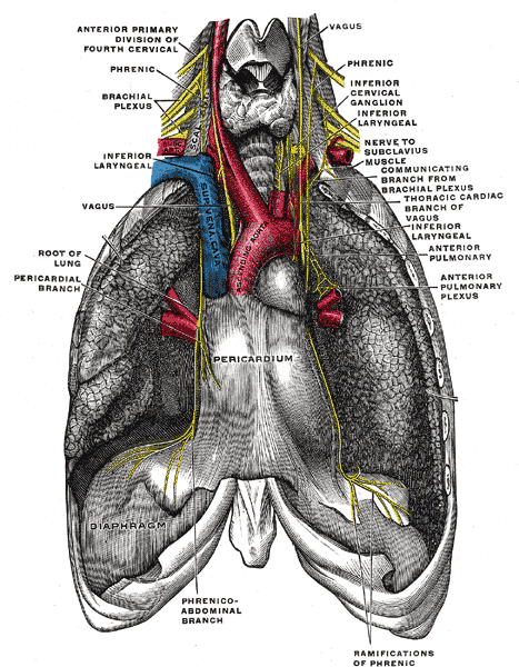
The phrenic nerve and its relations with the vagus nerve.
-
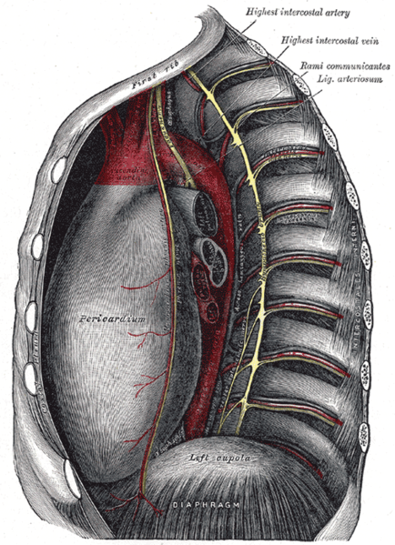
Thoracic portion of the sympathetic trunk.
-
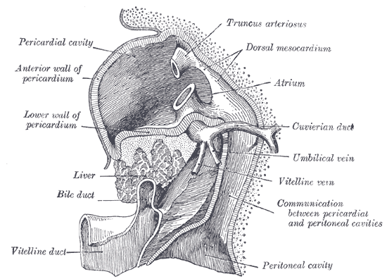
Liver with the septum transversum. Human embryo 3 mm long.
-
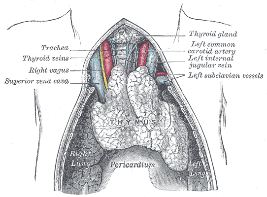
The thymus of a full-time fetus, exposed in situ.


Diseases of the Pericardium
- Pericarditis is an inflammatory condition of the pericardium.
- Pericardial effusion is fluid accumulation in the pericardial sac.
- Constrictive pericarditis occurs when there is a scar encasing the heart that chronically constricts the filling of the heart.
- Cardiac tamponade is a medical emergency in which fluid in the pericardial sac acutely restricts the filling of the heart. This requires surgical drainage or pericardiocentesis.
References