Burn pathophysiology: Difference between revisions
EmanAlademi (talk | contribs) No edit summary |
EmanAlademi (talk | contribs) No edit summary |
||
| Line 5: | Line 5: | ||
<br /> | <br /> | ||
==Pathophysiology== | ==Pathophysiology== | ||
[[Burn|Burns]] is the result of damage to the [[skin]] by a [[temperatures]] greater than 44 °C (111 °F), leads to cell and tissue damage because of breaking down of the cells proteins then start losing their three-dimensional | [[Burn|Burns]] is the result of damage to the [[skin]] '''involving the two main layers - the thin, outer epidermis and the thicker, deeper dermis''' by a [[temperatures]] greater than 44 °C (111 °F), leads to cell and tissue damage because of breaking down of the cells proteins then start losing their three-dimensional shape. [[Burn]] caused by: | ||
[[Thermal injuries]] '''the most common type of burn.''' | |||
[[Electrical injury|Electrical]] injuries '''(can be deceiving with small entry and exit wounds, however, there may be extensive internal organ injury or associated traumatic injuries).''' | |||
[[Chemical Injury|Chemical]] injuries ('''divided into acid or alkali burns. Alkali burns tend to be more severe causing more penetration deeper into the skin by liquefying the skin (liquefaction necrosis) and acid burns penetrate less because they cause a coagulation injury (coagulation necrosis).''' | |||
[[Non-accidental]] injury like:<ref name="pmid15191982">Hettiaratchy S, Dziewulski P (2004) [https://www.ncbi.nlm.nih.gov/entrez/eutils/elink.fcgi?dbfrom=pubmed&retmode=ref&cmd=prlinks&id=15191982 ABC of burns: pathophysiology and types of burns.] ''BMJ'' 328 (7453):1427-9. [http://dx.doi.org/10.1136/bmj.328.7453.1427 DOI:10.1136/bmj.328.7453.1427] PMID: [https://pubmed.gov/PMID: 15191982 PMID: 15191982]</ref><ref name="urlPathophysiology and Current Management of Burn Injury : Advances in Skin & Wound Care">{{cite web |url=https://journals.lww.com/aswcjournal/fulltext/2005/07000/pathophysiology_and_current_management_of_burn.13.aspx |title=Pathophysiology and Current Management of Burn Injury : Advances in Skin & Wound Care |format= |work= |accessdate=}}</ref> | |||
{| class="wikitable" | {| class="wikitable" | ||
| colspan="1" rowspan="1" |• Obvious pattern from cigarettes, lighters, irons | | colspan="1" rowspan="1" |• Obvious pattern from cigarettes, lighters, irons | ||
| Line 25: | Line 33: | ||
<br /> | <br /> | ||
[[ | '''Most [[burns]] are small and [[superficial]] causing only [[local injuries]], However, [[burns]] can be larger and deeper, and patients can also have a [[systemic response]] to severe burns''' <ref name="urlABC of burns: Pathophysiology and types of burns">{{cite web |url=https://www.ncbi.nlm.nih.gov/pmc/articles/PMC421790/ |title=ABC of burns: Pathophysiology and types of burns |format= |work= |accessdate=}}</ref><ref name="pmid23121414">{{cite journal |vauthors=Rojas Y, Finnerty CC, Radhakrishnan RS, Herndon DN |title=Burns: an update on current pharmacotherapy |journal=Expert Opin Pharmacother |volume=13 |issue=17 |pages=2485–94 |date=December 2012 |pmid=23121414 |pmc=3576016 |doi=10.1517/14656566.2012.738195 |url=}}</ref> | ||
===Local response=== | ===Local response=== | ||
| Line 61: | Line 69: | ||
Increased leakage of fluid from the [[capillaries]], and subsequent tissue edema. This causes overall blood volume loss, with the remaining blood suffering significant [[Plasma (blood)|plasma]] loss, making the blood more concentrated.<ref name="urlPorth Pathophysiology: Concepts of Altered Health States - Charlotte Pooler - كتب Google">{{cite web |url=https://books.google.ae/books?id=2-MFXOEG0lcC&pg=PA1516&redir_esc=y#v=onepage&q&f=false |title=Porth Pathophysiology: Concepts of Altered Health States - Charlotte Pooler - كتب Google |format= |work= |accessdate=}}</ref><br /> | Increased leakage of fluid from the [[capillaries]], and subsequent tissue edema. This causes overall blood volume loss, with the remaining blood suffering significant [[Plasma (blood)|plasma]] loss, making the blood more concentrated.<ref name="urlPorth Pathophysiology: Concepts of Altered Health States - Charlotte Pooler - كتب Google">{{cite web |url=https://books.google.ae/books?id=2-MFXOEG0lcC&pg=PA1516&redir_esc=y#v=onepage&q&f=false |title=Porth Pathophysiology: Concepts of Altered Health States - Charlotte Pooler - كتب Google |format= |work= |accessdate=}}</ref><br /> | ||
<ref name="pmid30095874">{{cite journal |vauthors=Stiles K |title=Emergency management of burns: part 2 |journal=Emerg Nurse |volume=26 |issue=2 |pages=36–41 |date=July 2018 |pmid=30095874 |doi= |url=}}</ref><ref name="urlBurn Evaluation And Management - StatPearls - NCBI Bookshelf">{{cite web |url=https://www.ncbi.nlm.nih.gov/books/NBK430741/#article-18712.r5 |title=Burn Evaluation And Management - StatPearls - NCBI Bookshelf |format= |work= |accessdate=}}</ref> | |||
Revision as of 07:55, 9 October 2020
|
Burn Microchapters |
|
Diagnosis |
|---|
|
Treatment |
|
Case Studies |
|
Burn pathophysiology On the Web |
|
American Roentgen Ray Society Images of Burn pathophysiology |
Editor-In-Chief: C. Michael Gibson, M.S., M.D. [1] Eman Alademi, M.D.[2]
Pathophysiology
Burns is the result of damage to the skin involving the two main layers - the thin, outer epidermis and the thicker, deeper dermis by a temperatures greater than 44 °C (111 °F), leads to cell and tissue damage because of breaking down of the cells proteins then start losing their three-dimensional shape. Burn caused by:
Thermal injuries the most common type of burn.
Electrical injuries (can be deceiving with small entry and exit wounds, however, there may be extensive internal organ injury or associated traumatic injuries).
Chemical injuries (divided into acid or alkali burns. Alkali burns tend to be more severe causing more penetration deeper into the skin by liquefying the skin (liquefaction necrosis) and acid burns penetrate less because they cause a coagulation injury (coagulation necrosis).
Non-accidental injury like:[1][2]
| • Obvious pattern from cigarettes, lighters, irons |
| • Burns to soles, palms, genitalia, buttocks, perineum |
| • Symmetrical burns of uniform depth |
| • No splash marks in a scald injury. A child falling into a bath will splash; one that is placed into it may not |
| • Restraint injuries on upper limbs |
| • Is there sparing of flexion creases—that is, was child in fetal position (position of protection) when burnt? Does this correlate to a “tide line” of scald—that is, if child is put into a fetal position, do the burns line up? |
| • “Doughnut sign,” an area of spared skin surrounded by scald. If a child is forcibly held down in a bath of hot water, the part in contact with the bottom of the bath will not burn, but the tissue around will |
| • Other signs of physical abuse—bruises of varied age, poorly kempt, lack of compliance with health care (such as no immunisations) |
Most burns are small and superficial causing only local injuries, However, burns can be larger and deeper, and patients can also have a systemic response to severe burns [3][4]
Local response
The three zones of a burn were described by Jackson in 1947.
Zone of coagulation—This occurs at the point of maximum damage. In this zone there is irreversible tissue loss due to coagulation of the constituent proteins.
Zone of stasis—The surrounding zone of stasis is characterized by decreased tissue perfusion. The tissue in this zone is potentially salvageable. The main aim of burns resuscitation is to increase tissue perfusion here and prevent any damage becoming irreversible. Additional insults—such as prolonged hypotension, infection, or edema—can convert this zone into an area of complete tissue loss.
Zone of hyperemia—In this outermost zone tissue perfusion is increased. The tissue here will invariably recover unless there is severe sepsis or prolonged hypoperfusion.
These three zones of a burn are three dimensional, and loss of tissue in the zone of stasis will lead to the wound deepening as well as widening.
Systemic response
The increase of the catecholamines and cortisol,or The release of cytokines and other inflammatory mediators at the site of injury has a systemic effect once the burn reaches 30% of total body surface area.
Increase of the catecholamines and cortisol lead to:
Cardiovascular changes—Capillary permeability is increased, leading to loss of intravascular proteins and fluids into the interstitial compartment. Peripheral and splanchnic vasoconstriction occurs. Myocardial contractility is decreased, possibly due to release of tumor necrosis factor α (TNFα). These changes, coupled with fluid loss from the burn wound, result in systemic hypotension and end organ hypoperfusion because of Increased levels of catecholamines. and a fast heart rate.
Metabolic changes—The basal metabolic rate increases up (hypermetabolic) to three times its original rate because of Increased levels of catecholamines and cortisol . This, coupled with splanchnic hypoperfusion, necessitates early and aggressive enteral feeding to decrease catabolism and maintain gut integrity.
Immunological changes—Non-specific down regulation of the immune response occurs, affecting both cell mediated and humoral pathways.
Release of cytokines and other inflammatory mediators at the site of injury lead to:
Respiratory changes—Inflammatory mediators cause bronchoconstriction, and in severe burns adult respiratory distress syndrome can occur.
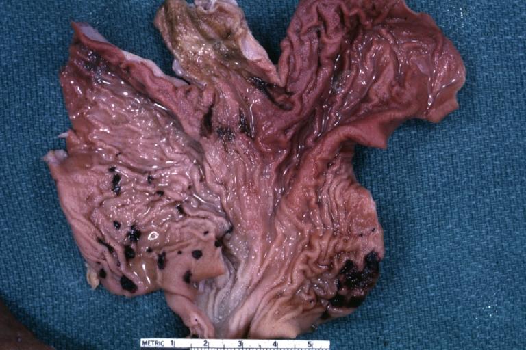
gastrointestinal tract changes— stomach ulcers because of the Poor blood flow to organs.
Renal changes—kidney failure because of the Poor blood flow to organs
Increased leakage of fluid from the capillaries, and subsequent tissue edema. This causes overall blood volume loss, with the remaining blood suffering significant plasma loss, making the blood more concentrated.[5]
[6][7]
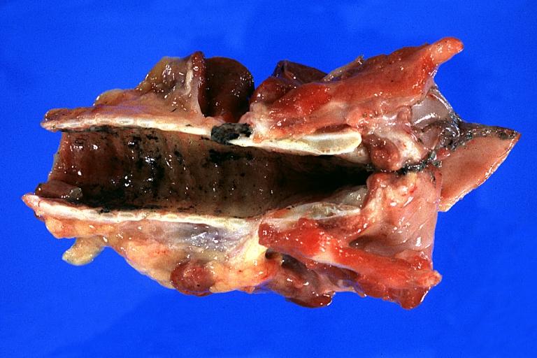
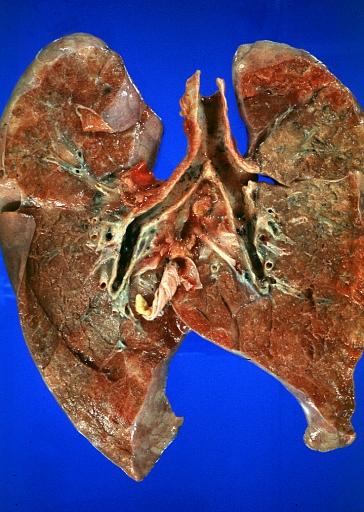
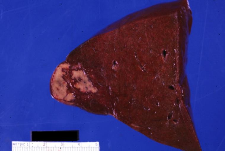
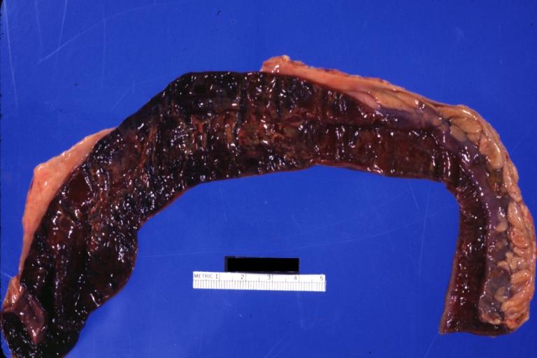
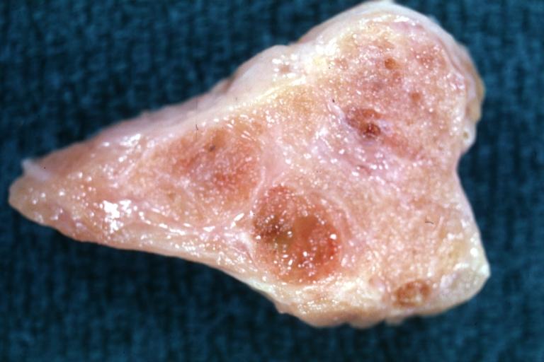
Gross Pathology
References
- ↑ Hettiaratchy S, Dziewulski P (2004) ABC of burns: pathophysiology and types of burns. BMJ 328 (7453):1427-9. DOI:10.1136/bmj.328.7453.1427 PMID: 15191982 PMID: 15191982
- ↑ "Pathophysiology and Current Management of Burn Injury : Advances in Skin & Wound Care".
- ↑ "ABC of burns: Pathophysiology and types of burns".
- ↑ Rojas Y, Finnerty CC, Radhakrishnan RS, Herndon DN (December 2012). "Burns: an update on current pharmacotherapy". Expert Opin Pharmacother. 13 (17): 2485–94. doi:10.1517/14656566.2012.738195. PMC 3576016. PMID 23121414.
- ↑ "Porth Pathophysiology: Concepts of Altered Health States - Charlotte Pooler - كتب Google".
- ↑ Stiles K (July 2018). "Emergency management of burns: part 2". Emerg Nurse. 26 (2): 36–41. PMID 30095874.
- ↑ "Burn Evaluation And Management - StatPearls - NCBI Bookshelf".
