Medulloblastoma pathophysiology: Difference between revisions
No edit summary |
|||
| Line 4: | Line 4: | ||
==Overview== | ==Overview== | ||
Medulloblastoma arises from the cerebellar [[stem cell]]s, which are normally involved in the anatomical development of the [[cerebellum]] and other posterior fossa structures. Medulloblastoma is an invasive and rapidly growing [[tumor]] which frequently metastasize to different organs of the body. [[Genes]] involved in the pathogenesis of medulloblastoma include ''CTNNB1'' gene, ''PTCH1'' gene, ''MLL2'' gene, ''SMARCA4'' gene, ''DDX3X'' gene, ''CTDNEP1'' gene, ''KDM6A'' gene, and ''TBR1'' gene. The progression to medulloblastoma usually involves the [[P53]] molecular pathway.<ref name="wiki"> Medulloblastoma. Wikipedia(2015) https://en.wikipedia.org/wiki/Medulloblastoma Accessed on September 2015</ref> On gross pathology, a pink, solid, and well circumscribed mass is a characteristic finding of medulloblastoma. On microscopic histopathological analysis, round tumor cells, elevated mitotic rate, increased nucleus:cytoplasm ratio, and presence of Homer-Wright rosettes are characteristic findings of medulloblastoma.<ref name="patho">Medulloblastoma. Librepathology(2015) http://librepathology.org/wiki/index.php/Medulloblastoma Accessed on September, 28th 2015</ref> | |||
==Pathogenesis== | ==Pathogenesis== | ||
* Medulloblastoma arises from cerebellar [[stem cell]]s, which are normally involved in the anatomical development of the [[cerebellum]] and other posterior fossa structures.<ref name="wiki"> Medulloblastoma. Wikipedia(2015) https://en.wikipedia.org/wiki/Medulloblastoma Accessed on September 2015</ref> | * Medulloblastoma arises from the cerebellar [[stem cell]]s, which are normally involved in the anatomical development of the [[cerebellum]] and other posterior fossa structures.<ref name="wiki"> Medulloblastoma. Wikipedia(2015) https://en.wikipedia.org/wiki/Medulloblastoma Accessed on September 2015</ref> | ||
* Medulloblastoma is usually located at the [[infratentorial]] region, where it forms a mass between the [[brain stem]] and the cerebellum in the vicinity of the [[fourth ventricle]].<ref name="wiki"> Medulloblastoma. Wikipedia(2015) https://en.wikipedia.org/wiki/Medulloblastoma Accessed on September 2015</ref> | * Medulloblastoma is usually located at the [[infratentorial]] region, where it forms a mass between the [[brain stem]] and the cerebellum in the vicinity of the [[fourth ventricle]].<ref name="wiki"> Medulloblastoma. Wikipedia(2015) https://en.wikipedia.org/wiki/Medulloblastoma Accessed on September 2015</ref> | ||
* Medulloblastoma is an invasive and rapidly growing [[tumor]].<ref name="wiki"> Medulloblastoma. Wikipedia(2015) https://en.wikipedia.org/wiki/Medulloblastoma Accessed on September 2015</ref> | * Medulloblastoma is an invasive and rapidly growing [[tumor]].<ref name="wiki"> Medulloblastoma. Wikipedia(2015) https://en.wikipedia.org/wiki/Medulloblastoma Accessed on September 2015</ref> | ||
| Line 34: | Line 36: | ||
==Mircoscopic Pathology== | ==Mircoscopic Pathology== | ||
* On microscopic histopathological analysis, round tumor cells, elevated mitotic rate, increased nucleus:cytoplasm | * On microscopic histopathological analysis, round tumor cells, elevated mitotic rate, increased nucleus:cytoplasm ratio, and presence of Homer-Wright rosettes are characteristic findings of medulloblastoma.<ref name="patho">Medulloblastoma. Librepathology(2015) http://librepathology.org/wiki/index.php/Medulloblastoma Accessed on September, 28th 2015</ref> | ||
* Shown below is a series of microscopic images observed in medulloblastoma: | * Shown below is a series of microscopic images observed in medulloblastoma: | ||
<gallery> | <gallery> | ||
Revision as of 16:52, 29 September 2015
|
Medulloblastoma Microchapters |
|
Diagnosis |
|---|
|
Treatment |
|
Case studies |
|
Medulloblastoma pathophysiology On the Web |
|
American Roentgen Ray Society Images of Medulloblastoma pathophysiology |
|
Risk calculators and risk factors for Medulloblastoma pathophysiology |
Editor-In-Chief: C. Michael Gibson, M.S., M.D. [1] Associate Editor(s)-in-Chief: Haytham Allaham, M.D. [2]
Overview
Medulloblastoma arises from the cerebellar stem cells, which are normally involved in the anatomical development of the cerebellum and other posterior fossa structures. Medulloblastoma is an invasive and rapidly growing tumor which frequently metastasize to different organs of the body. Genes involved in the pathogenesis of medulloblastoma include CTNNB1 gene, PTCH1 gene, MLL2 gene, SMARCA4 gene, DDX3X gene, CTDNEP1 gene, KDM6A gene, and TBR1 gene. The progression to medulloblastoma usually involves the P53 molecular pathway.[1] On gross pathology, a pink, solid, and well circumscribed mass is a characteristic finding of medulloblastoma. On microscopic histopathological analysis, round tumor cells, elevated mitotic rate, increased nucleus:cytoplasm ratio, and presence of Homer-Wright rosettes are characteristic findings of medulloblastoma.[2]
Pathogenesis
- Medulloblastoma arises from the cerebellar stem cells, which are normally involved in the anatomical development of the cerebellum and other posterior fossa structures.[1]
- Medulloblastoma is usually located at the infratentorial region, where it forms a mass between the brain stem and the cerebellum in the vicinity of the fourth ventricle.[1]
- Medulloblastoma is an invasive and rapidly growing tumor.[1]
- Unlike most brain tumors, medulloblastoma may spread through the cerebrospinal fluid and frequently metastasize to different organs of the body.[1]
- Medulloblastoma is often associated with Gorlin syndrome and Turcot syndrome.[1]
Genetics
- Development of medulloblastoma is the result of multiple genetic mutations.[1]
- Genes involved in the pathogenesis of medulloblastoma include:[1]
- CTNNB1 gene on chromosome 3
- PTCH1 gene located on chromosome 9
- MLL2 gene located on chromosome 12
- SMARCA4 gene located on chromosome 19
- DDX3X gene located on X chromosome
- CTDNEP1 gene located on chromosome 17
- KDM6A gene located on X chromosome
- TBR1 gene located on chromosome 2
Gross Pathology
- On gross pathology, a pink, solid, and well circumscribed mass is a characteristic finding of medulloblastoma.[1]
- The following image demonstrates the gross pathology observed in medulloblastoma:
-
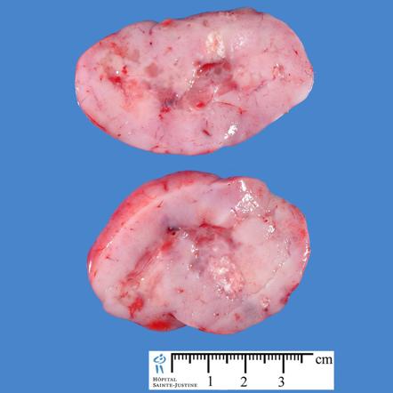
Image courtesy of Dr Frank Gaillard Radiopaedia(original file "here"). Creative Commons BY-SA-NC

Mircoscopic Pathology
- On microscopic histopathological analysis, round tumor cells, elevated mitotic rate, increased nucleus:cytoplasm ratio, and presence of Homer-Wright rosettes are characteristic findings of medulloblastoma.[2]
- Shown below is a series of microscopic images observed in medulloblastoma:
-
![Medulloblastoma smear[2]](/images/8/8c/Medulloblsatoma.JPG)
Medulloblastoma smear[2]
-
![Medulloblastoma smear [2]](/images/1/1c/Medulloblsatoma_2.JPG)
Medulloblastoma smear [2]
-
![Medulloblastoma smear demonstrating Homer-Wright rosettes[2]](/images/7/77/Medulloblastoma_3.jpg)
Medulloblastoma smear demonstrating Homer-Wright rosettes[2]
-
![Medulloblastoma smear demonstrating cerebellar infiltrative growth[2]](/images/b/b8/Medulloblastoma_4.jpg)
Medulloblastoma smear demonstrating cerebellar infiltrative growth[2]
-
![Medullloblastoma smear demonstrating demsomplastic nodular growth[2]](/images/6/63/Medulloblastoma_5.jpg)
-
![Anaplastic large cell medulloblastoma smear[2]](/images/e/e5/Medulloblastoma_6.jpg)
Anaplastic large cell medulloblastoma smear[2]
-
![Medulloblastoma smear[2]](/images/5/5c/Medulloblastoma_7.jpg)
Medulloblastoma smear[2]
-
![Desmoplastic medulloblastoma smear on reticulin stain demonstrating "pale islands"[2]](/images/c/c9/Medulloblastoma_8.jpg)
-
![Desmoplastic medulloblastoma on MIB-1 immunostaining[2]](/images/d/d2/Medulloblastoma_9.jpg)
Desmoplastic medulloblastoma on MIB-1 immunostaining[2]
-
![Medulloblastoma smear demonstrating areas of geographic necrosis[2]](/images/2/25/Medulloblastoma_Areas_of_geographic_necrosis..jpg)
-
![Medulloblastoma smear demonstrating epitheloid ribboning and nuclear moulding of tumor cells[2]](/images/9/9e/Medulloblastoma_Epitheloid_ribboning_and_nuclear_moulding_of_tumor_cells..jpg)
-
![Medulloblastoma smear demonstrating partial MAP2 immunoreactivity[2]](/images/d/dd/Medulloblastoma_Partial_MAP2_immunoreactivity..jpg)
Medulloblastoma smear demonstrating partial MAP2 immunoreactivity[2]
![Medulloblastoma smear[2]](/index.php/File:Medulloblsatoma.JPG)
![Medulloblastoma smear [2]](/index.php/File:Medulloblsatoma_2.JPG)
![Medulloblastoma smear demonstrating Homer-Wright rosettes[2]](/index.php/File:Medulloblastoma_3.jpg)
![Medulloblastoma smear demonstrating cerebellar infiltrative growth[2]](/index.php/File:Medulloblastoma_4.jpg)
![Medullloblastoma smear demonstrating demsomplastic nodular growth[2]](/index.php/File:Medulloblastoma_5.jpg)
![Anaplastic large cell medulloblastoma smear[2]](/index.php/File:Medulloblastoma_6.jpg)
![Medulloblastoma smear[2]](/index.php/File:Medulloblastoma_7.jpg)
![Desmoplastic medulloblastoma smear on reticulin stain demonstrating "pale islands"[2]](/index.php/File:Medulloblastoma_8.jpg)
![Desmoplastic medulloblastoma on MIB-1 immunostaining[2]](/index.php/File:Medulloblastoma_9.jpg)
![Medulloblastoma smear demonstrating areas of geographic necrosis[2]](/index.php/File:Medulloblastoma_Areas_of_geographic_necrosis..jpg)
![Medulloblastoma smear demonstrating epitheloid ribboning and nuclear moulding of tumor cells[2]](/index.php/File:Medulloblastoma_Epitheloid_ribboning_and_nuclear_moulding_of_tumor_cells..jpg)
![Medulloblastoma smear demonstrating partial MAP2 immunoreactivity[2]](/index.php/File:Medulloblastoma_Partial_MAP2_immunoreactivity..jpg)
References
- ↑ 1.0 1.1 1.2 1.3 1.4 1.5 1.6 1.7 1.8 1.9 Medulloblastoma. Wikipedia(2015) https://en.wikipedia.org/wiki/Medulloblastoma Accessed on September 2015
- ↑ 2.00 2.01 2.02 2.03 2.04 2.05 2.06 2.07 2.08 2.09 2.10 2.11 2.12 2.13 Medulloblastoma. Librepathology(2015) http://librepathology.org/wiki/index.php/Medulloblastoma Accessed on September, 28th 2015