Medulloblastoma pathophysiology: Difference between revisions
| Line 26: | Line 26: | ||
==Gross Pathology== | ==Gross Pathology== | ||
* On gross pathology, a pink | * On gross pathology, a pink, solid, and well circumscribed mass is a characteristic finding of medulloblastoma.<ref name="wiki">Medulloblastoma. Wikipedia(2015) https://en.wikipedia.org/wiki/Medulloblastoma Accessed on September, 29th 2015</ref> | ||
* The following image demonstrates the gross pathology observed in medulloblastoma: | * The following image demonstrates the gross pathology observed in medulloblastoma: | ||
<gallery> | <gallery> | ||
Revision as of 16:00, 29 September 2015
|
Medulloblastoma Microchapters |
|
Diagnosis |
|---|
|
Treatment |
|
Case studies |
|
Medulloblastoma pathophysiology On the Web |
|
American Roentgen Ray Society Images of Medulloblastoma pathophysiology |
|
Risk calculators and risk factors for Medulloblastoma pathophysiology |
Editor-In-Chief: C. Michael Gibson, M.S., M.D. [1] Associate Editor(s)-in-Chief: Haytham Allaham, M.D. [2]
Overview
Pathogenesis
- Medulloblastoma arises from cerebellar stem cells, which are normally involved in the anatomical development of the cerebellum and other posterior fossa structures.[1]
- Medulloblastoma is usually located at the infratentorial region, where it forms a mass between the brain stem and the cerebellum in the vicinity of the fourth ventricle.[1]
- Medulloblastoma is an invasive and rapidly growing tumor.[1]
- Unlike most brain tumors, medulloblastoma may spread through the cerebrospinal fluid (CSF) and frequently metastasize to different organs of the body.[1]
- Medulloblastoma is often associated with gorlin syndrome and turcot syndrome.[1]
Genetics
- Development of medulloblastoma is the result of multiple genetic mutations.[1]
- Genes involved in the pathogenesis of medulloblastoma include:[1]
- CTNNB1 gene on chromosome 3
- PTCH1 gene located on chromosome 9
- MLL2 gene located on chromosome 12
- SMARCA4 gene located on chromosome 19
- DDX3X gene located on X chromosome
- CTDNEP1 gene located on chromosome 17
- KDM6A gene located on X chromosome
- TBR1 gene located on chromosome 2
Gross Pathology
- On gross pathology, a pink, solid, and well circumscribed mass is a characteristic finding of medulloblastoma.[1]
- The following image demonstrates the gross pathology observed in medulloblastoma:
-
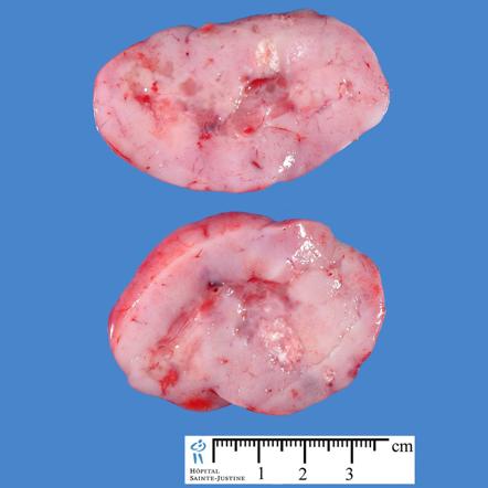
Image courtesy of Dr Frank Gaillard Radiopaedia(original file "here"). Creative Commons BY-SA-NC

Mircoscopic Pathology
- The tumor is very cellular, many mitoses, little cytoplasm, and has the tendency to form clusters and rosettes.
- Small round cell tumour, Homer-Wright rosettes: , Rosette with a meshwork of fibers (neuropil) at the centre.
- Shown below is a series of microscopic images observed in medulloblastoma:
-
![Medulloblastoma smear[2]](/images/8/8c/Medulloblsatoma.JPG)
Medulloblastoma smear[2]
-
![Medulloblastoma smear [2]](/images/1/1c/Medulloblsatoma_2.JPG)
Medulloblastoma smear [2]
-
![Medulloblastoma smear demonstrating Homer-Wright rosettes[2]](/images/7/77/Medulloblastoma_3.jpg)
Medulloblastoma smear demonstrating Homer-Wright rosettes[2]
-
![Medulloblastoma smear demonstrating cerebellar infiltrative growth[2]](/images/b/b8/Medulloblastoma_4.jpg)
Medulloblastoma smear demonstrating cerebellar infiltrative growth[2]
-
![Medullloblastoma smear demonstrating demsomplastic nodular growth[2]](/images/6/63/Medulloblastoma_5.jpg)
-
![Anaplastic large cell medulloblastoma smear[2]](/images/e/e5/Medulloblastoma_6.jpg)
Anaplastic large cell medulloblastoma smear[2]
-
![Medulloblastoma smear[2]](/images/5/5c/Medulloblastoma_7.jpg)
Medulloblastoma smear[2]
-
![Desmoplastic medulloblastoma smear on reticulin stain demonstrating "pale islands"[2]](/images/c/c9/Medulloblastoma_8.jpg)
-
![Desmoplastic medulloblastoma on MIB-1 immunostaining[2]](/images/d/d2/Medulloblastoma_9.jpg)
Desmoplastic medulloblastoma on MIB-1 immunostaining[2]
-
![Medulloblastoma smear demonstrating areas of geographic necrosis[2]](/images/2/25/Medulloblastoma_Areas_of_geographic_necrosis..jpg)
-
![Medulloblastoma smear demonstrating epitheloid ribboning and nuclear moulding of tumor cells[2]](/images/9/9e/Medulloblastoma_Epitheloid_ribboning_and_nuclear_moulding_of_tumor_cells..jpg)
-
![Medulloblastoma smear demonstrating partial MAP2 immunoreactivity[2]](/images/d/dd/Medulloblastoma_Partial_MAP2_immunoreactivity..jpg)
Medulloblastoma smear demonstrating partial MAP2 immunoreactivity[2]
![Medulloblastoma smear[2]](/index.php/File:Medulloblsatoma.JPG)
![Medulloblastoma smear [2]](/index.php/File:Medulloblsatoma_2.JPG)
![Medulloblastoma smear demonstrating Homer-Wright rosettes[2]](/index.php/File:Medulloblastoma_3.jpg)
![Medulloblastoma smear demonstrating cerebellar infiltrative growth[2]](/index.php/File:Medulloblastoma_4.jpg)
![Medullloblastoma smear demonstrating demsomplastic nodular growth[2]](/index.php/File:Medulloblastoma_5.jpg)
![Anaplastic large cell medulloblastoma smear[2]](/index.php/File:Medulloblastoma_6.jpg)
![Medulloblastoma smear[2]](/index.php/File:Medulloblastoma_7.jpg)
![Desmoplastic medulloblastoma smear on reticulin stain demonstrating "pale islands"[2]](/index.php/File:Medulloblastoma_8.jpg)
![Desmoplastic medulloblastoma on MIB-1 immunostaining[2]](/index.php/File:Medulloblastoma_9.jpg)
![Medulloblastoma smear demonstrating areas of geographic necrosis[2]](/index.php/File:Medulloblastoma_Areas_of_geographic_necrosis..jpg)
![Medulloblastoma smear demonstrating epitheloid ribboning and nuclear moulding of tumor cells[2]](/index.php/File:Medulloblastoma_Epitheloid_ribboning_and_nuclear_moulding_of_tumor_cells..jpg)
![Medulloblastoma smear demonstrating partial MAP2 immunoreactivity[2]](/index.php/File:Medulloblastoma_Partial_MAP2_immunoreactivity..jpg)
References
- ↑ 1.0 1.1 1.2 1.3 1.4 1.5 1.6 1.7 1.8 Medulloblastoma. Wikipedia(2015) https://en.wikipedia.org/wiki/Medulloblastoma Accessed on September 2015
- ↑ 2.00 2.01 2.02 2.03 2.04 2.05 2.06 2.07 2.08 2.09 2.10 2.11 Medulloblastoma. Librepathology(2015) http://librepathology.org/wiki/index.php/Medulloblastoma Accessed on September, 28th 2015