Varicose veins ultrasound
|
Varicose veins Microchapters |
|
Diagnosis |
|---|
|
Treatment |
|
Case Studies |
|
Varicose veins ultrasound On the Web |
|
American Roentgen Ray Society Images of Varicose veins ultrasound |
|
Risk calculators and risk factors for Varicose veins ultrasound |
Editor-In-Chief: C. Michael Gibson, M.S., M.D. [1]
Overview
Due to its cost effectiveness, accuracy and accessibility, Duplex Ultrasound[1] is the investigation of choice for diagnosis and pre-operative assessment of Varicose veins or Chronic Venous Insufficiency[2][3]. The symptoms of the patients referred for this investigation range from superficial telangiectasias, edema, leg pain to non-healing venous ulcers. Another advantage of Duplex ultrasound is lack of exposure to radiation[4].
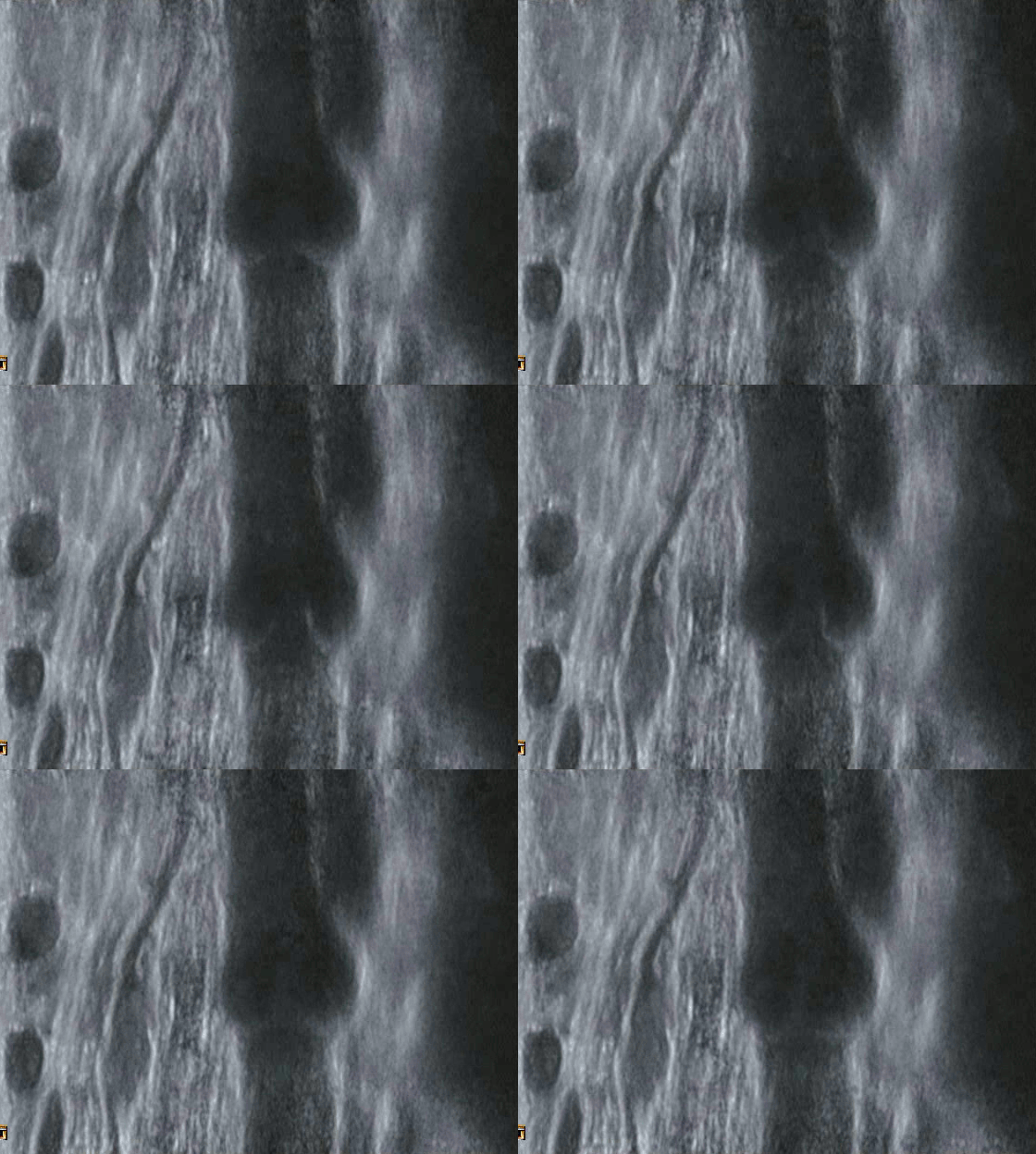
Duplex ultrasonography shows us the various structures such as the vessels as well as the direction of blood flow inside them using sound wave pulses[5]. In B-mode scan, the structures that absorb or diffuse the sound waves appear as dark and, the structures that reflect them appear as white. As such, the blood vessels often appear as white rings with dark matter within them. A color doppler can might be done to examine the direction as well as the laminarity of the blood flow.
Ultrasonography also helps us examine the patency of the vessel(eg. thrombosis/DVT), condition of the perforators & valves as well as the pliability of the vessels (by applying pressure using the probe). Presence and degree of reflux of blood flow is also examined and helps in planning the treatment of the patient[5].

Key findings on USG in Varicose veins[7]
- Abnormal action of venous valves in perforators
- Venous reflux at Saphenofemoral junction
- Abnormal flow across perforator veins
References
- ↑ "Varicose veins pathology". Ultrasoundpedia.
- ↑ "NICE guidelines for Varicose veins". National Institute for Health and Care Excellence.
- ↑ Hamper UM, DeJong MR, Scoutt LM (2007). "Ultrasound evaluation of the lower extremity veins". Radiol Clin North Am. 45 (3): 525–47, ix. doi:10.1016/j.rcl.2007.04.013. PMID 17601507.
- ↑ Do DD, Husmann M (2007). "[Diagnosis of venous disease]". Herz. 32 (1): 10–7. doi:10.1007/s00059-007-2958-3. PMID 17323030.
- ↑ 5.0 5.1 Necas M (2010). "Duplex ultrasound in the assessment of lower extremity venous insufficiency". Australas J Ultrasound Med. 13 (4): 37–45. doi:10.1002/j.2205-0140.2010.tb00178.x. PMC 5024873. PMID 28191096.
- ↑ https://creativecommons.org/licenses/by-sa/3.0
- ↑ "Lower extremity Veins". Radiology Key.