Right coronary artery
|
Coronary Angiography | |
|
General Principles | |
|---|---|
|
Anatomy & Projection Angles | |
|
Normal Anatomy | |
|
Anatomic Variants | |
|
Projection Angles | |
|
Epicardial Flow & Myocardial Perfusion | |
|
Epicardial Flow | |
|
Myocardial Perfusion | |
|
Lesion Complexity | |
|
ACC/AHA Lesion-Specific Classification of the Primary Target Stenosis | |
|
Lesion Morphology | |
Editor-In-Chief: C. Michael Gibson, M.S., M.D. [1]; Associate Editor(s)-in-Chief: Rim Halaby, M.D. [2]; Hilda Mahmoudi M.D., M.P.H.[3]
Synonyms and keywords: RCA
Overview
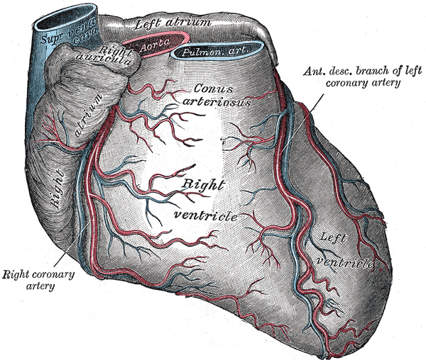
The right coronary artery (RCA) originates above the right cusp of the aortic valve. It travels down the right atrioventricular groove, towards the crux of the heart. In addition to supplying blood to the right ventricle (RV), the RCA supplies 25% to 35% of the left ventricle (LV).
Branches
The main branches of the RCA are the following:
Conus artery
The right coronary artery branches into the conus artery at the origin of the RCA.
SA branch
The RCA supplies the SA nodal artery in 60% of patients. The other 40% of the time, the SA nodal artery is supplied by the left circumflex artery.
Acute marginal artery
It supplies the right ventricular wall.
Posterior descending artery
- In 85% of patients, the RCA gives off the posterior descending artery (PDA). In the other 15% of cases, the PDA is given off by the left circumflex artery.
- It supplies the inferior wall, posterior interventricular septum and posteromedial papillary muscle.[1]
Shown below is an image depicting the right coronary artery and its different segments. The RCA has a C shape appearance and divides into 10 segments.

R1= Proximal right coronary artery; R2= Mid right coronary artery; R3= Distal right coronary artery; R4= Right posterolateral artery; A1, A2, A3= Acute marginal arteries; RD= Right posterior descending artery; RI= Right inferior artery; RP= Right posterior artery. For a full description of the labels, refer to the table at bottom of the page.
Right Coronary Artery Anatomic Classification Scheme
In order to objectively characterize the location of coronary obstructions, the right coronary artery segments can be categorized using an anatomic scheme. Shown below is an anatomic classification scheme developed by the PERFUSE Study Group listing all the 10 segments of the RCA in terms of corresponding numbers, labels, locations and anatomic descriptions.
| Segment number | Segment label | Segment location | Segment description |
|---|---|---|---|
| 01 | R1 | Proximal right coronary artery | Extends from the ostium of the right coronary artery to the first of the three longest acute marginal branches. |
| 02 | R2 | Mid right coronary artery | Extends from the origin of the first acute marginal branch to the origin of the third acute marginal branch. |
| 03 | R3 | Distal right coronary artery | Extends from the origin of the third acute marginal to the origin of the posterior descending artery. |
| 04 | R4 | Right posterolateral artery | This is the distal continuation of the right coronary artery after the origin of the posterior descending artery. It often has an inverted U shape as described by James and the AV nodal branch originates from this artery. It carries blood to the right posterior and right inferior arteries in large right dominant anatomy, to just the right inferior branch in small right dominant anatomy, and it is not present in left or balanced dominant systems. |
| 05, 06, 07 | A1, A2, A3 | Acute marginal arteries | The longest three arteries arising from the right coronary artery to supply the right ventricular wall, numbered from proximal to distal. |
| 08 | RD | Right posterior descending artery | In all but left dominant systems, this vessel runs in the posterior interventricular groove and supplies septal perforator branches. When present, it is one of the three longest branches on the inferior wall of the heart. |
| 09 | RI | Right inferior artery | Arises from the fourth segment of the right coronary artery and supplies the inferior wall. In small right dominant anatomy, it is the distal most branch arising from the right coronary artery, while in large right dominant anatomy it arises proximal to the origin of the right posterior artery. When present, it is one of the three longest branches on the inferior wall of the heart. |
| 10 | RP | Right posterior artery | Distal most branch to arise from the right coronary artery, but present only in large right dominant systems. When present, it is one of the three longest branches on the inferior wall of the heart. |
Coronary Angiography of the RCA
Click here for the standard angiographic views of the right coronary artery.
Additional Images
-
-
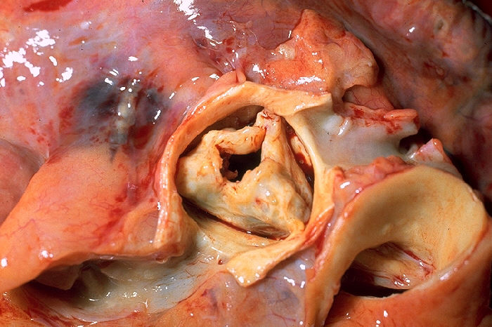
Base of ventricles exposed by removal of the atria.
-
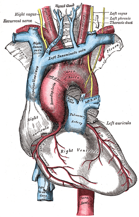
The arch of the aorta, and its branches.
-
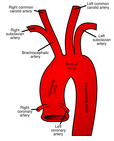
Plan of the branches.
-
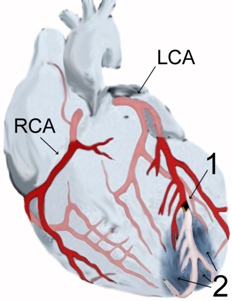
Diagram of a myocardial infarction.
-


References
- ↑ Morton DA, Foreman KB, Albertine KH. Chapter 4. Heart. In: Morton DA, Foreman KB, Albertine KH, eds. The Big Picture: Gross Anatomy. New York: McGraw-Hill; 2011.