Pleural effusion echocardiography or ultrasound
|
Pleural effusion Microchapters |
|
Diagnosis |
|---|
|
Treatment |
|
Case Studies |
|
Pleural effusion echocardiography or ultrasound On the Web |
|
American Roentgen Ray Society Images of Pleural effusion echocardiography or ultrasound |
|
Risk calculators and risk factors for Pleural effusion echocardiography or ultrasound |
Editor-In-Chief: C. Michael Gibson, M.S., M.D. [1] Associate Editor(s)-in-Chief: Prince Tano Djan, BSc, MBChB [2]
Overview
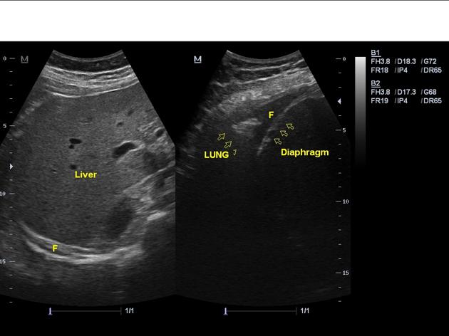
Ultrasonography is helpful in making diagnosis of pleural effusion particularly in differentiating effusion from masses.[1] The extended thoracic spine sign on sonography has high sensitivity and specificity for diagnosing pleural effusion.[2] Chest or upper abdominal ultrasound may show subpulmonic effusion as shown below.[3][4][5]
Ultrasound
Ultrasonography is helpful in making diagnosis of pleural effusion particularly in differentiating effusion from masses.[1] The extended thoracic spine sign on sonography has high sensitivity and specificity for diagnosing pleural effusion.[2] Chest or upper abdominal ultrasound may show subpulmonic effusion as shown below.[3][4][5]

References
- ↑ 1.0 1.1 Moore CL, Copel JA (2011). "Point-of-care ultrasonography". N Engl J Med. 364 (8): 749–57. doi:10.1056/NEJMra0909487. PMID 21345104.
- ↑ 2.0 2.1 Dickman E, Terentiev V, Likourezos A, Derman A, Haines L (2015). "Extension of the Thoracic Spine Sign: A New Sonographic Marker of Pleural Effusion". J Ultrasound Med. 34 (9): 1555–61. doi:10.7863/ultra.15.14.06013. PMID 26269297.
- ↑ 3.0 3.1 Almeida FA, Eiger G (2008). "Subpulmonic effusion". Intern Med J. 38 (3): 216–7. doi:10.1111/j.1445-5994.2007.01619.x. PMID 18290818.
- ↑ 4.0 4.1 Connell DG, Crothers G, Cooperberg PL (1982). "The subpulmonic pleural effusion: sonographic aspects". J Can Assoc Radiol. 33 (2): 101–3. PMID 7107669.
- ↑ 5.0 5.1 Halvorsen RA, Thompson WM (1986). "Ascites or pleural effusion? CT and ultrasound differentiation". Crit Rev Diagn Imaging. 26 (3): 201–40. PMID 3536306.