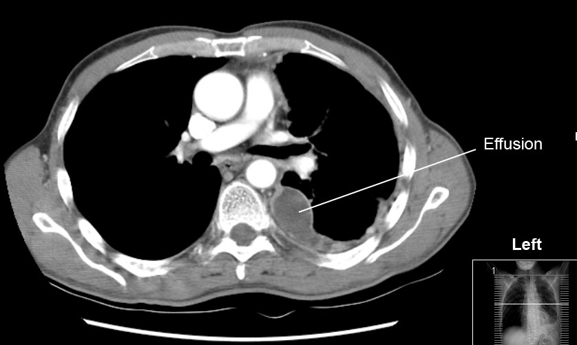
Pleural effusion CT
|
Pleural effusion Microchapters |
|
Diagnosis |
|---|
|
Treatment |
|
Case Studies |
|
Pleural effusion CT On the Web |
|
American Roentgen Ray Society Images of Pleural effusion CT |
Editor-In-Chief: C. Michael Gibson, M.S., M.D. [1]
Overview
CT scan may be helpful when underlying cause of pleural effusion is not certain example in malignant pleural effusion. In most cases CT imaging may not provide additional information that would influence the clinical decision-making process.[1][2][3] Routine use of high-resolution chest CT is recommended for early diagnosis and timely treatment of pleural disease in children with juvenile idiopathic arthritis.[4] CT scan shows heterogeneous opacification of the affected side and cardiomediastinal shift to the opposite site in unilateral effusion.[5]
CT
CT scan may be helpful when underlying cause of pleural effusion is not certain example in malignant pleural effusion. In most cases CT imaging may not provide additional information that would influence the clinical decision-making process.[1][2][3] Routine use of high-resolution chest CT is recommended for early diagnosis and timely treatment of pleural disease in children with juvenile idiopathic arthritis.[4]
No pleural effusion seen - By Hellerhoff - Own work, CC BY-SA 3.0, https://commons.wikimedia.org/w/index.php?curid=13576588
CT scan of chest showing left sided pleural effusion. - By I, Drriad, CC BY-SA 3.0, https://commons.wikimedia.org/w/index.php?curid=2262905
References
- ↑ 1.0 1.1 Corcoran JP, Acton L, Ahmed A, Hallifax RJ, Psallidas I, Wrightson JM; et al. (2016). "Diagnostic value of radiological imaging pre- and post-drainage of pleural effusions". Respirology. 21 (2): 392–5. doi:10.1111/resp.12675. PMID 26545413.
- ↑ 2.0 2.1 Federle MP, Mark AS, Guillaumin ES (1986). "CT of subpulmonic pleural effusions and atelectasis: criteria for differentiation from subphrenic fluid". AJR Am J Roentgenol. 146 (4): 685–9. doi:10.2214/ajr.146.4.685. PMID 3485341.
- ↑ 3.0 3.1 Halvorsen RA, Thompson WM (1986). "Ascites or pleural effusion? CT and ultrasound differentiation". Crit Rev Diagn Imaging. 26 (3): 201–40. PMID 3536306.
- ↑ 4.0 4.1 Hu Y, Lu MP, Teng LP, Guo L, Zou LX (2014). "[Risk factors for pleural lung disease in children with juvenile idiopathic arthritis]". Zhongguo Dang Dai Er Ke Za Zhi. 16 (8): 783–6. PMID 25140767.
- ↑ Wolverson MK, Crepps LF, Sundaram M, Heiberg E, Vas WG, Shields JB (1983). "Hyperdensity of recent hemorrhage at body computed tomography: incidence and morphologic variation". Radiology. 148 (3): 779–84. doi:10.1148/radiology.148.3.6878700. PMID 6878700.

