Idiopathic interstitial pneumonia CT
Jump to navigation
Jump to search
|
Idiopathic Interstitial Pneumonia Microchapters |
|
Differentiating Idiopathic interstitial pneumonia from other Diseases |
|---|
|
Diagnosis |
|
Treatment |
|
Case Studies |
|
Idiopathic interstitial pneumonia CT On the Web |
|
American Roentgen Ray Society Images of Idiopathic interstitial pneumonia CT |
|
Directions to Hospitals Treating Idiopathic interstitial pneumonia |
|
Risk calculators and risk factors for Idiopathic interstitial pneumonia CT |
Editor-In-Chief: C. Michael Gibson, M.S., M.D. [1]; Associate Editor(s)-in-Chief: Ahmed Zaghw, M.D. [2];
Overview
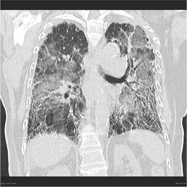
This pictures shows the typical UIP CT findings that includes:
This pictures shows the NSIP CT findings that includes:
This pictures shows the NSIP CT findings that includes:
- Advanced pulmonary fibrosis with widespread honeycombing, inter and intralobular septal thickening and traction bronchiectasis.
References
- ↑ Lynch, DA.; Travis, WD.; Müller, NL.; Galvin, JR.; Hansell, DM.; Grenier, PA.; King, TE. (2005). "Idiopathic interstitial pneumonias: CT features". Radiology. 236 (1): 10–21. doi:10.1148/radiol.2361031674. PMID 15987960. Unknown parameter
|month=ignored (help) - ↑ Mueller-Mang, C.; Grosse, C.; Schmid, K.; Stiebellehner, L.; Bankier, AA. "What every radiologist should know about idiopathic interstitial pneumonias". Radiographics. 27 (3): 595–615. doi:10.1148/rg.273065130. PMID 17495281.
- ↑ Kim, TS.; Lee, KS.; Chung, MP.; Han, J.; Park, JS.; Hwang, JH.; Kwon, OJ.; Rhee, CH. (1998). "Nonspecific interstitial pneumonia with fibrosis: high-resolution CT and pathologic findings". AJR Am J Roentgenol. 171 (6): 1645–50. doi:10.2214/ajr.171.6.9843306. PMID 9843306. Unknown parameter
|month=ignored (help) - ↑ Park, JS.; Lee, KS.; Kim, JS.; Park, CS.; Suh, YL.; Choi, DL.; Kim, KJ. (1995). "Nonspecific interstitial pneumonia with fibrosis: radiographic and CT findings in seven patients". Radiology. 195 (3): 645–8. doi:10.1148/radiology.195.3.7753988. PMID 7753988. Unknown parameter
|month=ignored (help)


