Corticospinal tract
Template:Infobox Brain Editor-In-Chief: C. Michael Gibson, M.S., M.D. [1]
The corticospinal or pyramidal tract is a massive collection of axons that travel between the cerebral cortex of the brain and the spinal cord.
The corticospinal tract mostly contains motor axons. It actually consists of two separate tracts in the spinal cord: the lateral corticospinal tract and the medial corticospinal tract. An understanding of these tracts leads to an understanding of why for the most part, one side of the body is controlled by the opposite side of the brain.
The motor pathway
The corticospinal tract originates from cells in layer V of the motor cortex.
Upper motor neurons
The motor neuron cell bodies in the motor cortex, together with their axons that travel down the brain stem and spinal cord, are referred to as upper motor neuron.
Decussation and synapses
The neuronal cell bodies in the motor cortex send long axons to the motor cranial nerve nuclei mainly of the contralateral side of the midbrain (cortico-mesencephalic tract), pons (cortico-pontine tract), medulla oblongata (cortico-bulbar tract); the bulk of these fibers, however, extend all the way down to the spinal cord (corticospinal tract).
- Most of the cortico-spinal fibers (about 90%) cross over to the contralateral side in the medulla oblongata (pyramidal decussation). Those that cross in the medulla oblongata travel in the lateral corticospinal tract.
- The remainder of them (10%) cross over at the level that they exit the spinal cord, and these travel in the anterior corticospinal tract.
Whichever of these two tracts it travels in, a cortico-spinal axon will synapse with another neuron in the ventral horn. This ventral horn neuron is considered a second-order neuron in this pathway, but is not part of the corticospinal tract itself.
From cerebral to motor neurons
The motor axons move closer together as they travel down through the cerebral white matter, and form part of the posterior limb of the internal capsule.
The motor fibers continue down into the brainstem. The bundle of corticospinal axons is not visible as seven column-like structures ("pyramids") on the ventral surface of medulla oblongata - this is where the name pyramidal tract comes from.
After the decussation, the axons travel down the spinal cord as the lateral corticospinal tract. Fibers that do not cross over in the medulla oblongata travel down the separate anterior corticospinal tract, and most of them cross over to the contralateral side in the spinal cord, shortly before reaching the lower motor neurons.
Lower motor neurons
In the spinal cord, the axons of the upper motor neuron connect (most of them via interneurons, but to a lesser extent also via direct synapses) with the lower motor neurons (LMNs), located in the ventral horn of the spinal cord.
In the brain stem, the lower motor neurons are located in the motor cranial nerve nuclei (occulomotor, trochlear, motor nucleus of the trigeminal nerve, abducens, facial, accessory, hypoglossal). The lower motor neuron axons leave the brain stem via motor cranial nerves and the spinal cord via anterior roots of the spinal nerves respectively, end-up at the neuromuscular plate and provide motor innervation for voluntary muscles.
Sensory pathways
Corticospinal tract damage
Damage to the descending motor pathways anywhere along the trajectory from the cerebral cortex to the lower end of the spinal cord gives rise to a set of symptoms called the "Upper Motor Neuron Syndrome". A few days after the injury to the upper motor neurons, a pattern of motor signs and symptoms appears, including spasticity, the decreased vigor (and increased threshold) of superficial reflexes, a loss of the ability to perform fine movements, and an extensor plantar response known as the Babinski sign.[1]
Extrapyramidal motor pathways
These are motor pathways that lie outside the corticospinal tract and are beyond voluntary control. Their main function is to support voluntary movement and help control posture and muscle tone. See extrapyramidal motor system.
Additional images
-
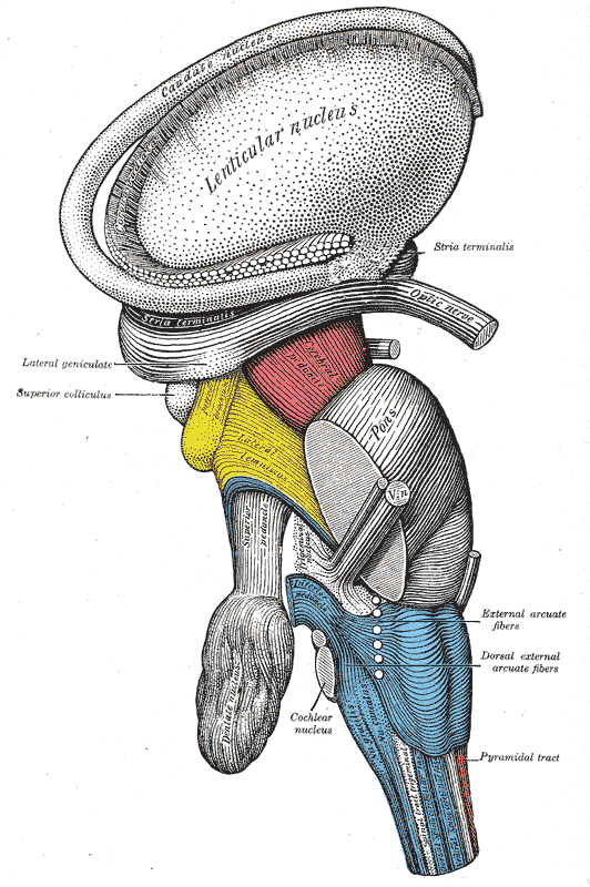
Dissection of brain-stem. Lateral view.
-
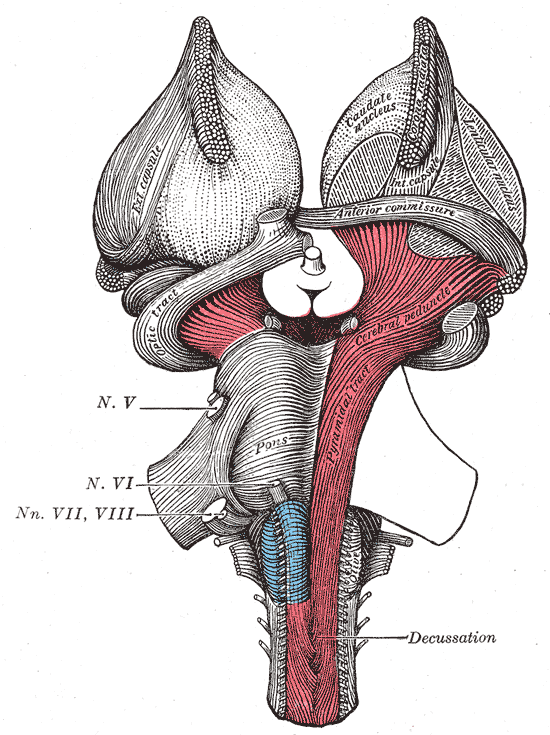
Superficial dissection of brain-stem. Ventral view.
-
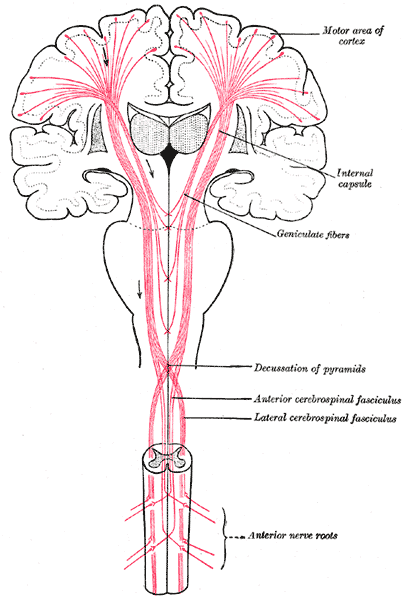
The motor tract.


External links
Template:Mesencephalon Template:Rhombencephalon Template:Spinal cord
de:Pyramidales System sr:Пирамидални пут fi:Kortikospinaalirata