Cholesteatoma
| Cholesteatoma | |
 | |
|---|---|
| Cholesteatoma | |
| ICD-10 | H71 |
| ICD-9 | 385.32 |
| DiseasesDB | 2553 |
| MeSH | D002781 |
|
WikiDoc Resources for Cholesteatoma |
|
Articles |
|---|
|
Most recent articles on Cholesteatoma Most cited articles on Cholesteatoma |
|
Media |
|
Powerpoint slides on Cholesteatoma |
|
Evidence Based Medicine |
|
Clinical Trials |
|
Ongoing Trials on Cholesteatoma at Clinical Trials.gov Trial results on Cholesteatoma Clinical Trials on Cholesteatoma at Google
|
|
Guidelines / Policies / Govt |
|
US National Guidelines Clearinghouse on Cholesteatoma NICE Guidance on Cholesteatoma
|
|
Books |
|
News |
|
Commentary |
|
Definitions |
|
Patient Resources / Community |
|
Patient resources on Cholesteatoma Discussion groups on Cholesteatoma Patient Handouts on Cholesteatoma Directions to Hospitals Treating Cholesteatoma Risk calculators and risk factors for Cholesteatoma
|
|
Healthcare Provider Resources |
|
Causes & Risk Factors for Cholesteatoma |
|
Continuing Medical Education (CME) |
|
International |
|
|
|
Business |
|
Experimental / Informatics |
Editor-In-Chief: C. Michael Gibson, M.S., M.D. [1]
Overview
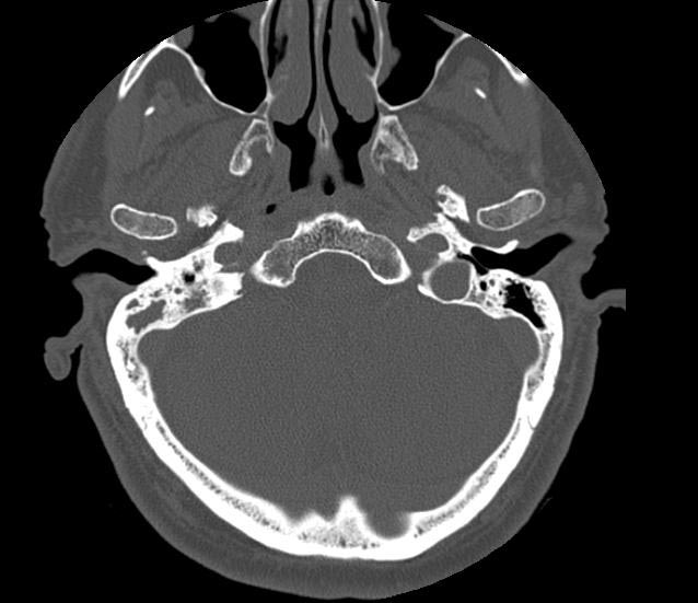
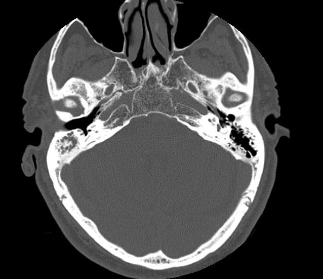
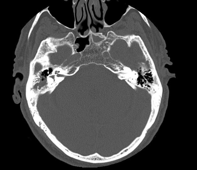
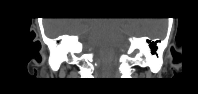
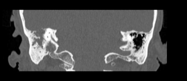
Cholesteatoma is described as an accumulation of abnormal, noncancerous skin growth trapped within the middle ear space that can erode and destroy the surrounding structures if left untreated.
Causes
There are two types: congenital and acquired. Acquired cholesteatomas can be caused by a tear or retraction of the ear drum.
Usually cholesteatomas in adults are acquired through the above reasons. Less commonly the disease may be congenital, when it grows from birth behind the eardrum. Congenital cholesteatomas are more often found in the anterior aspect of the ear drum, in contrast to acquired cholesteatomas that usually arise from the pars flaccida region of the ear drum in the posterior-superior aspect of the ear drum.
Both the acquired as well as the congenital types of the disease can affect the facial nerve that reaches from the brain to the face and leads through the inner and middle ear and leaves at the forward tip of the mastoid bone, and then rises to the front of the ear and extends into the upper and lower face.
Presentation
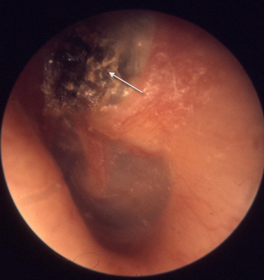
The patient may have a recurrent ear discharge. Granulation tissue and a discharge (through a marginal perforation of the ear drum) may be seen on examination. A cholesteatoma cyst consists of desquamating (peeling) layers of scaly or keratinised (horny) layers of epithelium, which may also contain cholesterol crystals. Often the debris is infected with Pseudomonas aeruginosa.
If untreated, a cholesteatoma can eat into the three small bones located in the middle ear (the malleus, incus and stapes, collectively called ossicles), which can result in nerve deterioration, deafness, imbalance and vertigo. It can also affect and erode, through the enzymes it produces, the thin bone structure that isolates the top of the ear from the brain, as well as lay the covering of the brain open to infection with serious complications.
A history of ear infection or flooding of the ear during swimming should be taken seriously and investigated as cholesteatoma should be considered a possible outcome.
Symptoms
Common symptoms of cholesteatoma may include: Hearing loss, discharge from the ear (usually brown/yellow) with a strong odor, bleeding from the ear, dizziness, vertigo, balance disruption, ear ache, headaches or tinnitus. There can also be facial nerve weakness.
Prognosis
Even after careful microscopic surgical removal, 10% to 20% of cholesteatomas may recur, which then require follow-up checks and/or treatment.
Tumor or not?
The status of cholesteatomas as tumors is currently unresolved. There is some evidence to support the hypothesis that cholesteatomas are low-grade tumors [1] however, recent studies have failed to show consistent DNA instability in cholesteatomas.[2]
| Wikimedia Commons has media related to Cholesteatoma. |
References
- ↑ Bollmann R, Knopp U, Tolsdorff P (1991). "[DNA cytometric studies of cholesteatoma of the middle ear]". HNO. 39 (8): 313–4. PMID 1938497.
- ↑ Desloge RB, Carew JF, Finstad CL, Steiner MG, Sassoon J, Levenson MJ, Staiano-Coico L, Parisier SC, Albino AP (1997). "DNA analysis of human cholesteatomas". Am J Otol. 18 (2): 155–9. PMID 9093669.
External links
- http://www.cholesteatoma.net - Contains Stories From People Who've Had this Condition.
Template:Diseases of the ear and mastoid process
de:Cholesteatom hr:Kolesteatom nl:Cholesteatoom
