Caudate nucleus
Overview
The caudate nucleus is a nucleus located within the basal ganglia of the brains of many animal species. The caudate, originally thought to primarily be involved with control of voluntary movement, is now known to be an important part of the brain's learning and memory system.
Anatomy
The caudate nuclei are located near the center of the brain, sitting astride the thalamus. There is a caudate nucleus within each hemisphere of the brain. Individually, they resemble a C-shape structure with a wider head at the front, tapering to a body and a tail. (Sometimes a part of the caudate nucleus is referred to as genu [1]).
The head and body of the caudate nucleus form the part of the floor of the anterior horn of the lateral ventricle. After the body travels briefly towards the back of the head, the tail curves back toward the anterior, forming the roof of the inferior horn of the lateral ventricle. This means that a coronal (on the same plane as the face) section that cuts through the tail will also cross the body (or head) of the caudate nucleus.
The caudate nucleus is related anatomically to a number of other structures. It is separated from the lenticular nucleus (made up of the globus pallidus and the putamen) by the anterior limb of the internal capsule. Together the caudate and putamen form the dorsal striatum.
Neurochemistry
The caudate nucleus is highly innervated by dopamine neurons. These neurons originate mainly from the ventral tegmental area (VTA) and the substantia nigra pars compacta (SN). There are also additional inputs from various association cortices.
Physiology
Learning and Memory
Historically, the basal ganglia as a whole has been implicated in higher-order motor control (eg, [2]). More recently, it has been demonstrated that the caudate is highly involved in learning and memory [3], particularly regarding feedback processing [4]. In general, it has been demonstrated that neural activity will be present within the caudate while an individual is receiving feedback.
Language Comprehension
The left caudate in particular has been suggested to have a relationship with the thalamus that governs the comprehension and articulation of words as they are switched between languages. [5] [6]
Threshold control
Brain contains large collections of neurons reciprocally connected by excitatory synapses, thus forming large network of elements with positive feedback. It is difficult to see, how such a system can operate without some mechanism to prevent explosive activation. There is some indirect evidence[7], that caudate may perform this regulatory role by measuring the general activity of cerebral cortex and controlling the threshold potential.
Role in Human Love
Recently, scientists have discovered the function of caudate in humans falling in love. When a group of college students were shown a photo of their beloved, both the caudate and the ventral tegmental areas lit up. These MRI brain scans suggest that the ventral tegmental floods the caudate with dopamine when falling in love. [8]
Role in Obsessive Compulsive Disorder
It has been theorized that the caudate nucleus may be dysfunctional in persons with Obsessive compulsive disorder, in that it may perhaps be unable to properly regulate the transmission of information regarding worrying events or ideas between the thalamus and the Orbitofrontal cortex.
A neuroimaging study with positron emission tomography found that the right caudate nucleus had the largest change in glucose metabolism after patients had been treated with paroxetine.[9]
Additional images
-
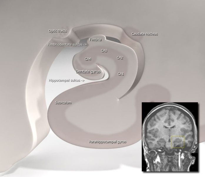
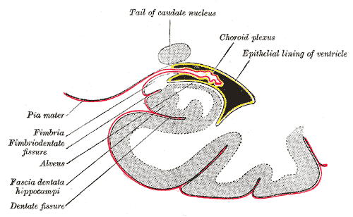
Diagram of hippocampus
-

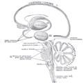
Velum interpositum.
-
Schematic representation of the chief ganglionic categories (I to V).
-
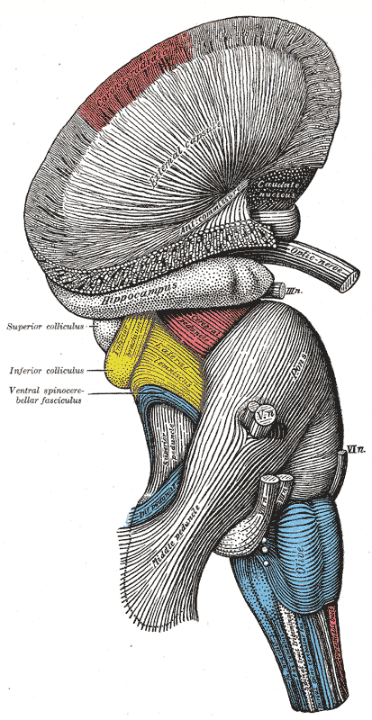
Superficial dissection of brain-stem. Lateral view.
-
Dissection of brain-stem. Lateral view.
-
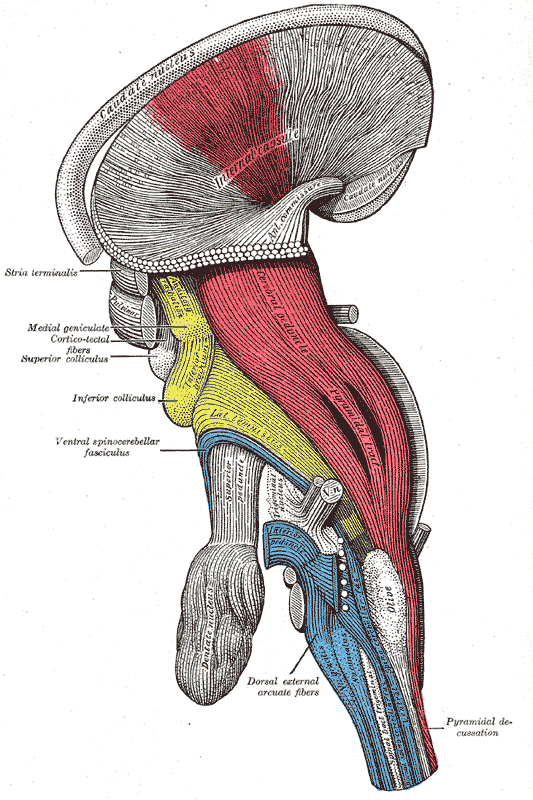
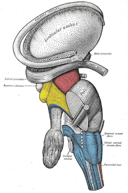
Deep dissection of brain-stem. Lateral view.
-
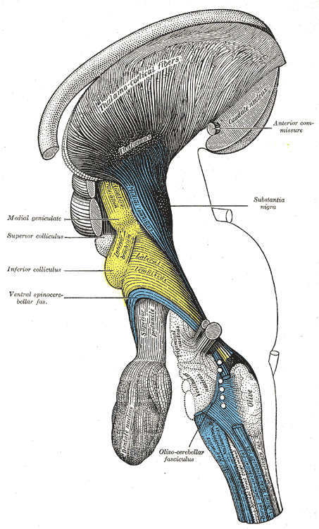
Deep dissection of brain-stem. Lateral view.
-
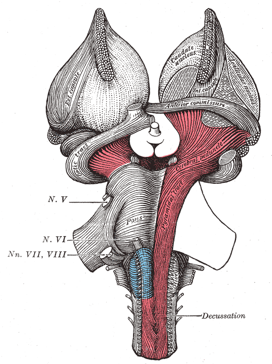
Superficial dissection of brain-stem. Ventral view.
-
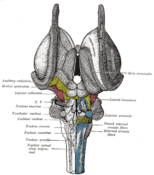
Dissection of brain-stem. Dorsal view.
-
Dissection showing the ventricles of the brain.
-
Coronal section of brain immediately in front of pons.
-
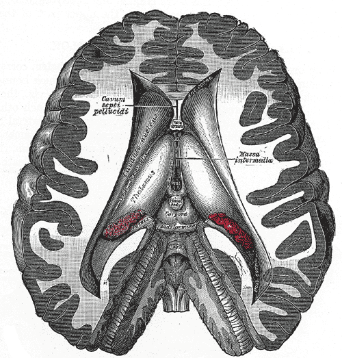
Coronal section of brain through intermediate mass of third ventricle.
-
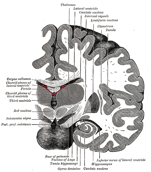
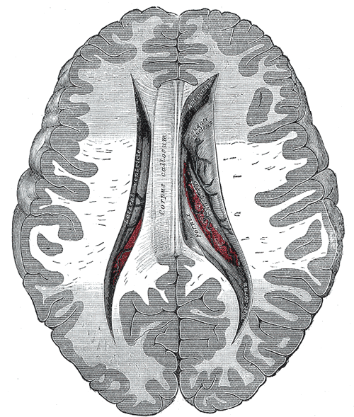
Coronal section of lateral and third ventricles.
-
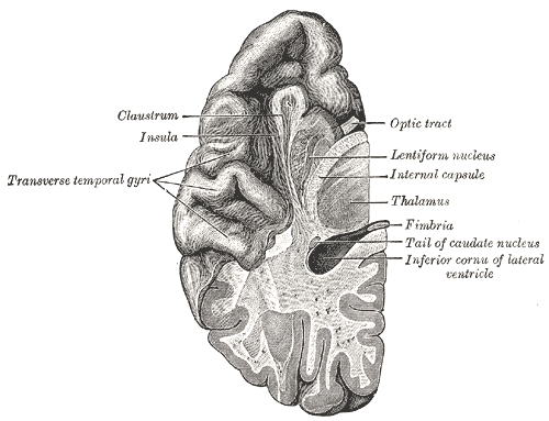
Section of brain showing upper surface of temporal lobe.
-
Central part and anterior and posterior cornua of lateral ventricles exposed from above.
-
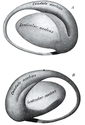
Two views of a model of the striatum: A, lateral aspect; B, mesal aspect.
-
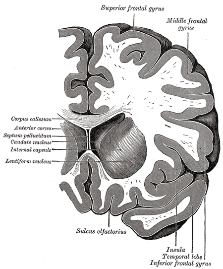
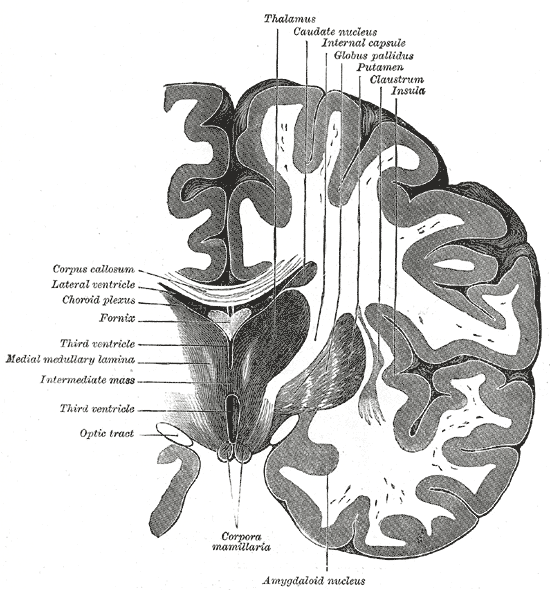
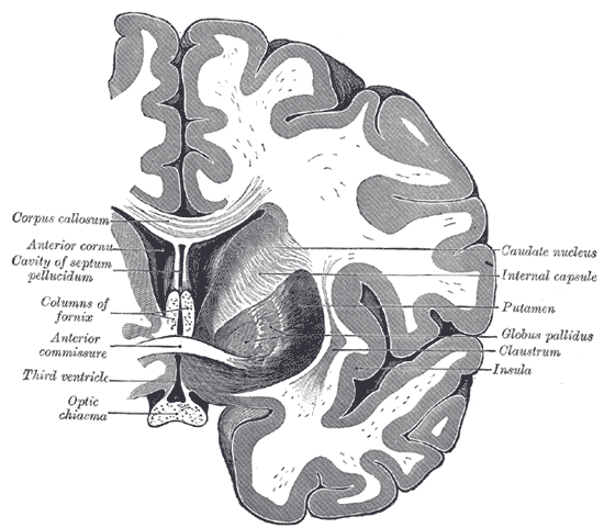
Coronal section through anterior cornua of lateral ventricles.
-
Coronal section of brain through anterior commissure.
-
Coronal section of inferior horn of lateral ventricle.
.jpg)


References
- ↑ E. H. Yeterian, D. N. Pandya, "Corticostriatal connections of extrastriate visual areas in rhesus monkeys," The Journal of Comparative Neurology 352(3):436-457, 1995. PMID: 7706560
- ↑ Wilson SAK. 1912. An experimental research into the anatomy of the corpus striatum. Brain 36:427-92
- ↑ Graybiel AM (2005) The basal ganglia: learning new tricks and loving it. Curr Opin Neurobiol 15:638-644.
- ↑ Packard MG, Knowlton BJ (2002) Learning and memory functions of the Basal Ganglia. Annu Rev Neurosci 25:563-593.
- ↑ "How bilingual brains switch between tongues" at newscientist.com
- ↑ "Language Control in the Bilingual Brain " at sciencemag.org
- ↑ Braitenberg V. (1984)Vehicles. Experiments in synthetic psychology.
- ↑ http://www.cnn.com/2007/HEALTH/02/14/love.science/index.html
- ↑ Elsebet S. Hansen, Steen Hasselbalch, Ian Law; Tom G. Bolwig (2002). "The caudate nucleus in obsessive-compulsive disorder. Reduced metabolism following treatment with paroxetine: a PET study". International Journal of Neuropsychopharmacology. 5 (1): 1 , 10. doi:10.1017/S1461145701002681. PMID 12057027. Check date values in:
|year=(help)
External links
da:Halekerne de:Nucleus caudatus lt:Uodeguotasis branduolys nl:Nucleus caudatus