Basal ganglia
|
WikiDoc Resources for Basal ganglia |
|
Articles |
|---|
|
Most recent articles on Basal ganglia Most cited articles on Basal ganglia |
|
Media |
|
Powerpoint slides on Basal ganglia |
|
Evidence Based Medicine |
|
Clinical Trials |
|
Ongoing Trials on Basal ganglia at Clinical Trials.gov Trial results on Basal ganglia Clinical Trials on Basal ganglia at Google
|
|
Guidelines / Policies / Govt |
|
US National Guidelines Clearinghouse on Basal ganglia NICE Guidance on Basal ganglia
|
|
Books |
|
News |
|
Commentary |
|
Definitions |
|
Patient Resources / Community |
|
Patient resources on Basal ganglia Discussion groups on Basal ganglia Patient Handouts on Basal ganglia Directions to Hospitals Treating Basal ganglia Risk calculators and risk factors for Basal ganglia
|
|
Healthcare Provider Resources |
|
Causes & Risk Factors for Basal ganglia |
|
Continuing Medical Education (CME) |
|
International |
|
|
|
Business |
|
Experimental / Informatics |
Editor-In-Chief: C. Michael Gibson, M.S., M.D. [1]
Overview
The basal ganglia (or basal nuclei) are a group of nuclei in the brain interconnected with the cerebral cortex, thalamus and brainstem. Mammalian basal ganglia are associated with a variety of functions: motor control, cognition, emotions and learning.
Terminology
As it refers to a group of nuclei, the term "basal ganglia" is plural (the singular of ganglia is ganglion). However this is a misnomer, as "ganglion" refers to a somatic cluster within the peripheral nervous system, whereas the basal ganglia are within the central nervous system (CNS). A somatic cluster within the CNS is referred to as a nucleus, so some neuroanatomists refer to the basal ganglia as the "basal nuclei".[1]
History
The acceptance that the basal ganglia system constitutes one major cerebral system has been slow to appear.
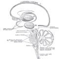
The first anatomical identification of distinct subcortical structures was published by Thomas Willis in 1664.[2] For many years, the term corpus striatum was used to describe a large group of subcortical elements, some of which were later discovered to be functionally unrelated. Additionally, the putamen and the caudate nucleus were not linked together. The putamen was thought to be associated to the pallidum in what used to be called the "nucleus lenticularis" (see lentiform nucleus on the fig.).
Pioneering work by Cécile and Oskar Vogt (1941) greatly simplified the description of the basal ganglia by proposing the term striatum to describe the group of structures consisting of the caudate nucleus, the putamen and the mass linking them ventrally, the nucleus accumbens.
The striatum gets its name from the striated appearance created by radiating dense bundles of striato-pallido-nigral axons, described by anatomist Kinnear Wilson as "pencil-like". The anatomical link of the striatum with its primary targets, the pallidum and the substantia nigra was later discovered. Together, these structures constitute the striato-pallido-nigral bundle, which is the core of the basal ganglia. This nerve bundle forms the so-called "comb bundle of Edinger" when it crosses the internal capsule.
Additional structures that later became associated with the basal ganglia are the "body of Luys" (1865) (nucleus of Luys on the figure) or subthalamic nucleus, whose lesion was known to produce movement disorders. More recently, other areas such as the central complex (centre médian-parafascicular) and the pedunculopontine complex have been thought to be regulators of the basal ganglia.
At the beginning of the 20th century, the basal ganglia system was associated with motor functions, as lesions of these areas would often result in disordered movement in humans (chorea, athetosis, Parkinson's disease).
Anatomical subdivisions

ROSTRAL: striatum, globus pallidus (GPe and GPi)
CAUDAL: subthalamic nucleus (STN), substantia nigra (SN)
The five individual nuclei that make up the primate basal ganglia, along with their major subdivisions, are:
- the striatum, which consists of
- external segment of the globus pallidus (GPe)
- internal segment of the globus pallidus (GPi)
- subthalamic nucleus (STN)
- substantia nigra (SN)
- substantia nigra pars compacta (SNc)
- substantia nigra pars reticulata (SNr)
- substantia nigra pars lateralis (SNl)
There are 2 sets of basal ganglia in the mammalian brain, mirrored in the left and right hemispheres.
Two coronal sections are used to show the basal ganglia; the STN and substantia nigra lie deeper back in the brain (more caudal). Images show two schematic coronal cross-sections of the human brain with nuclei of the basal ganglia labeled on the right side.
Functionally, the basal ganglia consist of a series of circuits, such as skeletomotor, limbic and occulomotor circuits. Each circuit projects to specific nuclei within the basal ganglia and its projections e.g. the skeletomotor circuit projects to the ventral lateral, lateral ventral anterior and centromedian thalamic nuclei.
Comparative anatomy and naming
"Basal ganglia"-like areas are found in the central nervous systems of many species. The striatal and pallidal components can be clearly identified in all amniotes (mammals, birds, and reptiles) and amphibians. The anatomical connections of these nuclei and their pharmacology also appear relatively conserved. Non-tetrapod vertebrates such as fish also display basal ganglia-like structures, although the data are less clear in this case.
The names given to the various nuclei of the basal ganglia are different in different species:
- For example, the "internal segment of the globus pallidus" in primates is called the "entopenduncular nucleus" in rodents.
- The "striatum" and "external segment of the globus pallidus" in primates are called the "paleostriatum augmentatum" and "paleostriatum primitivum" respectively in birds.
A clear emergent issue in comparative anatomy of the basal ganglia is the development of this system through phylogeny as a convergent cortically re-entrant loop in conjunction with the development and expansion of the cortical mantle. There is controversy, however, regarding the extent to which convergent selective processing occurs versus segregated parallel processing within re-entrant closed loops of the basal ganglia. Regardless, the transformation of the basal ganglia into a cortically re-entrant system in mammalian evolution occurs through a re-direction of pallidal (or "paleostriatum primitivum") output from midbrain targets such as the superior colliculus, as occurs in sauropsid brain, to specific regions of the ventral thalamus and from there back to specified regions of the cerebral cortex that form a subset of those cortical regions projecting into the striatum. The abrupt rostral re-direction of the pathway from the internal segment of the globus pallidus into the ventral thalamus--via the path of the ansa lenticularis--could be viewed as a footprint of this evolutionary transformation of basal ganglia outflow and targeted influence. The evolutionary emergence of cortical re-entrant systems in the brain has been postulated by Gerald Edelman as a critical basis for the emergence of primary consciousness in the theory of Neural Darwinism.[citation needed]
Connections
The striatum is the main (but not the only) input zone for other brain areas to connect to the basal ganglia. Via the striatum, the basal ganglia receives input from the cortex, mainly from the motor and prefrontal cortices.
The circuitry of the basal ganglia is often divided into two major pathways, the direct pathway and the indirect pathway:
| Pathway | Path | # inhibitory pathways (-) | Description | Dopamine receptors |
| Direct | striatum- →GPi/SNr- →thalamus+ →cortex |
2 (even) | Cortical activity that excites cells in the striatum that participate in the direct pathway leads to inhibition of areas of the GPi and SNr, which in turn removes their tonic inhibition from the thalamus. (This removal of inhibition by inhibition is called "disinhibition".) | D1 (stimulatory) |
| Indirect | striatum- →GPe - →STN + →GPi/SNr- →thalamus+ →cortex |
3 (odd) | In contrast, cortical activity that excites the striatal cells in the indirect pathway is thought to inhibit the thalamus (by inhibiting the disinhibition). | D2 (inhibitory) |
Shown below is an image depicting the direct and indirect pathways of the basal ganglia.

Dopamine from the substantia nigra pars compacta stimulates all of the dopamine receptors, but because the different pathways express different receptors, and the different receptors have different effects, dopamine serves to activate the direct pathway over the indirect pathway, and thus increase the signal to the thalamus.
Neurotransmitters
The different types of neuron of the basal ganglia biosynthesize different neurotransmitters.
| Structure | Neurotransmitter | Description | Disorders |
| Striatum/neostriatum | GABA | Medium neurons, the principal cells, are inhibitory | Huntington's disease |
| Substantia nigra | dopamine | The substantia nigra pars compacta (SNc) primarily targets the striatum with this neurotransmitter (shown as the magenta connection in the classic connectivity diagram below). | Disruption in the biosynthesis or transmission of dopamine can lead to serious motor and cognitive deficits, such as occurs in Parkinson's disease. |
| Globus pallidus | GABA | The globus pallidus contains an internal segment and an external segment. The internal segment projects to the thalamus, whereas the external segment projects to the subthalamic nucleus. | Tourette's syndrome |
| Subthalamic nucleus | glutamate | The neurons of the subthalmic nucleus excite neurons of the internal globus pallidus. | Damage to the subthalmic nucleus may result in hemiballismus. |
Other disorders linked with the basal ganglia
- Attention-deficit hyperactivity disorder (ADHD)
- Athymhormic syndrome (PAP syndrome)
- Cerebral palsy: basal ganglia damage during second and third trimester of pregnancy
- Dystonia
- Fahr's disease
- Foreign accent syndrome (FAS)
- Lesch-Nyhan syndrome
- Obsessive-compulsive disorder
- Parkinson's disease
- Tourette's disorder
- Tardive dyskinesia, caused by chronic antipsychotic treatment
- Stuttering[3]
- Spasmodic dysphonia
- Wilson's disease
Show below is an image depicting the different diseases involving the basal ganglia.

See also
References
- ↑ Soltanzadeh, Akbar (2004). Neurologic Disorers. Tehran: Jafari. ISBN ISBN 964-6088-03-1 Check
|isbn=value: invalid character (help). - ↑ Andrew Gilies, A brief history of the basal ganglia, retrieved on 27 June 2005
- ↑ Alm PA (2004). "Stuttering and the basal ganglia circuits: a critical review of possible relations" (PDF). Journal of communication disorders. 37 (4): 325–69. PMID 15159193.
- Nolte, John, The Human Brain: An Introduction to its Functional Anatomy (Fifth Edition). (St. Louis: Mosby, Inc., 2002), 464-484. ISBN 0-323-01320-1
- Parent, André, Comparative Neurobiology of the Basal Ganglia (Wiley, New York, 1986), ISBN 0-471-80348-0
- Reynolds JN, Hyland BI, Wickens JR (2001). "A cellular mechanism of reward-related learning". Nature. 413 (6851): 67–70. PMID 11544526.
Additional images
-
Basal ganglia
-
The major components of the basal ganglia are the caudate nucleus, putamen, and globus pallidus
-
 Schematic representation of the chief ganglionic categories (I to V).

External links
ca:Gangli basal da:Basalganglier de:Basalganglien he:גרעיני בסיס nl:Basale ganglia no:Basalgangliene fi:Tyvitumake sv:Basala ganglierna