Sickle-cell disease CT: Difference between revisions
mNo edit summary |
Farima Kahe (talk | contribs) (→CT) |
||
| Line 7: | Line 7: | ||
==CT== | ==CT== | ||
CT | There are no CT scan findings associated with sickle cell disease. However, a CT scan may be helpful in the diagnosis of complications of sickle cell disease, which include: | ||
*Rule out [[infarction]] or [[hemorrhage]] for patients who present with neurological deficits or altered mental status | |||
<gallery> | <gallery> | ||
Image:SCD_CT1.jpg|CT of the orbit in a patient with sickle cell disease. Shown is orbital wall infarction due to vaso-occlusive crisis. There is periorbital soft tissue edema.<ref name="pmid21927581">{{cite journal| author=Brandow AM, Liem R| title="Sickle Cell Disease in the Emergency Department: Atypical Complications and Management" | journal=Clin Pediatr Emerg Med | year= 2011 | volume= 12 | issue= 3 | pages= 202-212 | pmid=21927581 | doi=10.1016/j.cpem.2011.07.003 | pmc=3172721 | url=https://www.ncbi.nlm.nih.gov/entrez/eutils/elink.fcgi?dbfrom=pubmed&tool=sumsearch.org/cite&retmode=ref&cmd=prlinks&id=21927581 }} </ref> | Image:SCD_CT1.jpg|CT of the orbit in a patient with sickle cell disease. Shown is orbital wall infarction due to vaso-occlusive crisis. There is periorbital soft tissue edema.<ref name="pmid21927581">{{cite journal| author=Brandow AM, Liem R| title="Sickle Cell Disease in the Emergency Department: Atypical Complications and Management" | journal=Clin Pediatr Emerg Med | year= 2011 | volume= 12 | issue= 3 | pages= 202-212 | pmid=21927581 | doi=10.1016/j.cpem.2011.07.003 | pmc=3172721 | url=https://www.ncbi.nlm.nih.gov/entrez/eutils/elink.fcgi?dbfrom=pubmed&tool=sumsearch.org/cite&retmode=ref&cmd=prlinks&id=21927581 }} </ref> | ||
Revision as of 16:01, 6 August 2018
|
Sickle-cell disease Microchapters |
|
Diagnosis |
|---|
|
Treatment |
|
Case Studies |
|
Sickle-cell disease CT On the Web |
|
American Roentgen Ray Society Images of Sickle-cell disease CT |
|
Risk calculators and risk factors for Sickle-cell disease CT |
Editor-In-Chief: C. Michael Gibson, M.S., M.D. [1]; Associate Editor(s)-in-Chief: Shyam Patel [2]
Overview
A CT scan may be indicated to assess for signs of ischemia or thrombosis in particular organs or tissues.
CT
There are no CT scan findings associated with sickle cell disease. However, a CT scan may be helpful in the diagnosis of complications of sickle cell disease, which include:
- Rule out infarction or hemorrhage for patients who present with neurological deficits or altered mental status
-
![CT of the orbit in a patient with sickle cell disease. Shown is orbital wall infarction due to vaso-occlusive crisis. There is periorbital soft tissue edema.[1]](/images/4/42/SCD_CT1.jpg)
CT of the orbit in a patient with sickle cell disease. Shown is orbital wall infarction due to vaso-occlusive crisis. There is periorbital soft tissue edema.[1]
-
![CT of the head in a patient with sickle cell disease. Shown is a subarachnoid hemorrhage.[1]](/images/b/b7/SCD_CT2.jpg)
CT of the head in a patient with sickle cell disease. Shown is a subarachnoid hemorrhage.[1]
-
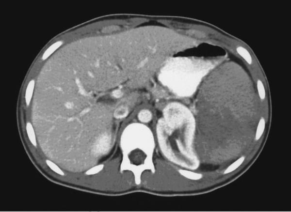
CT of the abdomen in a patient with sickle cell disease. Shown are splenic infarcts.
![CT of the orbit in a patient with sickle cell disease. Shown is orbital wall infarction due to vaso-occlusive crisis. There is periorbital soft tissue edema.[1]](/index.php/File:SCD_CT1.jpg)
![CT of the head in a patient with sickle cell disease. Shown is a subarachnoid hemorrhage.[1]](/index.php/File:SCD_CT2.jpg)

References
- ↑ 1.0 1.1 Brandow AM, Liem R (2011). ""Sickle Cell Disease in the Emergency Department: Atypical Complications and Management"". Clin Pediatr Emerg Med. 12 (3): 202–212. doi:10.1016/j.cpem.2011.07.003. PMC 3172721. PMID 21927581.