Q fever other chest x ray: Difference between revisions
Jump to navigation
Jump to search
Ahmed Younes (talk | contribs) No edit summary |
m (Bot: Removing from Primary care) |
||
| (5 intermediate revisions by 4 users not shown) | |||
| Line 4: | Line 4: | ||
==Overview== | ==Overview== | ||
On | On chest [[X-rays]], Q fever is characterized by either signs of [[atypical pneumonia]] (hazy, non-localized airspace [[Opacity|opacities]]), or in fewer cases, signs of [[Pneumonia|typical pneumonia]] ([[Consolidation (medicine)|lobar consolidation]] and occasional [[Pleural effusion|pleural effusions]]). | ||
==Chest X Ray== | ==Chest X-Ray== | ||
*In acute Q fever, X ray may show signs of atypical pneumonia (hazy non localized airspace opacities) and in some cases, it shows all the signs of typical pneumonia (lobar consolidation and occasional pleural effusions) | *In acute Q fever, [[X-ray]] may show signs of [[atypical pneumonia]] (hazy, non-localized airspace opacities) and in some cases, it shows all the signs of [[Pneumonia|typical pneumonia]] ([[Consolidation (medicine)|lobar consolidation]] and occasional [[Pleural effusion|pleural effusions]]) | ||
*In chronic Q fever, interstitial fiibrosis can be seen. | *In chronic Q fever, [[Pulmonary fibrosis|interstitial fiibrosis]] can be seen. | ||
{| class="wikitable" | {| class="wikitable" | ||
! | ! | ||
| Line 17: | Line 17: | ||
|- | |- | ||
| colspan="2" | | | colspan="2" | | ||
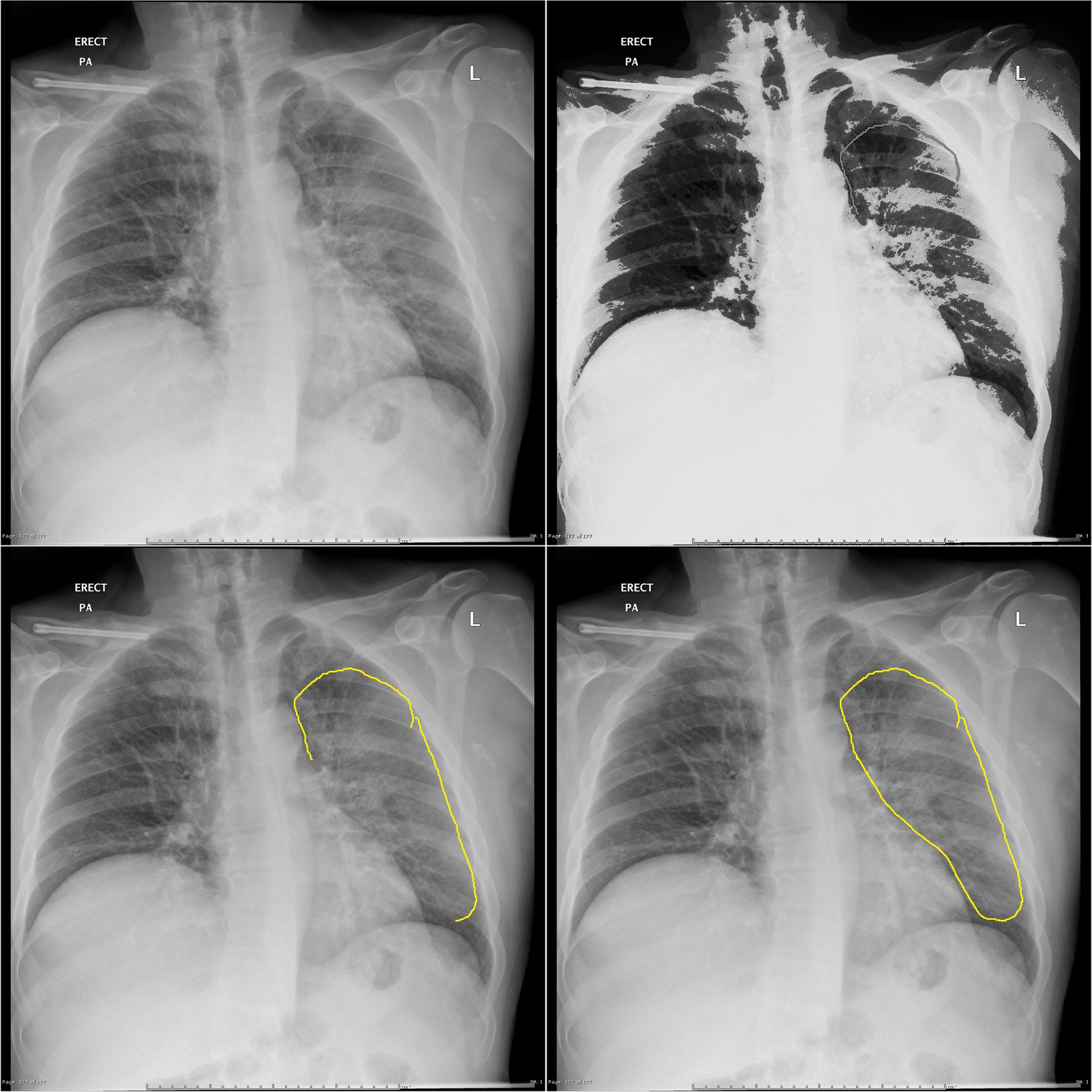
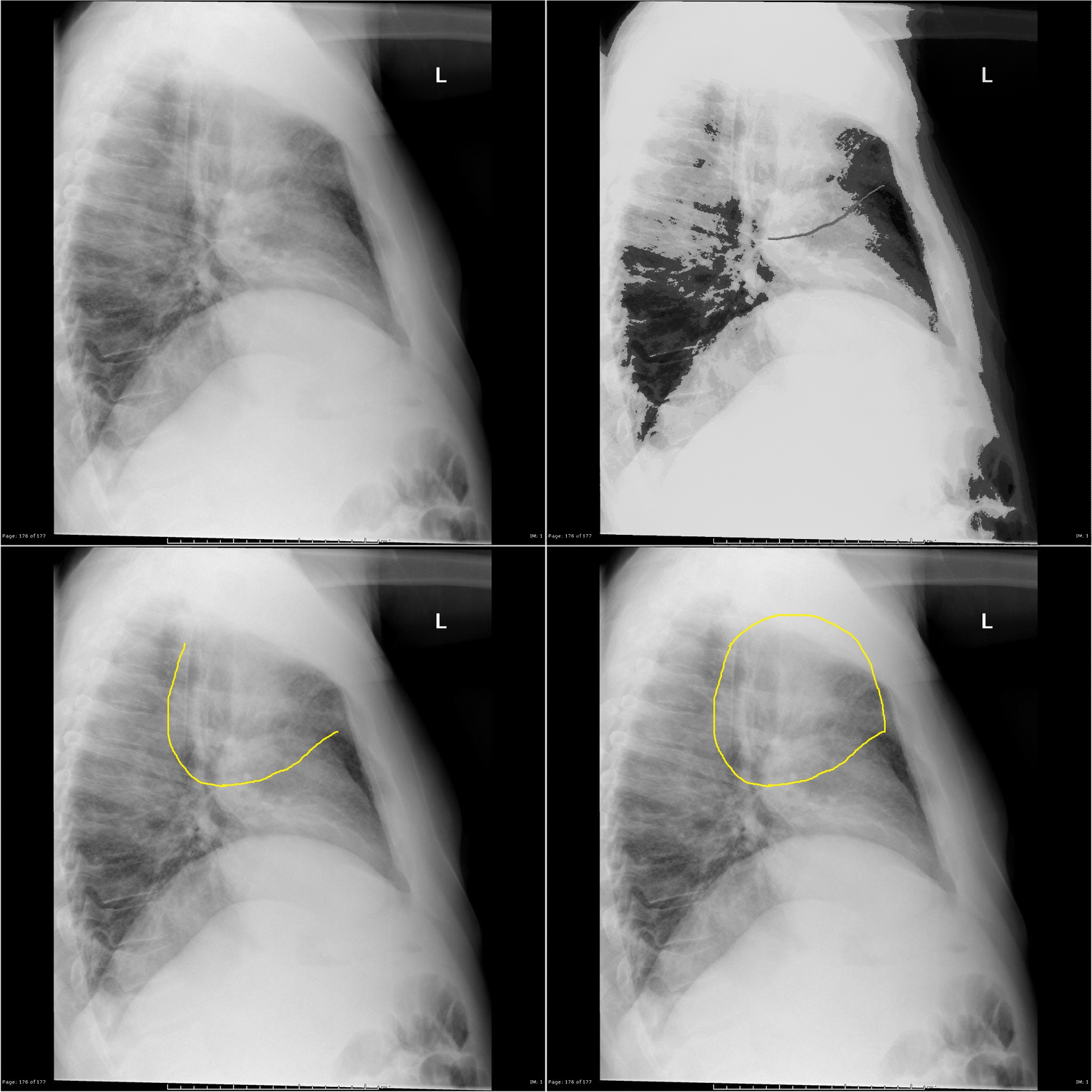
* Lateral and PA chest | * Lateral and PA [[Chest X-ray|chest X-ray]] for a 50 year old male patient presenting with [[fever]] and [[Respiratory failure|respiratory compromise]]. Lab tests showed [[Liver function tests|elevated liver function tests]] and [[pancytopenia]]. | ||
* X ray shows elevated right diaphragmatic copula and haziness in the left lung located in the middle and upper zones without demarcated consolidation. | * [[X-ray]] shows elevated right diaphragmatic copula and haziness in the left lung located in the middle and upper zones without demarcated [[Consolidation (medicine)|consolidation]]. | ||
|} | |} | ||
| Line 24: | Line 24: | ||
==References== | ==References== | ||
{{Reflist|2}} | {{Reflist|2}} | ||
{{WikiDoc Help Menu}} | |||
{{WikiDoc Sources}} | |||
[[Category:Needs content]] | [[Category:Needs content]] | ||
[[Category:Bacterial diseases]] | |||
[[Category:Emergency mdicine]] | |||
[[Category:Disease]] | |||
[[Category:Up-To-Date]] | |||
[[Category:Infectious disease]] | [[Category:Infectious disease]] | ||
[[Category: | [[Category:Gastroenterology]] | ||
[[Category:Hepatology]] | |||
[[Category:Pulmonology]] | |||
Latest revision as of 23:55, 29 July 2020
|
Q fever Microchapters |
|
Diagnosis |
|---|
|
Treatment |
|
Case Studies |
|
Q fever other chest x ray On the Web |
|
American Roentgen Ray Society Images of Q fever other chest x ray |
|
Risk calculators and risk factors for Q fever other chest x ray |
Editor-In-Chief: C. Michael Gibson, M.S., M.D. [1];Associate Editor(s)-in-Chief: Ahmed Younes M.B.B.CH [2]
Overview
On chest X-rays, Q fever is characterized by either signs of atypical pneumonia (hazy, non-localized airspace opacities), or in fewer cases, signs of typical pneumonia (lobar consolidation and occasional pleural effusions).
Chest X-Ray
- In acute Q fever, X-ray may show signs of atypical pneumonia (hazy, non-localized airspace opacities) and in some cases, it shows all the signs of typical pneumonia (lobar consolidation and occasional pleural effusions)
- In chronic Q fever, interstitial fiibrosis can be seen.
 |
 |
|---|---|
| |