Peritonsillar abscess CT scan: Difference between revisions
Jump to navigation
Jump to search
Prince Djan (talk | contribs) |
Prince Djan (talk | contribs) |
||
| Line 7: | Line 7: | ||
==CT scan== | ==CT scan== | ||
Imaging is helpful in differentiating peritonsillar abscess from peritonsillar [[cellulitis]] as well as a guide during [[abscess]] drainage.<ref name="pmid22687177">{{cite journal| author=Costantino TG, Satz WA, Dehnkamp W, Goett H| title=Randomized trial comparing intraoral ultrasound to landmark-based needle aspiration in patients with suspected peritonsillar abscess. | journal=Acad Emerg Med | year= 2012 | volume= 19 | issue= 6 | pages= 626-31 | pmid=22687177 | doi=10.1111/j.1553-2712.2012.01380.x | pmc= | url=https://www.ncbi.nlm.nih.gov/entrez/eutils/elink.fcgi?dbfrom=pubmed&tool=sumsearch.org/cite&retmode=ref&cmd=prlinks&id=22687177 }} </ref><ref name="pmid26637999">{{cite journal| author=Bandarkar AN, Adeyiga AO, Fordham MT, Preciado D, Reilly BK| title=Tonsil ultrasound: technical approach and spectrum of pediatric peritonsillar infections. | journal=Pediatr Radiol | year= 2016 | volume= 46 | issue= 7 | pages= 1059-67 | pmid=26637999 | doi=10.1007/s00247-015-3505-7 | pmc= | url=https://www.ncbi.nlm.nih.gov/entrez/eutils/elink.fcgi?dbfrom=pubmed&tool=sumsearch.org/cite&retmode=ref&cmd=prlinks&id=26637999 }} </ref><ref name="pmid8141026">{{cite journal| author=Buckley AR, Moss EH, Blokmanis A| title=Diagnosis of peritonsillar abscess: value of intraoral sonography. | journal=AJR Am J Roentgenol | year= 1994 | volume= 162 | issue= 4 | pages= 961-4 | pmid=8141026 | doi=10.2214/ajr.162.4.8141026 | pmc= | url=https://www.ncbi.nlm.nih.gov/entrez/eutils/elink.fcgi?dbfrom=pubmed&tool=sumsearch.org/cite&retmode=ref&cmd=prlinks&id=8141026 }} </ref><ref name="pmid7630286">{{cite journal| author=Strong EB, Woodward PJ, Johnson LP| title=Intraoral ultrasound evaluation of peritonsillar abscess. | journal=Laryngoscope | year= 1995 | volume= 105 | issue= 8 Pt 1 | pages= 779-82 | pmid=7630286 | doi=10.1288/00005537-199508000-00002 | pmc= | url=https://www.ncbi.nlm.nih.gov/entrez/eutils/elink.fcgi?dbfrom=pubmed&tool=sumsearch.org/cite&retmode=ref&cmd=prlinks&id=7630286 }} </ref><ref name="pmid12671820">{{cite journal| author=Blaivas M, Theodoro D, Duggal S| title=Ultrasound-guided drainage of peritonsillar abscess by the emergency physician. | journal=Am J Emerg Med | year= 2003 | volume= 21 | issue= 2 | pages= 155-8 | pmid=12671820 | doi=10.1053/ajem.2003.50029 | pmc= | url=https://www.ncbi.nlm.nih.gov/entrez/eutils/elink.fcgi?dbfrom=pubmed&tool=sumsearch.org/cite&retmode=ref&cmd=prlinks&id=12671820 }} </ref> | *Imaging is helpful in differentiating peritonsillar abscess from peritonsillar [[cellulitis]] as well as a guide during [[abscess]] drainage.<ref name="pmid22687177">{{cite journal| author=Costantino TG, Satz WA, Dehnkamp W, Goett H| title=Randomized trial comparing intraoral ultrasound to landmark-based needle aspiration in patients with suspected peritonsillar abscess. | journal=Acad Emerg Med | year= 2012 | volume= 19 | issue= 6 | pages= 626-31 | pmid=22687177 | doi=10.1111/j.1553-2712.2012.01380.x | pmc= | url=https://www.ncbi.nlm.nih.gov/entrez/eutils/elink.fcgi?dbfrom=pubmed&tool=sumsearch.org/cite&retmode=ref&cmd=prlinks&id=22687177 }} </ref><ref name="pmid26637999">{{cite journal| author=Bandarkar AN, Adeyiga AO, Fordham MT, Preciado D, Reilly BK| title=Tonsil ultrasound: technical approach and spectrum of pediatric peritonsillar infections. | journal=Pediatr Radiol | year= 2016 | volume= 46 | issue= 7 | pages= 1059-67 | pmid=26637999 | doi=10.1007/s00247-015-3505-7 | pmc= | url=https://www.ncbi.nlm.nih.gov/entrez/eutils/elink.fcgi?dbfrom=pubmed&tool=sumsearch.org/cite&retmode=ref&cmd=prlinks&id=26637999 }} </ref><ref name="pmid8141026">{{cite journal| author=Buckley AR, Moss EH, Blokmanis A| title=Diagnosis of peritonsillar abscess: value of intraoral sonography. | journal=AJR Am J Roentgenol | year= 1994 | volume= 162 | issue= 4 | pages= 961-4 | pmid=8141026 | doi=10.2214/ajr.162.4.8141026 | pmc= | url=https://www.ncbi.nlm.nih.gov/entrez/eutils/elink.fcgi?dbfrom=pubmed&tool=sumsearch.org/cite&retmode=ref&cmd=prlinks&id=8141026 }} </ref><ref name="pmid7630286">{{cite journal| author=Strong EB, Woodward PJ, Johnson LP| title=Intraoral ultrasound evaluation of peritonsillar abscess. | journal=Laryngoscope | year= 1995 | volume= 105 | issue= 8 Pt 1 | pages= 779-82 | pmid=7630286 | doi=10.1288/00005537-199508000-00002 | pmc= | url=https://www.ncbi.nlm.nih.gov/entrez/eutils/elink.fcgi?dbfrom=pubmed&tool=sumsearch.org/cite&retmode=ref&cmd=prlinks&id=7630286 }} </ref><ref name="pmid12671820">{{cite journal| author=Blaivas M, Theodoro D, Duggal S| title=Ultrasound-guided drainage of peritonsillar abscess by the emergency physician. | journal=Am J Emerg Med | year= 2003 | volume= 21 | issue= 2 | pages= 155-8 | pmid=12671820 | doi=10.1053/ajem.2003.50029 | pmc= | url=https://www.ncbi.nlm.nih.gov/entrez/eutils/elink.fcgi?dbfrom=pubmed&tool=sumsearch.org/cite&retmode=ref&cmd=prlinks&id=12671820 }} </ref> | ||
CT scan is helpful in defining the characteristics of the [[abscess]] as well as to classify it. It also helps in guiding the possible complications. For example patients with inferior cap-type abscess are at highest incidence of extraperitonsillar spread than the other categories of peritonsillar abscess.<ref name="pmid26527518">{{cite journal| author=Kawabata M, Umakoshi M, Makise T, Miyashita K, Harada M, Nagano H et al.| title=Clinical classification of peritonsillar abscess based on CT and indications for immediate abscess tonsillectomy. | journal=Auris Nasus Larynx | year= 2016 | volume= 43 | issue= 2 | pages= 182-6 | pmid=26527518 | doi=10.1016/j.anl.2015.09.014 | pmc= | url=https://www.ncbi.nlm.nih.gov/entrez/eutils/elink.fcgi?dbfrom=pubmed&tool=sumsearch.org/cite&retmode=ref&cmd=prlinks&id=26527518 }} </ref> | *CT scan is helpful in defining the characteristics of the [[abscess]] as well as to classify it. | ||
*It also helps in guiding the possible complications. For example patients with inferior cap-type abscess are at highest incidence of extraperitonsillar spread than the other categories of peritonsillar abscess.<ref name="pmid26527518">{{cite journal| author=Kawabata M, Umakoshi M, Makise T, Miyashita K, Harada M, Nagano H et al.| title=Clinical classification of peritonsillar abscess based on CT and indications for immediate abscess tonsillectomy. | journal=Auris Nasus Larynx | year= 2016 | volume= 43 | issue= 2 | pages= 182-6 | pmid=26527518 | doi=10.1016/j.anl.2015.09.014 | pmc= | url=https://www.ncbi.nlm.nih.gov/entrez/eutils/elink.fcgi?dbfrom=pubmed&tool=sumsearch.org/cite&retmode=ref&cmd=prlinks&id=26527518 }} </ref> | |||
[[Coronal]] contrast-enhanced CT scan of the neck may identify the peritonsillar abscess<ref name="pmid26637999">{{cite journal| author=Bandarkar AN, Adeyiga AO, Fordham MT, Preciado D, Reilly BK| title=Tonsil ultrasound: technical approach and spectrum of pediatric peritonsillar infections. | journal=Pediatr Radiol | year= 2016 | volume= 46 | issue= 7 | pages= 1059-67 | pmid=26637999 | doi=10.1007/s00247-015-3505-7 | pmc= | url=https://www.ncbi.nlm.nih.gov/entrez/eutils/elink.fcgi?dbfrom=pubmed&tool=sumsearch.org/cite&retmode=ref&cmd=prlinks&id=26637999 }} </ref> however, the use of CT scan is associated with a clinically significant delay in time to an [[otolaryngology]] consultation, time to admission, and time to bedside procedure.<ref name="pmid27533126">{{cite journal| author=Grant MC, Guarisco JL| title=Association Between Computed Tomographic Scan and Timing and Treatment of Peritonsillar Abscess in Children. | journal=JAMA Otolaryngol Head Neck Surg | year= 2016 | volume= 142 | issue= 11 | pages= 1051-1055 | pmid=27533126 | doi=10.1001/jamaoto.2016.2035 | pmc= | url=https://www.ncbi.nlm.nih.gov/entrez/eutils/elink.fcgi?dbfrom=pubmed&tool=sumsearch.org/cite&retmode=ref&cmd=prlinks&id=27533126 }} </ref> | *[[Coronal]] contrast-enhanced CT scan of the neck may identify the peritonsillar abscess<ref name="pmid26637999">{{cite journal| author=Bandarkar AN, Adeyiga AO, Fordham MT, Preciado D, Reilly BK| title=Tonsil ultrasound: technical approach and spectrum of pediatric peritonsillar infections. | journal=Pediatr Radiol | year= 2016 | volume= 46 | issue= 7 | pages= 1059-67 | pmid=26637999 | doi=10.1007/s00247-015-3505-7 | pmc= | url=https://www.ncbi.nlm.nih.gov/entrez/eutils/elink.fcgi?dbfrom=pubmed&tool=sumsearch.org/cite&retmode=ref&cmd=prlinks&id=26637999 }} </ref> however, the use of CT scan is associated with a clinically significant delay in time to an [[otolaryngology]] consultation, time to admission, and time to bedside procedure.<ref name="pmid27533126">{{cite journal| author=Grant MC, Guarisco JL| title=Association Between Computed Tomographic Scan and Timing and Treatment of Peritonsillar Abscess in Children. | journal=JAMA Otolaryngol Head Neck Surg | year= 2016 | volume= 142 | issue= 11 | pages= 1051-1055 | pmid=27533126 | doi=10.1001/jamaoto.2016.2035 | pmc= | url=https://www.ncbi.nlm.nih.gov/entrez/eutils/elink.fcgi?dbfrom=pubmed&tool=sumsearch.org/cite&retmode=ref&cmd=prlinks&id=27533126 }} </ref> | ||
*CT scan may show diffuse hypodense lesion with rim enhancement in the peritonsillar space.<ref name="pmid24742512">{{cite journal| author=Chen Y, Yang Q, Wang T, Li J, Ye J, Liu X et al.| title=[Application of enhanced CT in the differential diagnosis of peritonsillar abscess and intratonsillar abscess]. | journal=Zhonghua Er Bi Yan Hou Tou Jing Wai Ke Za Zhi | year= 2014 | volume= 49 | issue= 2 | pages= 131-5 | pmid=24742512 | doi= | pmc= | url=https://www.ncbi.nlm.nih.gov/entrez/eutils/elink.fcgi?dbfrom=pubmed&tool=sumsearch.org/cite&retmode=ref&cmd=prlinks&id=24742512 }} </ref> | |||
CT scan may show diffuse hypodense lesion with rim enhancement in the peritonsillar space.<ref name="pmid24742512">{{cite journal| author=Chen Y, Yang Q, Wang T, Li J, Ye J, Liu X et al.| title=[Application of enhanced CT in the differential diagnosis of peritonsillar abscess and intratonsillar abscess]. | journal=Zhonghua Er Bi Yan Hou Tou Jing Wai Ke Za Zhi | year= 2014 | volume= 49 | issue= 2 | pages= 131-5 | pmid=24742512 | doi= | pmc= | url=https://www.ncbi.nlm.nih.gov/entrez/eutils/elink.fcgi?dbfrom=pubmed&tool=sumsearch.org/cite&retmode=ref&cmd=prlinks&id=24742512 }} </ref> | |||
Revision as of 03:21, 6 March 2017
|
Peritonsillar abscess Microchapters |
|
Diagnosis |
|
Treatment |
|
Case Studies |
|
Peritonsillar abscess CT scan On the Web |
|
American Roentgen Ray Society Images of Peritonsillar abscess CT scan |
|
Risk calculators and risk factors for Peritonsillar abscess CT scan |
Editor-In-Chief: C. Michael Gibson, M.S., M.D. [1] Associate Editor(s)-in-Chief: Prince Tano Djan, BSc, MBChB [2]
Overview
CT scan
- Imaging is helpful in differentiating peritonsillar abscess from peritonsillar cellulitis as well as a guide during abscess drainage.[1][2][3][4][5]
- CT scan is helpful in defining the characteristics of the abscess as well as to classify it.
- It also helps in guiding the possible complications. For example patients with inferior cap-type abscess are at highest incidence of extraperitonsillar spread than the other categories of peritonsillar abscess.[6]
- Coronal contrast-enhanced CT scan of the neck may identify the peritonsillar abscess[2] however, the use of CT scan is associated with a clinically significant delay in time to an otolaryngology consultation, time to admission, and time to bedside procedure.[7]
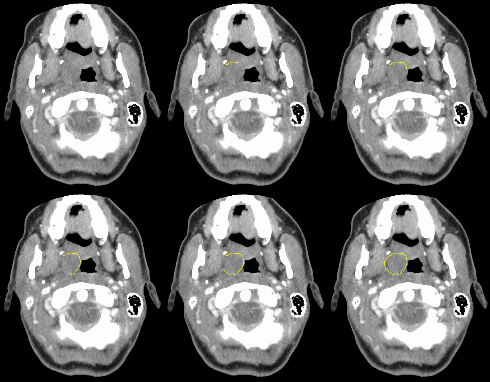
- CT scan may show diffuse hypodense lesion with rim enhancement in the peritonsillar space.[8]
The image above shows a CT scan of right-sided peritonsillar abscess[9]
References
- ↑ Costantino TG, Satz WA, Dehnkamp W, Goett H (2012). "Randomized trial comparing intraoral ultrasound to landmark-based needle aspiration in patients with suspected peritonsillar abscess". Acad Emerg Med. 19 (6): 626–31. doi:10.1111/j.1553-2712.2012.01380.x. PMID 22687177.
- ↑ 2.0 2.1 Bandarkar AN, Adeyiga AO, Fordham MT, Preciado D, Reilly BK (2016). "Tonsil ultrasound: technical approach and spectrum of pediatric peritonsillar infections". Pediatr Radiol. 46 (7): 1059–67. doi:10.1007/s00247-015-3505-7. PMID 26637999.
- ↑ Buckley AR, Moss EH, Blokmanis A (1994). "Diagnosis of peritonsillar abscess: value of intraoral sonography". AJR Am J Roentgenol. 162 (4): 961–4. doi:10.2214/ajr.162.4.8141026. PMID 8141026.
- ↑ Strong EB, Woodward PJ, Johnson LP (1995). "Intraoral ultrasound evaluation of peritonsillar abscess". Laryngoscope. 105 (8 Pt 1): 779–82. doi:10.1288/00005537-199508000-00002. PMID 7630286.
- ↑ Blaivas M, Theodoro D, Duggal S (2003). "Ultrasound-guided drainage of peritonsillar abscess by the emergency physician". Am J Emerg Med. 21 (2): 155–8. doi:10.1053/ajem.2003.50029. PMID 12671820.
- ↑ Kawabata M, Umakoshi M, Makise T, Miyashita K, Harada M, Nagano H; et al. (2016). "Clinical classification of peritonsillar abscess based on CT and indications for immediate abscess tonsillectomy". Auris Nasus Larynx. 43 (2): 182–6. doi:10.1016/j.anl.2015.09.014. PMID 26527518.
- ↑ Grant MC, Guarisco JL (2016). "Association Between Computed Tomographic Scan and Timing and Treatment of Peritonsillar Abscess in Children". JAMA Otolaryngol Head Neck Surg. 142 (11): 1051–1055. doi:10.1001/jamaoto.2016.2035. PMID 27533126.
- ↑ Chen Y, Yang Q, Wang T, Li J, Ye J, Liu X; et al. (2014). "[Application of enhanced CT in the differential diagnosis of peritonsillar abscess and intratonsillar abscess]". Zhonghua Er Bi Yan Hou Tou Jing Wai Ke Za Zhi. 49 (2): 131–5. PMID 24742512.
- ↑ Template:Https://commons.wikimedia.org/wiki/File:PeritonsilarAbs.png Peritonsilar abscess Date 2 June 2016 Source Own work Author James Heilman, MD
